New entities in soft tissue pathology
***FET-CREB fusion sarcomas***
Variable histology with epithelioid/round cells showing an odd combination of multiple aberrant markers (CK, EMA, MUC4, ALK, CD30, synapto , etc.) is valuable for screening FET-CREB sarcomas
Half of cases are intra-abdominal
Wide age range
Highly aggressive
Will be included in the upcoming BST WHO book
FET-CREB is an umbrella term for fusions between FET genes (mainly EWSR1, sometimes FUS) and CREB family transcription factors (CREB1, ATF1, or CREM)
Pitfall: Angiomatoid fibrohistiocytoma (AFH) and FET-CREB sarcomas have a similar genotype, but morphology differs and, most importantly, AFH mortality is >1% versus >50% in FET-CREB sarcomas
Dr. Agaimy #USCAP2026 #pathology #PathX #PathTwitter
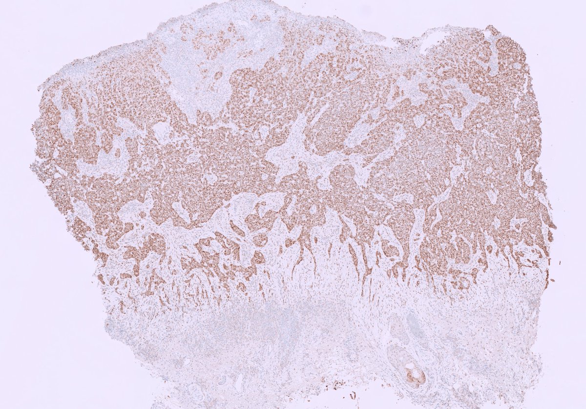
Our Gynecologic Pathology cases @UMiamiPathology are fascinating! Here’s a striking example of pilomatrix-like high-grade endometrial carcinoma (PiMHEC), a recently described entity mostly driven by CTNNB1 mutations. Look for solid basaloid growth with conspicuous central tumor cell necrosis, and pilomatrix-like keratinization with the hallmark ghost cells. This case also had a focal conventional FIGO grade 1 endometrioid component. IHC shows diffuse nuclear β-catenin with loss of PAX8 and ER in the pilomatrix-like component. These tumors are believed to behave aggressively.
2/ Dx: mammary Paget disease ... the name does not convey the histological picture of an in situ adenocarcinoma (DCIS) that is speading in the epidermis...
CK7 and p53 IHCs👇👇
Clinical implication: the patient has breast cancer, mostly DCIS, that has to be found and treated
Read this editorial titled "Extranodal extension in head and neck cancer: why HN-CLEAR matters and what still needs proof" by the authors - Dr Vikram Deshpande, Dr Munita Bal for an indepth understanding and clarity.
Link to the editorial https://t.co/5jP8eqCyg7
What’s inside the latest issue of JCP?
From evolving diagnostic dilemmas to rare case insights, the May 2026 issue brings together key reads across pathology.
Open Access Highlight: Autoimmune conditions & CMML outcomes.
If you read one paper today, make it this one!
#Pathology #HemePath #OncPath #OpenAccess #JCP #MedTwitter
@MaiElzieny@parthisandy 20.
Two fungal hyphae you might see in the lung are Aspergillus (green circle, more common) and Mucor (red circle, less conmon). Aspergillus has septa and branches at acute angles, Mucor has few or no septa and has more irregular shapes.
Not always easy to tell them apart!
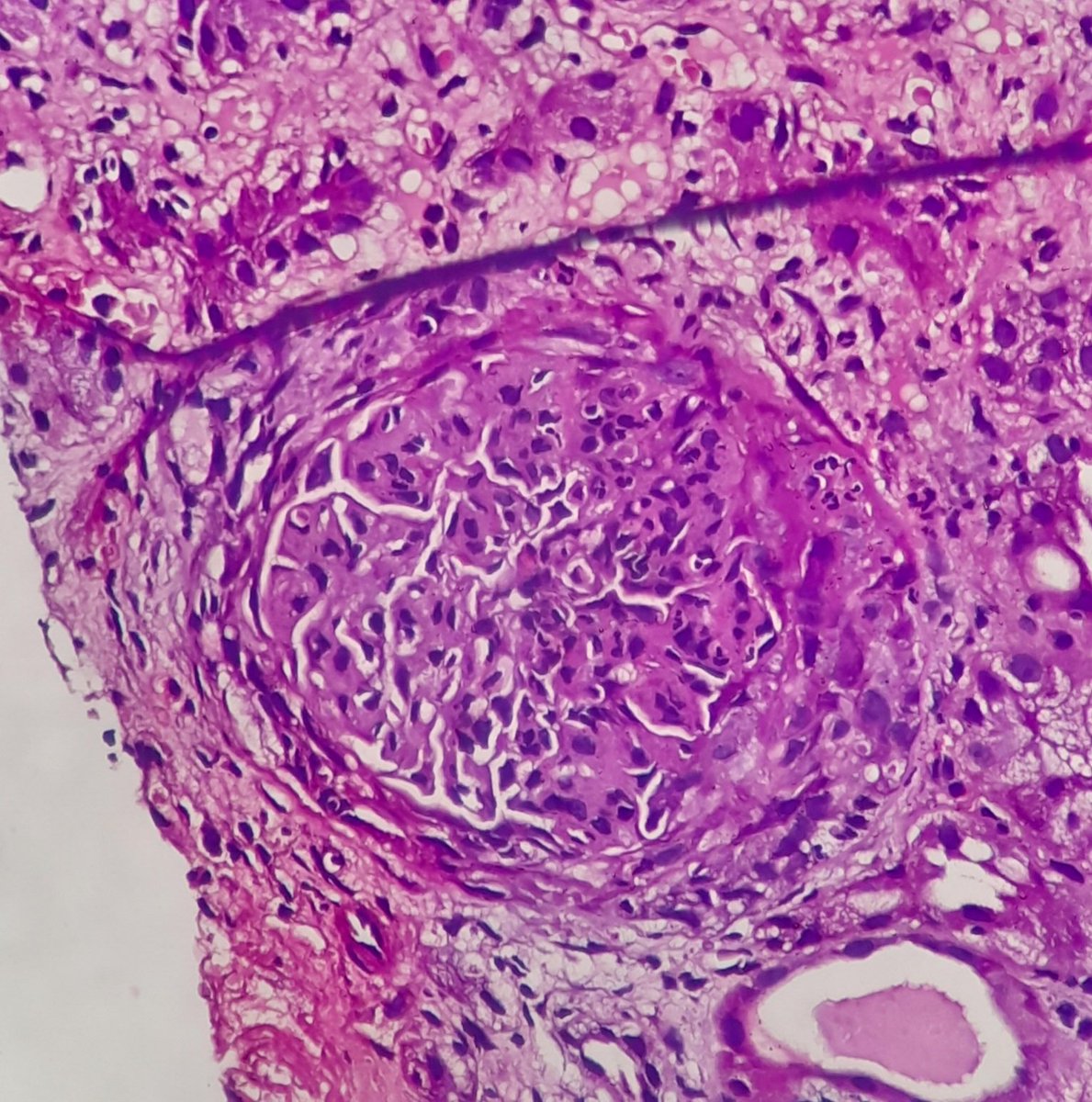
Difficult but good recent teaching case.
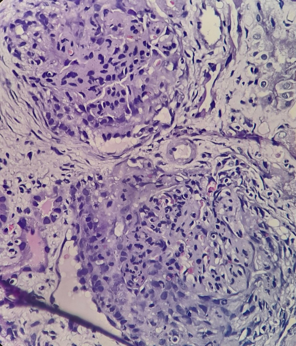
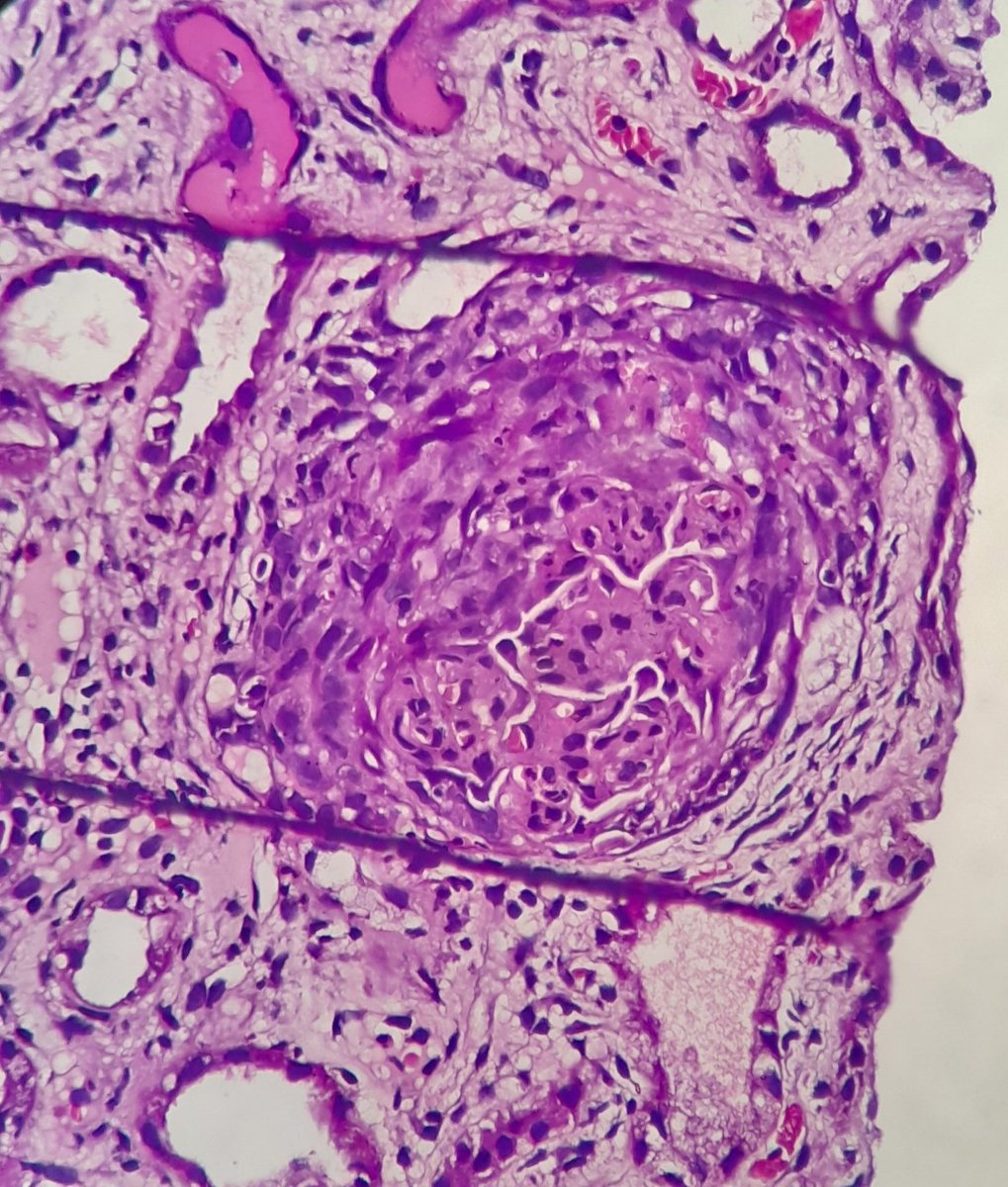
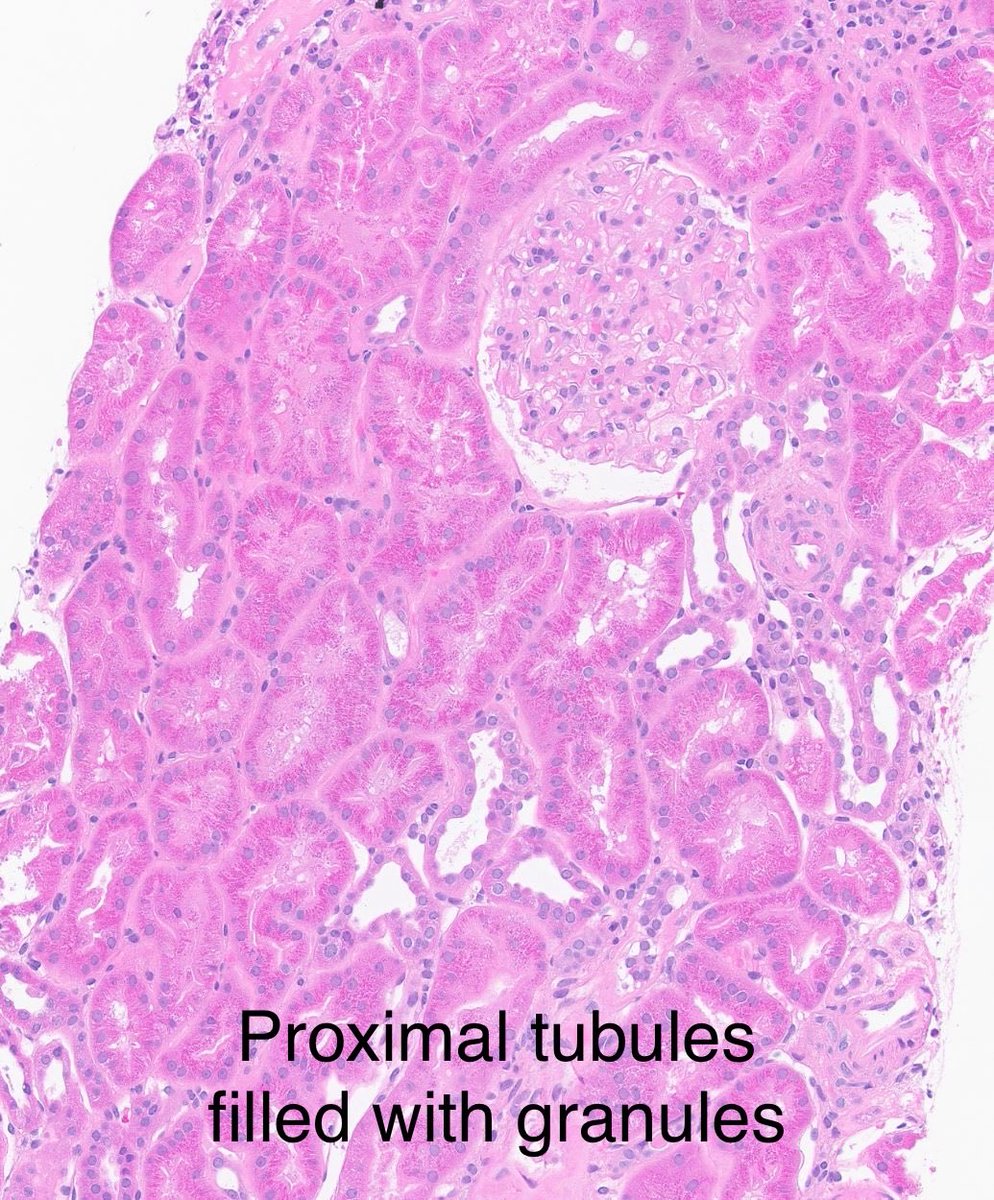
LM- normal appearing glomeruli but proximal tubules filled with eosinophilic granules.
IF- negative.
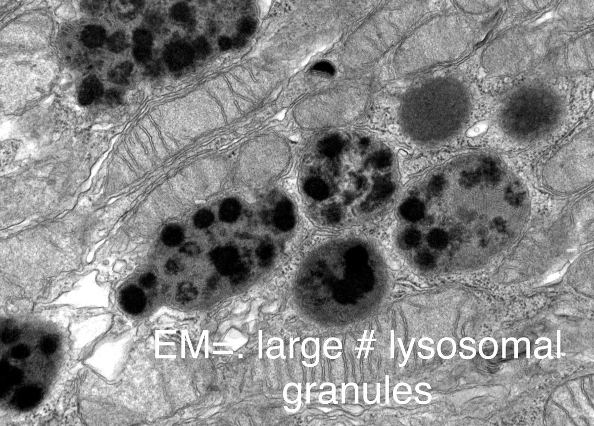
EM- large number of lysosomes, with degenerative changes.
Lysozyme stain 3+ positive in proximal tubules.
Dx: Lysozyme associated-tubulopathy (myelofibrosis/clinical).
Usually see this in patients with CML.
D/D: light chain proximal tubulopathy, but IF & pronase IF are negative.
60-yr old with chronic kidney disease, pulmonary nodules, and JAK2+ myelofibrosis.
Evaluation showed high levels of serum lysozyme, and lysozymuria.
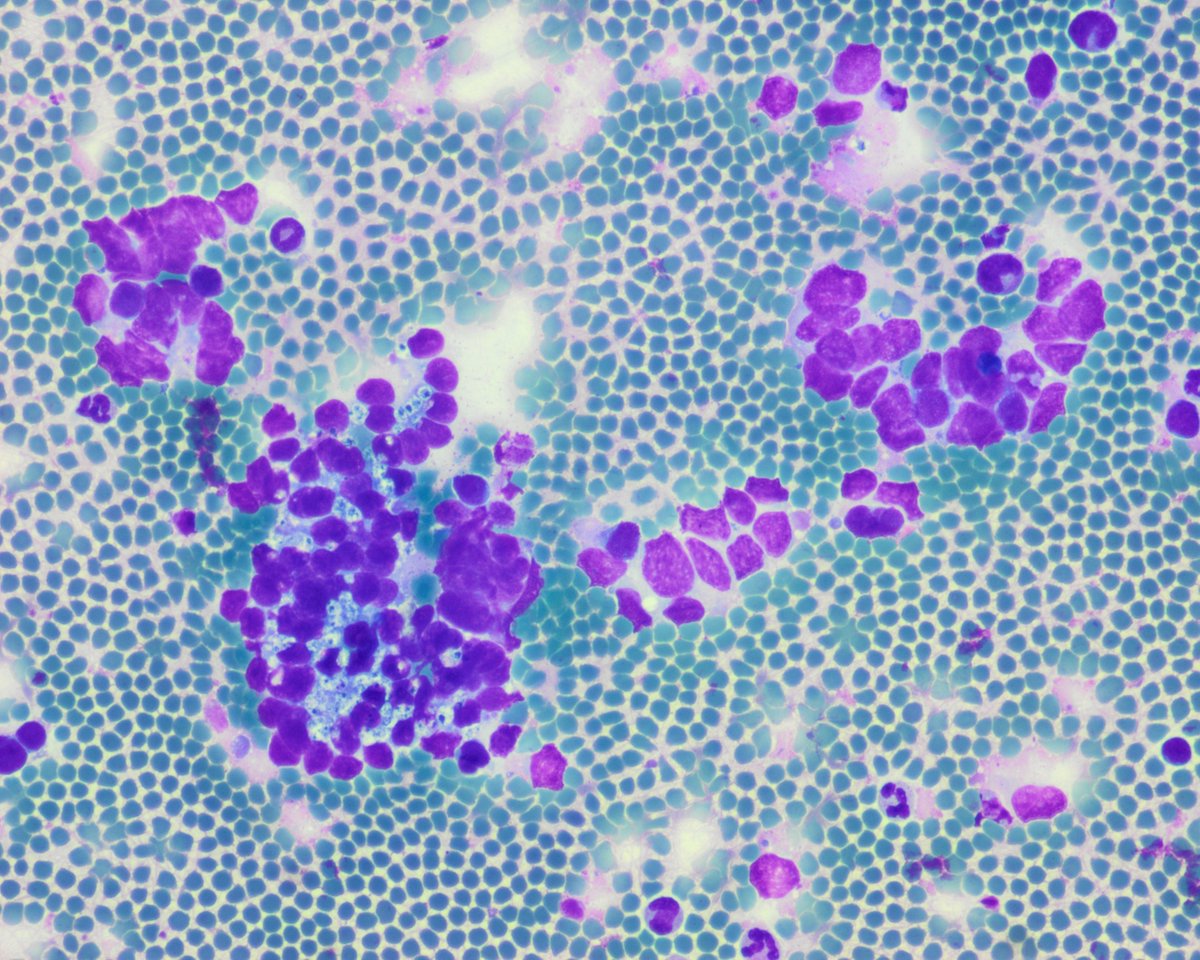
1st impression diagnosis?
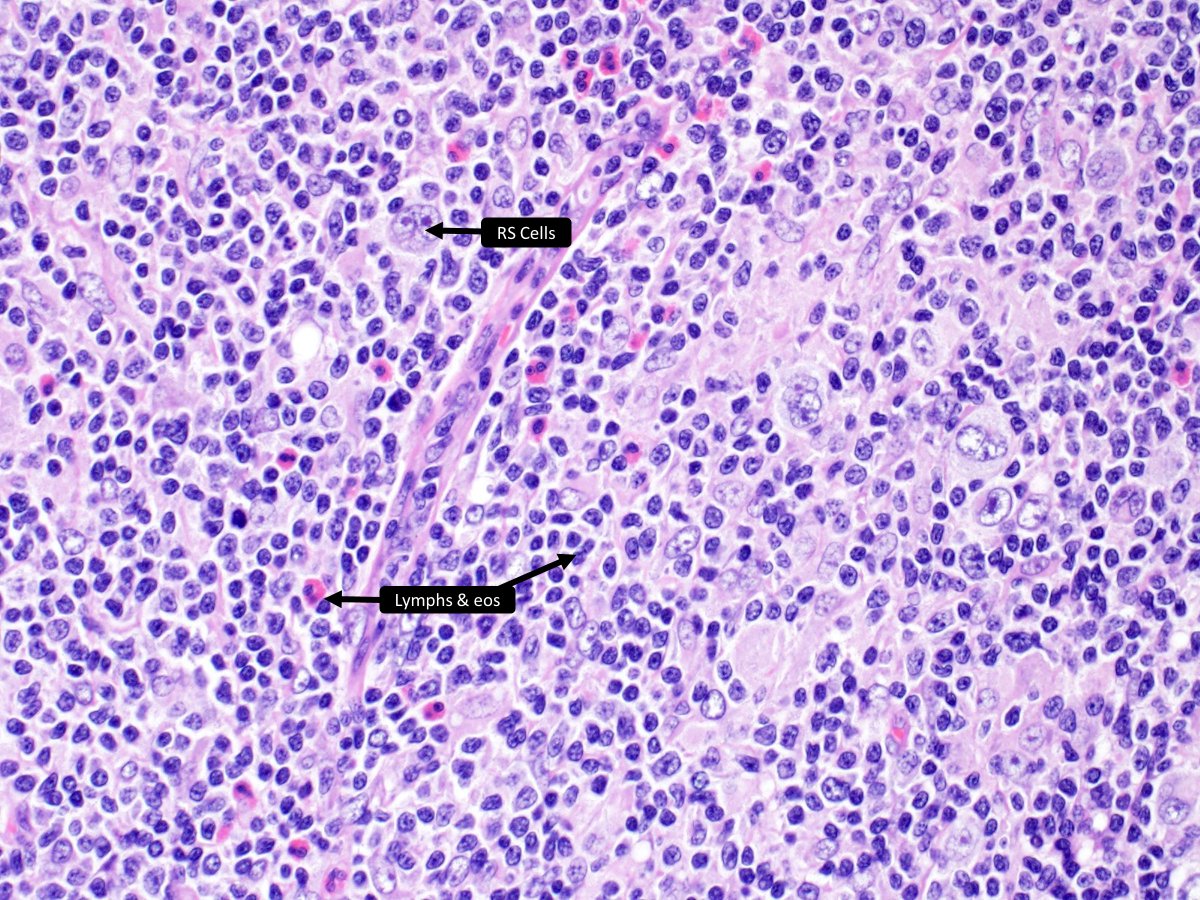
Mediastinal _____
a) nodular sclerosis CHL
b) NLP Hodgkin lymphoma
c) large B-cell lymphoma
d) grey zone lymphoma
#HemePath#PathTwitter#pathology
🔬https://t.co/PFtF9r22GP
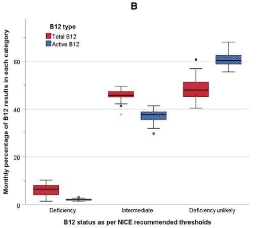
Better test ≠ better system.
This study shows that active B12:
🔍 Classifies more patients as deficiency unlikely
🔍 Reduces the grey “indeterminate” zone
Here’s the catch:
💸 It comes at a significantly higher cost ~26× more expensive !
💡 Take-home:
Better diagnostics don’t exist in isolation.
They live in real systems, with cost, access, and scale.
A great read if you’re thinking about lab medicine: https://t.co/bpMMsHMlvl.
#Pathology #LabMedicine #JCP