📢 New paper out in @NatureGenet!
🧬 A robust synthetic lethal interaction between CDS1 & CDS2 provides a therapeutic opportunity in uveal melanoma - as well as in other cancer types.
🔗 https://t.co/EV0Q7xVA75
👁️🧪⚔️ #CRISPR#SyntheticLethality#CancerResearch#UvealMelanoma
Huge congrats to Anjan for getting this out. Compensation between paralogs allows cancer cells to tolerate mutations that might otherwise be lethal. But what does this compensation look like? What are the proteins involved doing?
TP53:
- Discovered 45 years ago, most cited gene all time
- No therapies
- 500 million people currently living will die of TP53 mutant cancers without new therapies
Our Preprint:
- A general strategy for TP53 missense mutant cancers (majority) with prototype small molecules
It is an honour to share our @LawleyLab@sangerinstitute@ONJCRI work describing a high-resolution microbiome signature for combo immune checkpoint blockade (CICB) response, published today in @NatureMedicine! https://t.co/h7QjzBrWx9
A 🧵...
@nevillesanjana@danielevanbauer@zhangf thanks! I have to give a talk to non-CRISPR folk about my screen and this is a great example of how pooled CRISPR screens are fantastic for identifying therapeutic targets!
My job as a research fellow at one of the world's best universities (a requirement of which is a doctorate) pays less than this and so, on this definition, I am an unskilled worker who wouldn't be allowed into the country. This system is utterly ridiculous..
delighted to see @dekegelb's work out in @MolSystBiol! Tumors tolerate astonishing numbers of genetic alterations, including homozygous deletion of entire protein coding genes. How? Here we explored one potential explanation – paralog dispensability! https://t.co/yhQxPKBH0r
📢📢 Less than a week to go until #ESMO23⏳
@myESMO asked the Upper GI track chair 👩⚕️ to share some upcoming highlights below ⬇️
Whether you're on a ✈️, 🚝 or watching 💻 Friday 2pm CET is when the transformative Upper GI starts...see you in Madrid....
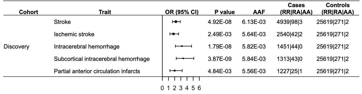
A new exciting preprint from my colleagues (Rodriguez-Flores et al.) at Regeneron Genetics Center reports the discovery of a major genetic risk factor of stroke in South Asians—a missense variant (Arg1231Cys) in NOTCH3 that is seen in ~1% of the Pakistanis but rare elsewhere in the world.
https://t.co/kHNKn45Skr
NOTCH3 is well known a well-known Mendelian gene for stroke. Pathogenic mutations in NOTCH3 cause an autosomal dominant stroke syndrome called CADASIL (Cerebral Autosomal Dominant Arteriopathy with Subcortical Infacts and Leukoencephalopathy).
NOTCH3 is one of those genes where our knowledge of the disease (CADASIL) is older than our knowledge of the gene itself. It is through the families that suffered from CADASIL we came to know of the existence of NOTCH3 in humans.
History of CADASIL
CADASIL has a fascinating history. The earliest case reports of CADASIL date back to the 1970s when multiple case reports of families suffering from a new type of stroke syndrome surfaced one by one. Each of the research groups came up with their own names for the syndrome based on the clinical features they observed: "hereditary multi-infarct dementia", "chronic familial vascular encephalopathy" etc. (https://t.co/UpPa8qrBsS).
Then they realized that all of these isolated reports were pointing to the same condition. All the patients suffered from early-onset recurrent strokes damaging the brain gradually resulting in dementia, and sensory and motor deficits resulting in death before 60 yrs of age. Looking at the postmortem brain tissues it became clear it's a disease of small blood vessels in the white matter.
Discovery of NOTCH3
In 1993, studying two French families with CADASIL, Tournier-Lasserve et al. triangulated the genomic region (19q12) holding the gene responsible for CADASIL and also coined the acronym CADASIL (https://t.co/QMLbWiVCsJ). Three years later, the same group successfully cloned the causative gene and realized it was a homologue of mouse Notch3 (https://t.co/cBDq5Sy5Ud).
Genetic architecture of CADASIL mutations
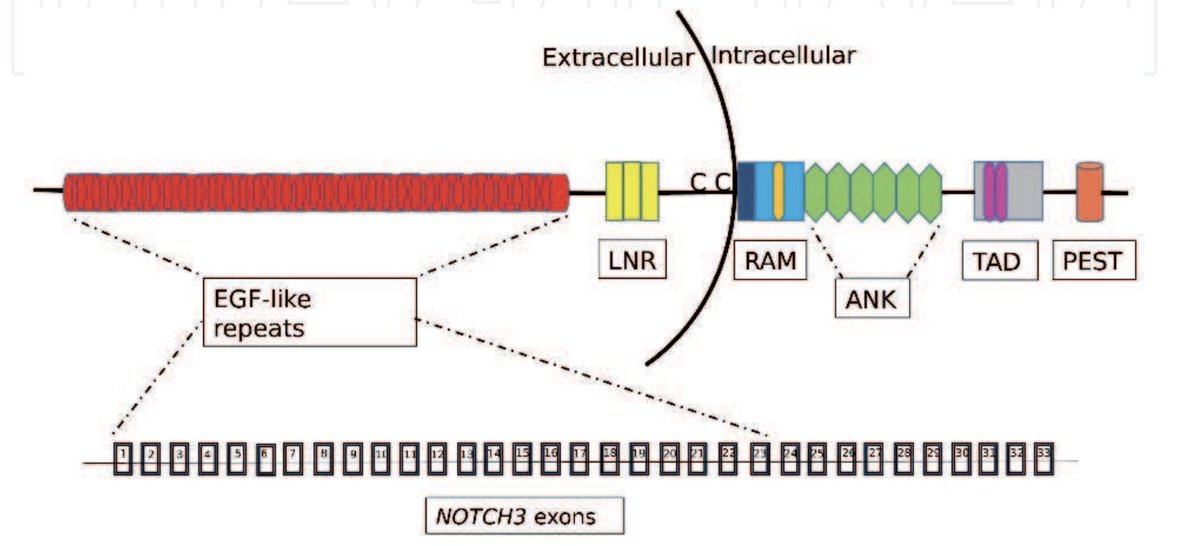
NOTCH3 is a huge protein with 2321 amino acids that gets chopped multiple times over its molecular life course. The most characteristic of NOTCH3 is its extracellular domain which contains 34 repeat units, each comprising six cysteine residues. Almost all the CADASIL mutations identified to date fall within these domains, either adding or deleting cysteine residues (cys-altering mutations) that interfere with dimerization resulting in a dominant negative pathology.(https://t.co/ckZz7W6CRZ).
What is exciting about the new discovery?
So far almost all the reported CADASIL mutations have been rare. The recent GWAS of stroke in more than 1 million individuals of predominantly Europeans found no signal around NOTCH3 (https://t.co/bSmZwgVNK2). But studying just 70k Pakistanis, my colleagues found a clear GWAS signal in NOTCH3 driven by a missense variant seen in as much as 1% of the Pakistanis with a large effect size (3.4 fold risk) suggesting this is probably the major genetic risk factor of stroke in South Asians.
If we had not looked at the South Asians and simply used European results to predict the stroke risk in South Asians, we would have missed the contribution of this important variant. This is a great demonstration of the value of studying non-European populations and a reminder that there are many more discoveries to be made in largely under-explored populations like South Asians.
This discovery was possible due to the RGC collaboration with Pakistani Genomic Resource built by Danish Saleheen and his team which has made tremendous contributions towards the advancement of South Asian genetics (https://t.co/cB2VFWAHDN).
Some recent posts:
1. Effect of consanguinuity on the risk of common diseases in South Asians (https://t.co/dTkPsl935K)
2. Discovery of the first genetic locus (ADRA2A) for Reynaud's phenomenon (https://t.co/zC7vGRmerb).
2. Gene by sex interactions of PNPLA3 I148M variant (https://t.co/RFegHHRS2z).
A fascinating work is published today @Nature by Zhang & Zhang et al. on a serendipitous discovery of haemoglobin expression outside the RBC cells--in chondrocytes--and their critical role in keeping our cartilage alive under an oxygen-deprived environment.
https://t.co/DPhuZN1JGE
Our school teachers taught that haemoglobin (Hb) is an oxygen-transporting protein expressed exclusively in the red blood cells. Although there exist occasional sporadic reports of Hb expression outside the RBCs (e.g. neurons, retinal cells etc.), no one would have guessed Hb would play a critical role (required for survival) outside the RBCs.
The discovery
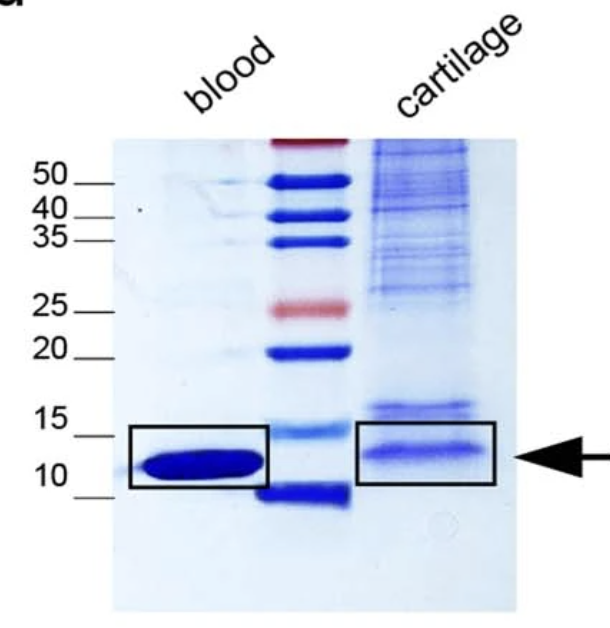
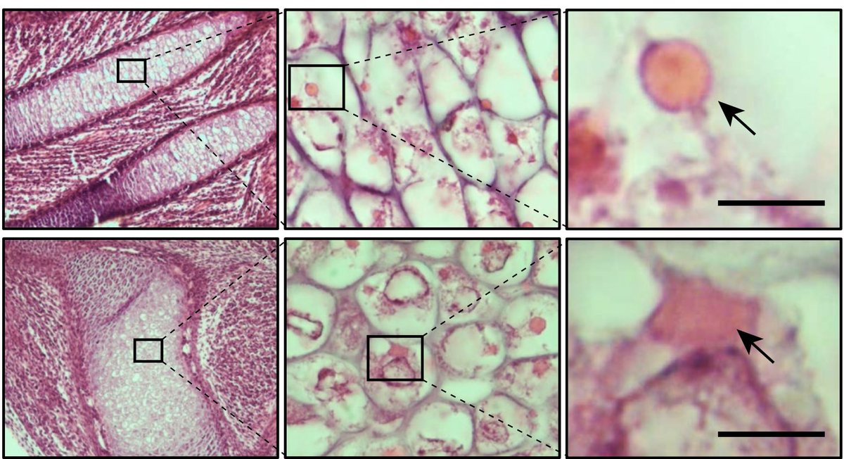
When studying the cartilage growth plate of neonatal mice, the authors noticed eosin-positive structures in the chondrocytes (cartilage cells) that resembled structures seen in RBCs. Out of curiosity, the researchers went on to stain and examine the chondrocytes of different cartilage tissue types both from mice and humans. They realized that no matter the source or species, the cells always displayed eosin-positive structures under the microscope.
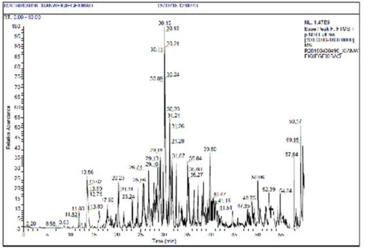
The curious researchers were determined to find out what these structures are made of. They carefully dissected these structures out and studied the protein components using mass spectrometry and were surprised to learn the results: the top hits were Hb proteins.
Unable to believe the results, they went on to study the proteins using different methods--western blotting, immunohistochemistry--and every time they ended up with the same results: the cytoplasm of cells was loaded with organelles-like bodies made of haemoglobin proteins. Finally, they came to the realization that Hb is abundantly produced in chondrocytes. They named these cytoplasmic Hb bodies as "Hedy".
Structure of Hedy
The authors studied the structure and formation of Hb bodies floating in the cytoplasm. Are they like an organelle? Do they have a membrane? Through various experiments, the researchers found that the Hedy structures do not have a membrane. The Hb proteins condense together by phase separation to form organelle-like structures in the cytoplasm. This condensation is itself an evolved process, requiring specific sequence structures of the Hb protein.
Globin switching
We know that there are different forms of Hb each expressed during different developmental stages: embryonic, fetal and adult Hb. There exists a sophisticated molecular machinery (which was believed to be RBC-specific) that switches one Hb type to the other at appropriate times. Using gene silencing experiments, the authors were further awestruck to find that the chondrocytes too switched their Hb types from embryonic to fetal to adult stages, just like RBCs!
Regulation of chondrocyte Hb production
It is well known that hypoxia induces Hb production via upregulating hypoxia-inducible factors (HIFs), an evolutionarily conserved molecular mechanism (Nobel Prize 2019; https://t.co/Sifi8LuYIX). But it turned out that chondrocytes have evolved to increase their Hb expression not via HIF proteins, but through a different protein, the same protein that is required for fetal to adult Hb switching: KLF1 (https://t.co/Zn0X8GpYdq).
How important is Hb for chondrocytes?
Such a high Hb expression in chondrocytes with similar globin switching behaviour as RBCs would mean that this Hb is critical for chondrocytes' survival. By deleting the Hbb gene specifically in the chondrocytes, the authors found that without Hb the chondrocytes die killing the animal a few days after birth.
Conclusion
Continuous oxygen supply is a prerequisite for the survival of cells in all tissues. The only way the cells can receive oxygen is through RBCs in the blood, which requires the tissue to be highly vascularized. When demand exceeds the supply, the cells evolve to survive an oxygen-depriving environment.
Muscles evolved to produce their own globin--myoglobin which has a higher affinity to oxygen than Hb thereby withholding O2 during oxygen excess states and releasing it back during oxygen-deprived state (during exercise). Likewise, the brain has its own globin: neuroglobin (https://t.co/6v78zvHajP).
Today, we are learning that cartilage (an avascular tissue), too, has its own globin. But unlike muscle and brain, have evolved to store oxygen not by making a new type of globin but by making just the same type as the ones in RBCs, but with a higher affinity than RBC Hb.
When it comes to fundamental biology, we often assume that we have found everything and then one day a discovery like this drops, hitting us on the head to make us realize that there is a whole universe of hidden biological secrets waiting to be discovered.
Some recent posts:
1. Gene x sex interaction of PNPLA3 I148M variant (https://t.co/RFegHHRS2z)
2. Effect of consanguineous marriage on the risk of common diseases in offsprings (https://t.co/dTkPsl935K)
3. Whole genome vs. Whole exome sequencing. Which is more cost-effective for genetic association studies? (https://t.co/BCwMzw4g16)