@MirunaPopescu13 Thank you very much for your feedback and the example. Maybe I am obsessing over the location of the nuclei, books generally show the perfect images that in the daily routine may vary.
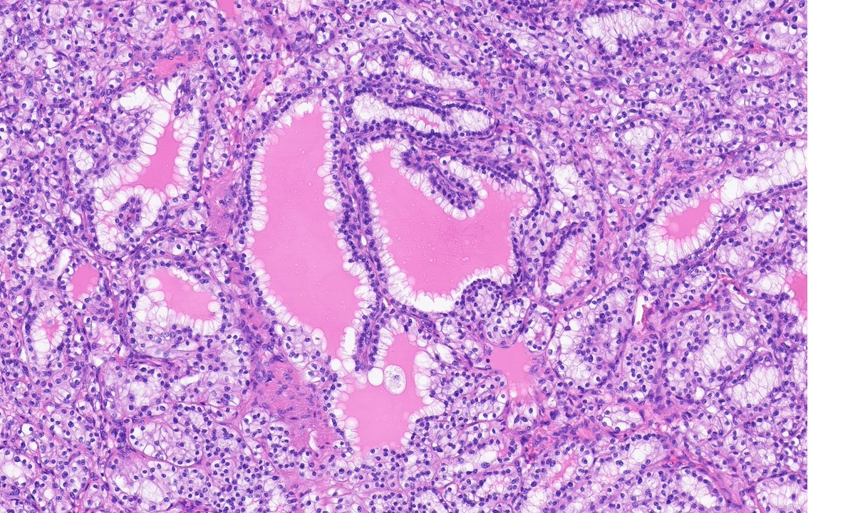
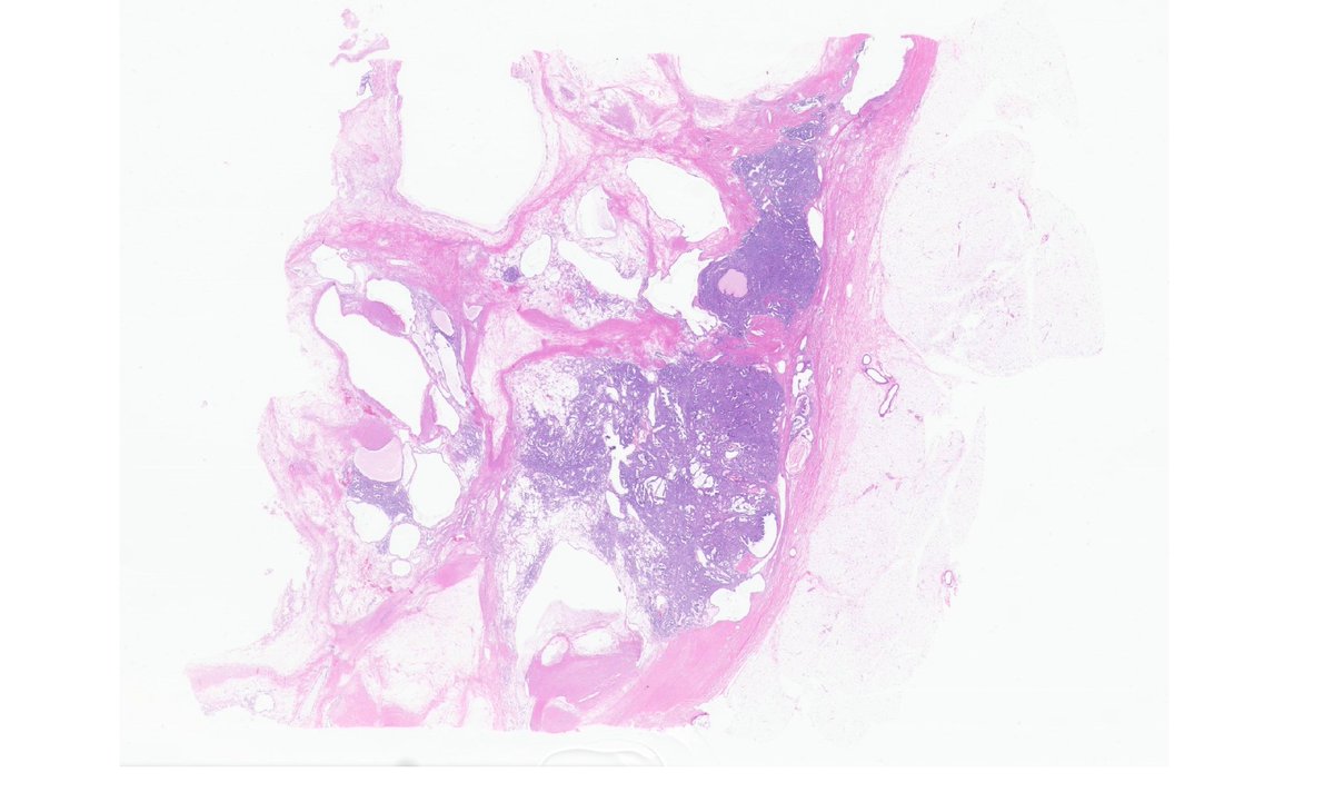
Need some help. 64yo woman with a solid-cystic encapsulated renal mass limited to kidney. Tubular and nested-cord-like architecture with clear cells basally/centrally located - not luminal (G2 max). Here some representative pics. IHQ on thread #GUpath#PathTwitter
IHQ favors a Clear cell papillary renal cell tumour over a CCRCC, but nucleus are not luminal but basally located. I do not see any high grade features. What are your thoughts?
IHQ (I do not have cathepsin K or TFE3):
- CAIX: diffuse cup-like (my interpretation)
- CK7: strongly positive, more heterogeneous in nested areas
- Vimentin: positive.
- Negative: CD10, AMACR, CK34BE12 and CD117.
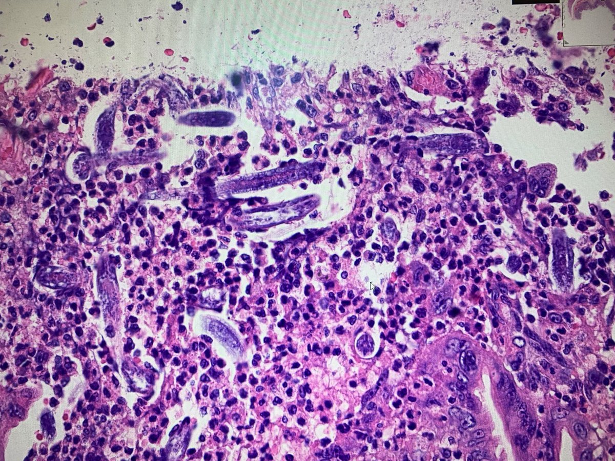
Ulcerative pancolitis in a 23 year-old male under infliximab and corticosteroid therapy. These are the reasons why his symptomatology exacerbated. Any guesses? #infectiouspathology#pathology#GIpath
2)
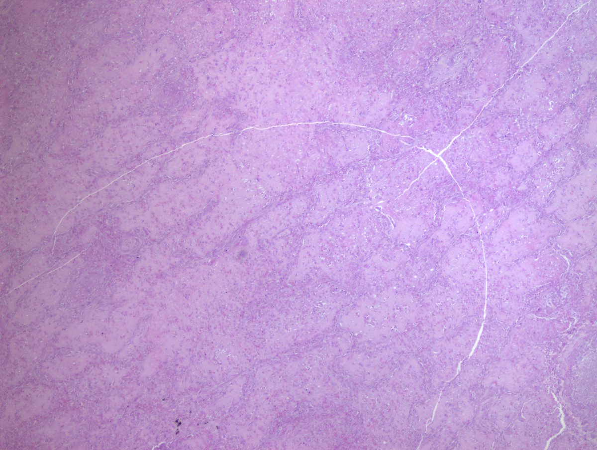
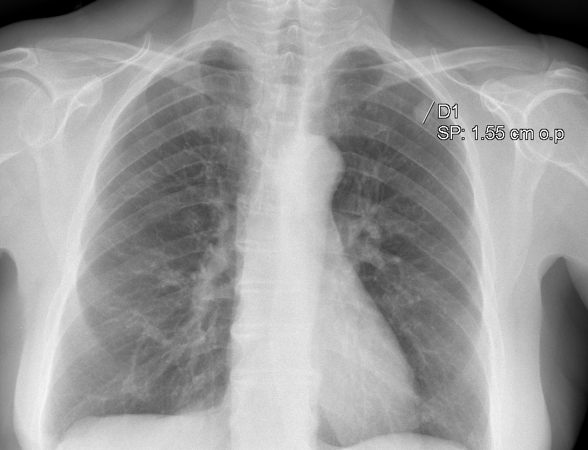
Coin lesions on chest X-ray) (1) under the microscope reveal infarct-like necrosis (2) with a rim of fibrosis, chronic inflammation with/out granulomatous reaction and MGCs (3)
Inside, a pulmonary artery with the worm in the lumen, better or worse preserved/calcified (4)
@nucleololailo@Quique_rs Yesss! Thank you carlos! I’m having quite a lot of infectious cases lately. Last week for example a case of anisakis in the jejunum.
Not everything in #GIpath is about adenomatous or serrated polyps. Following the last tweet’s infectious pathology theme, here a nice example of intestinal spirochetosis.
✅ #Bioaraba-ko ikerlariek umetoki-kanpoko estroma endometrialaren sarkoma ezohiko kasu bat aurkezten dute: eguneroko erronka txikiak ➡️ https://t.co/wy3c6YcUfL @igorostixe@alvaropr87
✅ Presentación de un caso inusual de sarcoma del estroma endometrial extrauterino por investigadores de #Bioaraba: pequeños retos diarios ➡️ https://t.co/wy3c6YcUfL @igorostixe@alvaropr87
I am putting together a talk on PD-L1 and I have developed some cartoons to help describe the different scoring systems and provide a visual easy to understand picture of how #Pathologists interpret PD-L1