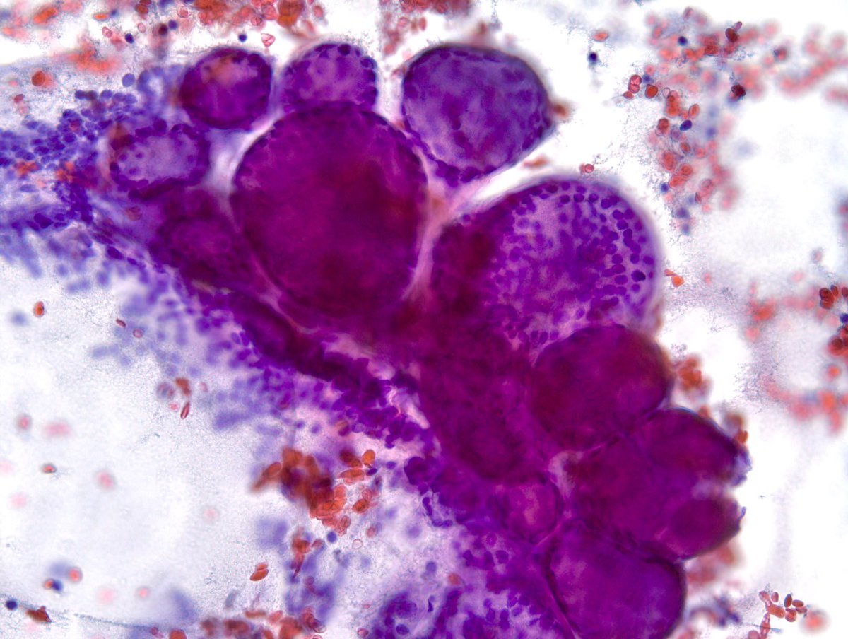
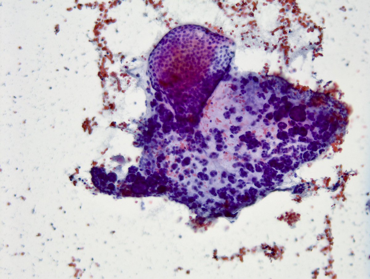
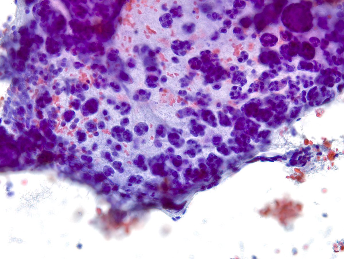
IMPORTANT Teaching Point - It’s NOT uncommon to have focal microfollicular (MF) proliferation (image#1) with rare colloid in “Benign” Thyroid FNAs. This “Adenomatoid Nodule” on another pass clearly shows macrofollicles. I look for at least 50% MFs for FN/SFN diagnosis.
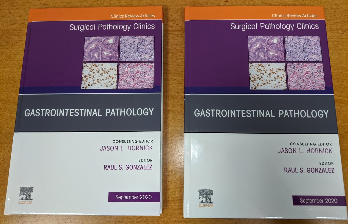
I have 2 copies of this GI issue of Surgical Pathology Clinics I'm giving away for FREE. Anyone in the world can enter! Just LIKE this post, SHARE this post, and REPLY with your favorite GI pathology diagnosis. I'll pick the winners randomly on Tuesday, 11/17. #pathology#gipath
#SocHemepath#SHQuickbyte
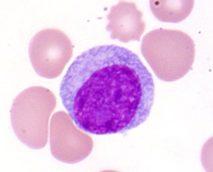
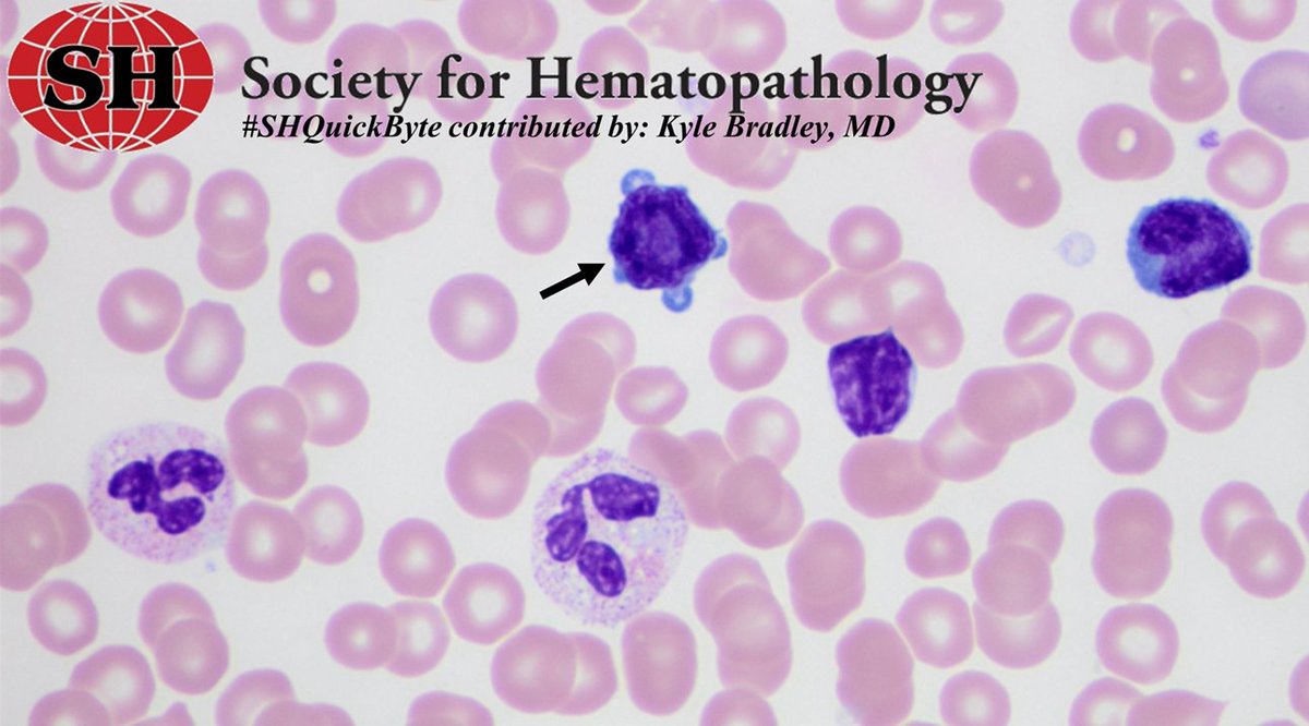
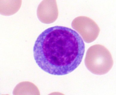
T-cell prolymphocytic leukemia is a rare, aggressive leukemia.
Cells often show cytoplasmic blebs on the blood smear (arrow)
CD4+ (60%), CD4+CD8+ (25%), CD8+ (15%) TCL-1 (usually +)
inv(14) is seen in 80% of cases
#hemepath#leusm#lymsm
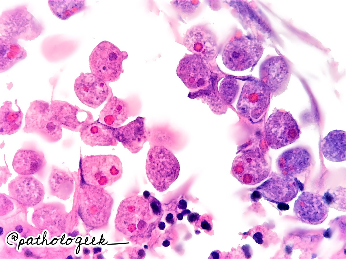
Look who I found today!
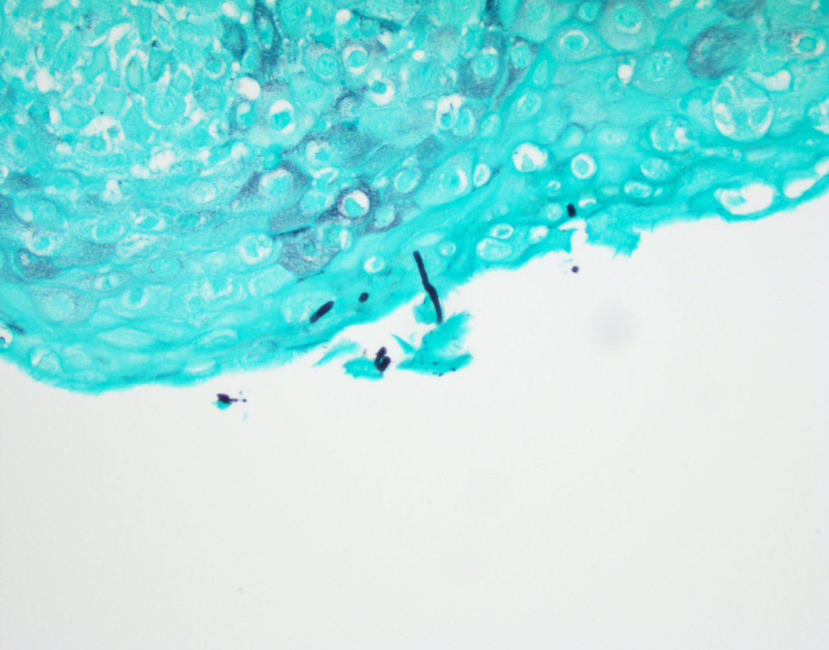
Perianal skin biopsy from a 68yo man with bloody diarrhea, abdominal pain and anal fissures. Clinical diagnosis: IBD- Crohn's disease.
Pathologists! Check out this case. Patient has history of plasma cell myeloma, now with elevated WBC. By flow, 40% of cells are clonal plasma cells (>6000 cells/microL), thus fulfilling criteria for plasma cell leukemia. #hemepath
Marrows are kaleidoscopic to begin with. Full of many hues.
Then you stumble across one of these.
Makes diffing an IgA MGUS worthwhile!
#PathTwitter#Hemepath
We have an opening for a Transfusion Medicine fellow in July 2021. A rich environment for cutting edge clinical practice, teaching and research. email [email protected] with your inquiry please. Thanks!