🇿🇦 Radiology Resident | South Africa 🎯 Future U.S. ABR Alternate Pathway Aspirant 🇺🇸 | Diagnostic Enthusiast 📸 Global Rad Collab | 💡 Gal 6:14 • John 3:3
I developed a distinctive medical-learning visual system: The Navy Recall Visual System - dark navy cinematic educational infographics with silver-white exam-focused text, designed to improve retention for USMLE and radiology learners. I believe this format has major commercial potential for AI-assisted education platforms and would be open to licensing, partnership, or acquisition discussions.
I developed a distinctive medical-learning visual system: The Navy Recall Visual System - dark navy cinematic educational infographics with silver-white exam-focused text, designed to improve retention for USMLE and radiology learners. I believe this format has major commercial potential for AI-assisted education platforms and would be open to licensing, partnership, or acquisition discussions.
@OpenAI and @ChatGPT stealing my ideas? Plagiarism.??
Over the last year, since early conception in March 2025, I’ve been developing a consistent study-visual format: dark navy medical infographics with silver-white text, designed to help retain high-yield USMLE/ABR content.
I’m now seeing very similar “navy background + white text” infographic concepts being promoted and marketed as suggested image styles in ChatGPT’s image interface. I’m not claiming certainty about how this happened, but I do believe if distinctive educational workflows influence platform-level tools and utilisation, thereby increasing revenue, that creators of these ideas and concepts should receive acknowledgement, and fair compensation of intellectual property.
@elonmusk Help??
#AIethics #MedicalEducation #USMLE #Radiology #ABRCore #CreatorRights #EdTech #AITransparency #Infographics
🗓️ Wednesday, December 3rd, 2025
✨ Wits Radiology Research Gala Evening ✨
Still completely overwhelmed.
“When we stand together, God multiplies what we can do.” — Max Lucado
Last week Friday, at our Wits Radiology Department Research Gala Awards, I stood among some of the smartest, hardest-working, kindest, and most inspiring humans I know… and somehow walked away with awards I truly never expected.
To be honest — I’m still trying to process it.
Receiving First Prize for Senior Registrar Academic Excellence, being assessed with the highest marks in the year, is an honour I never imagined.
Being voted tied First Runner-Up for Registrar of the Year alongside my brilliant friend Dr James Nutt — behind our phenomenal Registrar Representative Dr Mapule Mlawuli — meant even more. These are colleagues I look up to daily… colleagues who continually lift the standard and inspire me to raise my own.
Standing in that room, celebrating everyone’s achievements, handing out Spoof Awards, and recognising the incredible humans who walk this registrar journey with all its challenges… reminded me again why I love this department.
We carry each other.
We learn from each other.
We become better because of each other.
But the truth is — I didn’t achieve any of this alone.
Not even close.
My greatest strength, my anchor, my fuel, and my quiet daily miracle… is my family — my parents, my siblings.
My children, my pride and joy, who remind me why perseverance matters.
And my wife — my beautiful bride — who stands beside me daily, always looking absolutely breathtaking. I’ll go anywhere in this world, face anything, climb any mountain… as long as she’s beside me. Her support is beyond measure. Her belief in me is the reason I can keep giving my best, even on the hardest days.
I am humbled.
I am grateful.
And I am determined to become an even better version of myself — for my family, for my department, and for the patients we serve.
#WitsRadiology #ResearchGala2025 #GratefulHeart #HumbledAndHonoured #RadiologyLife #AcademicExcellence #RegistrarOfTheYear #TeamWits #MedicalJourney #SouthAfricanDoctors #FamilyFirst #MyBrideMyStrength #BecomingBetter #ExcellenceWithGrace
“The Lord bless you and keep you;
The Lord make His face shine upon you,
And be gracious to you;
The Lord lift up His countenance upon you,
And give you peace.” ’
Numbers 6:24-26 NKJV
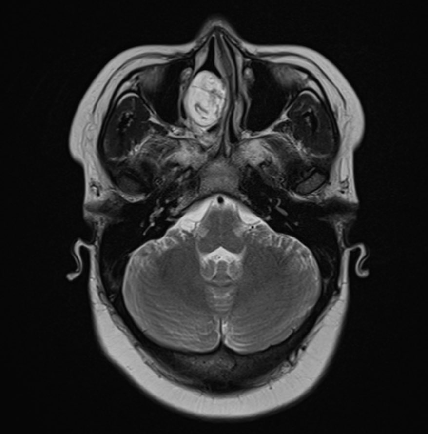
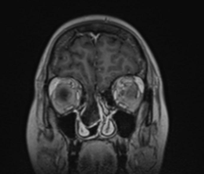
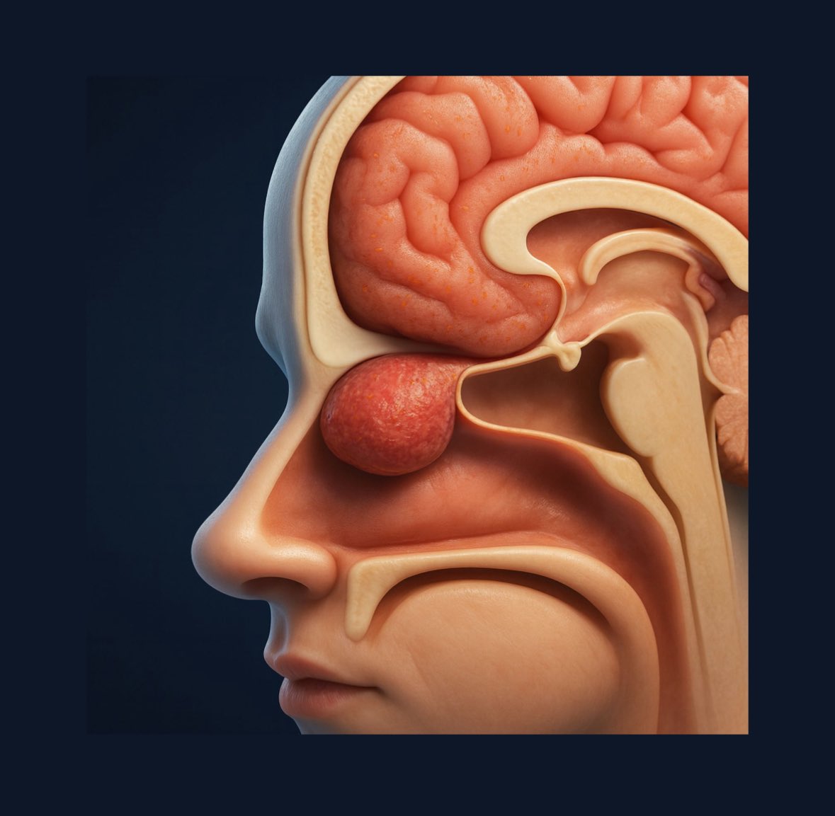
🔍 Case of the Day | The Value of MRI in Recurrent Meningitis
A 29-year-old female presents with recurrent episodes of meningitis, without prior trauma or surgery. Clinical suspicion: occult anterior skull base defect.
🩻 MRI Brain:
• Cribriform plate defect measuring 9.8 × 18.5 mm
• Herniation of medial right frontal lobe parenchyma
• Associated reactive pachymeningeal thickening and enhancement
• No diffusion restriction, hydrocephalus, nor venous sinus abnormality
• Brain is otherwise normal
📍 Diagnosis: Right nasoethmoidal encephalocele
⚕️ Neurosurgical consultation advised
📸 Artist-rendered anatomical illustration, as well as Axial T2w and Sagittal/Coronal T1w Post-Gad images included
🧠 CT may find it. MRI delineates it.
💡 Teaching Pearl:
In the delicate spaces beneath the frontal lobe lies a region easily overlooked. Yet in cases of recurrent meningitis, the humble cribriform plate may whisper its secrets.
While CT may hint, High-resolution MRI reveals what is unmistakably visible: encephaloceles extending through tiny skull base defects.
❓Question:
When does a defect qualify as a nasofrontal encephalocele rather than nasoethmoidal—and why is this entity vanishingly rare in adulthood?
📚 Key Imaging References:
• Abele TA et al. Radiographics. 2010;30(2):441–456 – “Imaging of Transcranial Skull Base Defects”
• Lloyd KM, DelGaudio JM. AJNR. 2008;29:1201–1206 – “Spontaneous CSF Leaks: A Radiologic Review”
#MRIbrain #SkullBase #CSFleak #Encephalocele #RecurrentMeningitis #DuraDefect #CribriformPlate #NeuroRad #RadiologyCase #RadTwitter #FOAMRad #AcademicRadiology #Neuroanatomy #USMLE #ABRAlternatePathway #IMGjourney #EmergencyRadiology #GlobalRadiology #WitsRadiology #DiagnosticRadiology #MedTwitter #FellowshipAspirant #IMGsInRadiology #MedEd #RadiologyAfrica
👩🦳 49-year-old woman
👃💉 Recurrent nosebleeds
❓🤔 What is the diagnosis?
We’ll post the answer in 24h. Share companion cases with us using #CookyBites #233. We will RT the best cases!
#RGphx@cookyscan1@RadioGraphics
“Have I not commanded you? Be strong and of good courage; do not be afraid, nor be dismayed, for the Lord your God is with you wherever you go.”
Joshua 1:9 NKJV
24-year-old man with known right testicular tumor before and after treatment.
What is the diagnosis🔍🧠❓
We’ll post the answer in 24h. Share companion cases with us using #CookyBites #232. We will RT the best cases!
#RGphx@cookyscan1@RadioGraphics
🔍 Case of the Day | Subtle Clue to Prior CVA
73M with HTN + DMT2, presenting outside the thrombolytic window with left-sided hemiplegia and dysarthria.
🧠 CT Brain Angiogram:
• Focal encephalomalacia in the right anterior cerebral peduncle — distal territory of the anterior choroidal artery
• No acute infarct or hemorrhage
• Patent Circle of Willis + normal intracranial vessels
📍 Working Dx: Chronic right anterior choroidal artery infarct → prior cerebral peduncular stroke, explaining isolated contralateral left hemiplegia via decussation of corticospinal tracts at the pyramids
📸 Axial CT reconstructions + 3D anatomical illustration of corticospinal decussation shown
💡 Teaching Pearl:
Anterior cerebral peduncle lesions → contralateral hemiparesis
A small, subtle, yet diagnostic finding — particularly important in crowded stroke units where “old infarct” = current deficits can be easily missed
🧠 Think cross-midline motor deficit clues, especially in patients with prior vascular risk factors.
📚 References:
📖 Yim et al. AJNR Am J Neuroradiol 2009;30:127–131
📖 Blumenfeld H. Neuroanatomy Through Clinical Cases, 2nd Ed
#NeuroRad #Stroke #CTAngiogram #RadTwitter #FOAMRad #AcademicRadiology #Neuroanatomy #AnteriorChoroidalArtery #CerebralPeduncle #USMLE #ABRAlternatePathway #IMGjourney #EmergencyRadiology #GlobalRadiology #RadiopaediaStyle #CHBAH #WitsRadiology
@CasesCookyJar@cookyscan1@RadioGraphics Anterior sacral meningocele and sacral osseous defect, that is sometimes referred to as Currarino syndrome even in the event of no associated anorectal malformation that would complete the classic Currarino triad. Careful assessment for occult teratoma advised
48M presents with intermittent pain 🤕 in the 👉 right upper quadrant. Diagnosis? ❓🩺
We’ll post the answer in 24h. Share companion cases with us using #CookyBites #226. We will RT the best cases!
#RGphx@cookyscan1@RadioGraphics
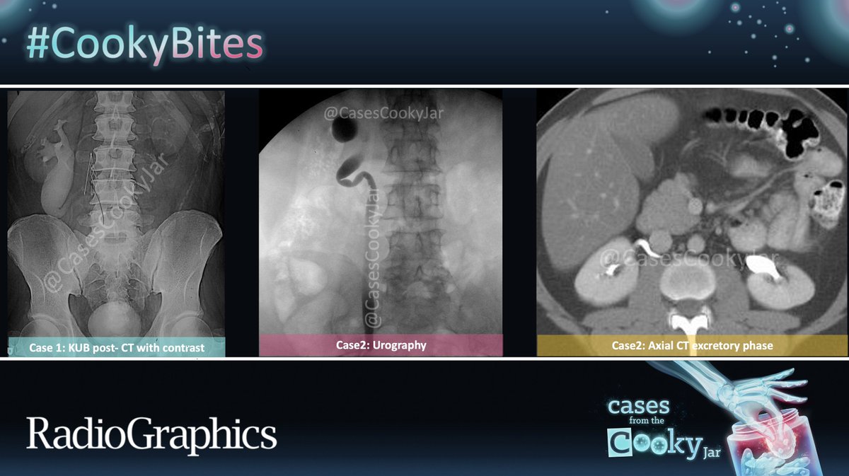
@CasesCookyJar@cookyscan1@RadioGraphics Kinked upper third of the right ureter with retrocaval course leading to intermittent functional ureteric extrinsic obstruction, with hydronephrosis, not appreciated at the time of CT IVP imaging acquisition.