PhD in Computational and Systems Biology | Postdoctoral Research Fellow | Chambers Lab - Nanyang Technological University| Human and Population Genetics
Pakistan Genomic Resource (PGR), founded by Danish Saleheen, is the world's largest genetic database of human knockouts. It has been quietly building since 2017, from 10,000 individuals (Saleheen et al. Nature 2017) to nearly 200,000 today.
A new paper in Nature by Koch et al. reports a new analysis of 173,303 Pakistanis from PGR, offering genetic insights from natural inbreeding experiments in humans.
Koch et al. Nature 2026
https://t.co/Uk9YaeN1Ev
In biology, to understand what a gene does, you delete it, a standard experiment. Knock out the gene in a mouse and see what breaks: organ fails, behaviour changes, animal dies. Thirty years of biology built this way. It works, until it doesn't. Because, you know, mice are not humans.
To know the function of a gene in humans, you need a similar experiment. But that's unethical, you cannot deliberately delete a gene in a human. Except you don't have to, because nature has already been doing that experiment.
Occasionally, a person inherits a broken copy of a gene from both parents and is born with no working version at all--a human knockout. The problem is finding them. In most of the world's populations, where mating is largely random, these events are nearly invisible. For a gene-inactivating variant at 0.1% frequency, you'd expect one homozygous individual in every million people.
That math collapses in populations like Pakistan, where consanguineous marriage has been practised for centuries. In first-cousin marriages, the odds of observing a human knockout for the same 0.1% variant rise to roughly 1 in 16,000, a 63-fold enrichment. And the rarer the variant, the larger the advantage: ~630-fold for a 0.01% variant, ~6,300-fold for a 0.001% variant. The variants too rare to ever be seen in European biobanks become findable here.
Sequencing more than 170,000 individuals from highly consanguineous communities, the authors report a mind blowing statistic: at least one living human knockout was observed for 6,476 genes, which is nearly 1/3rd of the entire protein-coding genome!
What do we find when we finally have the human knockouts?
Studying the phenotypes in the human knockouts helps us confirm or refute our understanding of the gene's function based on animal studies. A few examples I highlight below.
PRDM9
PRDM9 might be one of the most popular genes among animal biologists. It encodes a protein that controls where chromosomes break and recombine during sperm and egg formation. Deleting the gene has caused infertility in every animal. PRDM9 was classified as the first hybrid sterility gene in vertebrates, so fundamental that crosses between mouse species with different PRDM9 alleles can't produce a fertile offspring. PGR now has 4 human PRDM9 knockouts : three women, one man. All fertile, with 2 to 7 children each. A 14-year biological fact, overturned by four families in Pakistan.
LRRK2
LRRK2 is a well-established Parkinson's disease risk gene. Activating mutations in LRRK2 are among the most common risk factors for Parkinson's. LRRK2 is a therapeutic target with many companies exploring ways to switch off this gene in the brain to treat Parkinson's. Large-scale sequencing studies have found individuals with partial loss of LRRK2, who did not show any concerning health issues, predicting adverse effects of LRRK2 inhibition in humans. Animal knockouts though warned of kidney damage. Now PGR has two LRRK2 knockouts, both with kidney disease, confirming animal studies.
RXFP1
RXFP1 encodes the receptor for a pregnancy hormone called relaxin, which has long been studied in rodents. Animal studies suggested it played a critical role in cardiovascular adaptation and connective tissue remodelling, fuelling relaxin-targeted drug development, which failed in late-stage trials. PGR found 16 RXFP1 knockouts, expanded to 26 via recall-by-genotype, all tested with cardiac imaging. None had consistent cardiovascular or reproductive deficits, retrospectively explaining the failure of relaxin-targeted drug programmes that might have spent millions of dollars. Mouse physiology failed to inform humans in the case of relaxin.
Gene constraint insights
Existing large-scale biobanks are predominantly European-based outbred populations, which shaped our understanding of gene constraints largely based on intolerance to partial loss of function. Now PGR, a South Asian-based cohort enriched for consanguineous communities, is beginning to offer insights into gene constraints based on intolerance to complete loss of function.
PGR showed that nearly 1/3rd of human genes tolerate complete loss of function. As much as the genes for which knockouts were found, the genes for which knockouts weren't found can offer biological insights.
The authors find genes depleted for knockouts in PGR are enriched for genes essential for cell survival, known Mendelian disease genes (both dominant and recessive) and genes broadly expressed across human tissues.
A fascinating insight is significant enrichment for knockouts in tissue-specific genes (OR=2.39). Human knockouts confirm what drug developers have always thought: tissue-specific genes are much safer therapeutic targets than broadly expressed genes.
The above insight should be read with caveats. The sample size of PGR is small, hence not saturated for human knockouts. It's likely the number of genes will increase as the sample size grows. There is a survivorship bias, like any other volunteer-based cohort. Absence of a gene knockout here doesn't mean biological impossibility. It means incompatibility with being a 'healthy' adult volunteer. If you build a cohort based on a hospital-based pediatric rare disease South Asian cohort, you'd expect to see knockouts that never appeared in PGR.
South Asian populations represent nearly a quarter of humanity, yet they have been largely absent from the genomic revolution. PGR shows what absence has been costing the field: overturned biological assumptions, failed trials, missed targets. The biology was always there. We just weren't looking in the right place.
PERSPECTIVE | TH Mina, JC Chambers et al (@NTUsg)
In this Perspective, the authors discuss reasons for the increased cardiometabolic disease risk observed in Asian populations, including nutrition, genetic factors and environmental adversity.
https://t.co/fOXVc1EO0B
These comments in Nature Genetics on a GWAS signal for cerebral amyloid angiopathy at APOE are a great case study in the challenges of interpreting genetic associations even with abundant data.
Worth reading for anyone interested in translating GWAS hits to drug targets👇
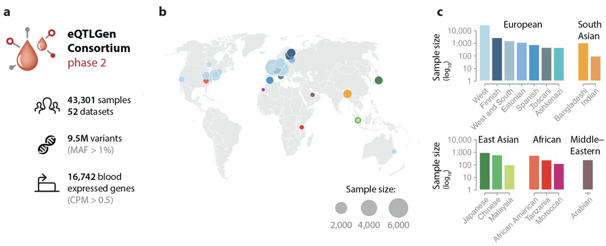
eQTLGen is for years the largest eQTL resource out there.
They now released the second phase of this initiative from 43,301 samples, revealing cis-eQTLs for 94% and trans-eQTLs for 56% of human genes.
Amazing resource for following-up on GWAS signals👇
🧵Super excited to share our new paper in @NatureGenet ⭐️
We apply a new multiple traits and ancestry approach to improve genetic discovery and polygenic prediction in lung diseases and traits 🫁
An interesting preprint from Alkes Price's lab shows that GWAS effect sizes' relationship with minor allele frequency (MAF) is better explained by African MAF than European MAF.
We know that rare variants have larger effect sizes, which is a reflection of negative selection. Disease-causing deleterious variants are kept at lower frequencies by natural selection.
However, allele frequency of a variant is influenced not only by natural selection, but also by other factors like genetic drift caused bottleneck events.
MAFs of all non-African populations are influenced by out-of-Africa bottleneck effects. Such frequency changes reflect drift, not disease biology.
In this work, Rossen et al. empirically show that in fact African MAF estimates better explain GWAS effect sizes than European MAF, even when the GWAS is performed in European populations.
These results suggest that many downstream GWAS analyses—such as heritability estimation, fine-mapping, and polygenic risk prediction—may be systematically misspecified when relying solely on European-derived MAFs. Incorporating African MAFs is likely to yield more accurate effect-size modeling, even for GWAS performed entirely in European populations.
Rossen et al. medRxiv 2026
https://t.co/pHyVYqHK0p
A new preprint reports one of the most fascinating and in-depth investigations of a GWAS locus that I’ve seen.
Locus 9p21.3 is the first robust GWAS signal linked to coronary artery disease (CAD), discovered in 2007 by multiple research groups. The risk variants show the largest effect sizes known for common variants influencing CAD.
One of the most interesting aspects of this locus is its ancestral heterogeneity. The signal appears sharply in GWAS of CAD performed in Europeans, East Asians, and Admixed Americans, but is absent in African populations—despite many of the top risk variants being common in African ancestries.
In this preprint, the authors attempt to solve this puzzle by diving deep into this locus across multiple ancestries and arrive at a compelling conclusion.
The most likely reason African GWAS of CAD were unable to capture the signal at 9p21.3 is not lack of power, rare variants absent in Africans, or gene–environment interactions. Instead, it reflects differences in linkage disequilibrium (LD) and haplotype structure between African and other populations.
The 9p21.3 locus spans a relatively large regulatory region (~0.4–0.5 Mb) and contains a complex haplotype structure. It is a noncoding locus with multiple regulatory variants contributing to risk independently. These variants are distributed differently across LD blocks in African ancestries compared to others.
In European genomes, the risk variants tend to sit on shared, longer LD blocks, effectively amplifying the GWAS signal. In African genomes, the same variants are spread across multiple shorter LD blocks, diluting the signal.
The takeaway: absence of 9p21.3 CAD locus isn’t missing biology in Africans, it’s rather shared biology hidden by greater haplotype diversity.
Alkhairo et al. medRxiv
https://t.co/9AkUblxVkm
My first Postdoc paper is now out on medRxiv. We performed the largest Epigenome wide association study (EWAS) of Type 2 Diabetes in an Asian Population. Happy to receive feedback and comments!!
https://t.co/3tvun7XJJg
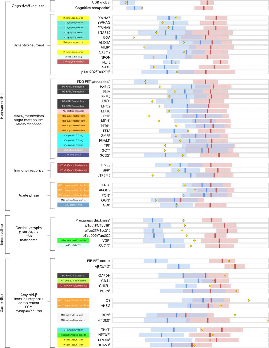
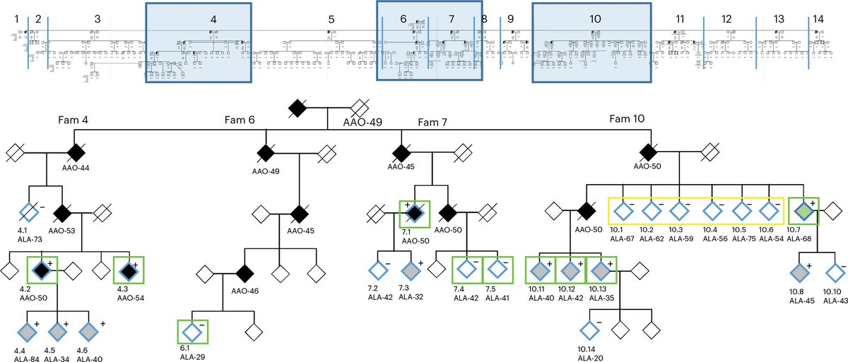
A fascinating new study just published online today in @NatureMedicine reports an extraordinary case of an individual carrying the PSEN2 (p.Asn141Ile) mutation—a known driver of dominantly inherited Alzheimer’s disease (DIAD)—who remained cognitively healthy well into his mid-70s. This is nearly 18 years past the expected age of onset. The study combines longitudinal clinical assessments, neuroimaging, genetic analysis, and proteomics to uncover the mechanisms underlying his remarkable resistance to Alzheimer’s (https://t.co/MKBBWo4zcC) 🎉🧬🧠🔍
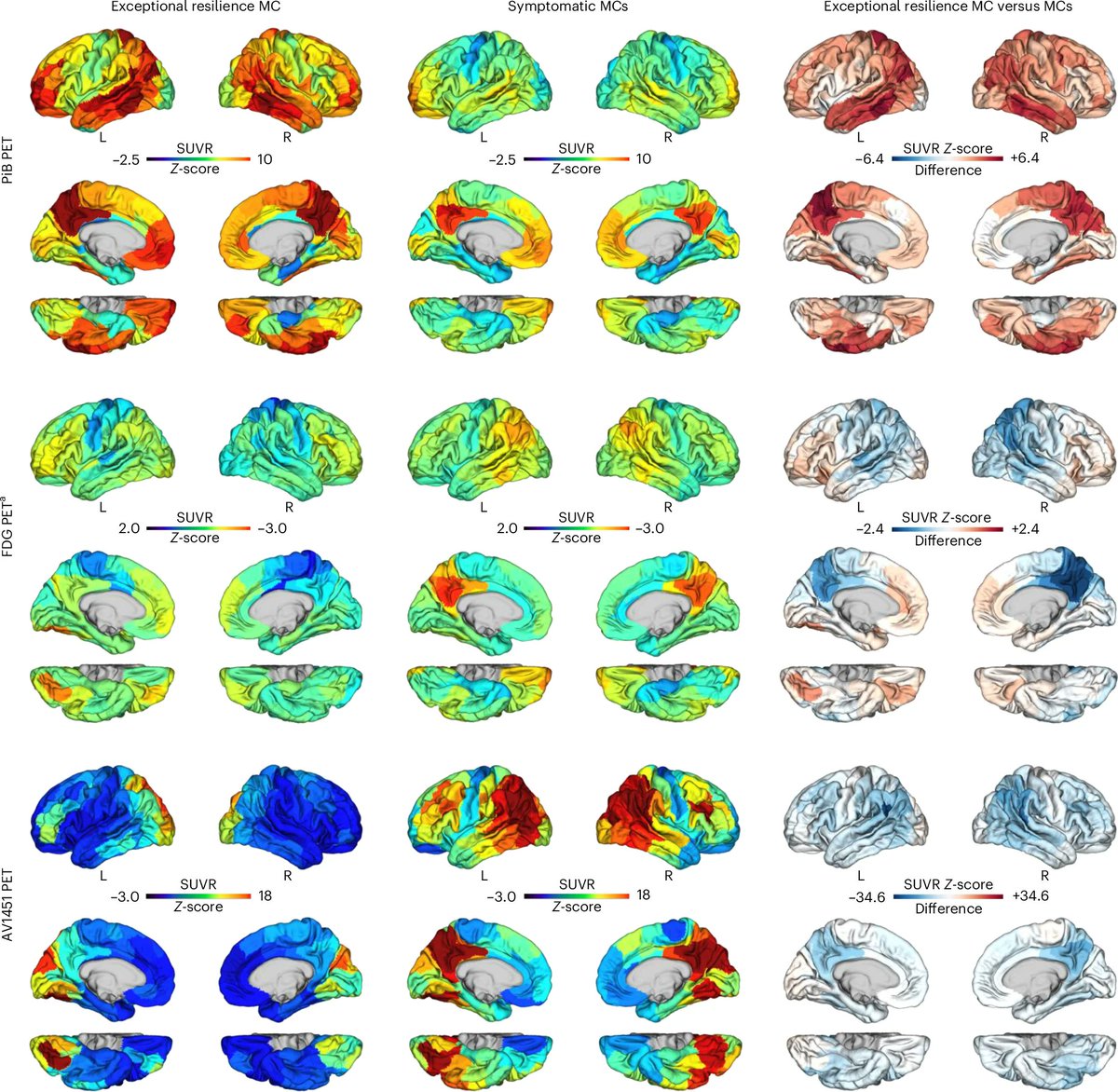
The PSEN2 p.Asn141Ile mutation causes excessive production of amyloid-beta, resulting in amyloid plaques—one of the hallmarks of Alzheimer’s disease. While this individual’s brain was filled with amyloid deposits, the expected cognitive decline and dementia never materialized. Tau pathology, another key feature of Alzheimer’s, was restricted to the occipital lobe, a brain region associated with visual processing but not typically involved in Alzheimer’s-related cognitive decline. This rare localized tau pattern may explain the individual’s cognitive preservation and highlights the potential of tau-targeted therapies.
Over the course of 10 years of follow-up, the individual’s Cognitive Dementia Rating (CDR) and Mini-Mental State Examination (MMSE) scores remained normal (CDR = 0; MMSE = 30) for the subsequent four (2014, 2016, 2018 and 2021) assessments performed over 10 years, including the most recent evaluation in 2021 at age 71.
Genetic analysis in this study utilized whole-genome sequencing (WGS) and whole-exome sequencing (WES) on family members, including the exceptional resilience mutation carrier, to identify rare and common coding variants associated with resilience to Alzheimer’s disease. Segregation analysis prioritized variants that were exclusive to the resilience carrier or specific subgroups within the family. Several unique variants were identified within the 17q21.31 MAPT-H1 haplotype region, along with potential protective variants in AD-associated genes such as TREM2 and MAPT.
Proteomic profiling in this study identified an abundance of heat shock proteins, suggesting a potential adaptive stress response. Remarkably, this individual had prolonged exposure to high temperatures while working as a mechanic on a naval ship, requiring frequent cooling with water to prevent overheating. The heat-shock response triggered by such extreme conditions may have contributed to his resistance to neurodegeneration. These results suggest that a combination of genetic factors, environmental exposures, and enriched biological pathways may contribute to the delayed onset of AD in this individual.
The imaging data tell a powerful story. PET scans showed elevated amyloid burden with a PiB cortical SUVR (Z-scored relative to non-carriers aged 50 years old and above) of ~3.6, far above the threshold for abnormal amyloid deposition. However, tau PET imaging revealed that tau pathology remained focal and asymmetric, concentrated in the occipital lobe, rather than spreading to key regions like the medial temporal lobe or parietal cortex, which are typically affected in Alzheimer’s disease. This restricted tau spread contrasts sharply with the usual progression seen in mutation carriers, where tau accumulation is tightly linked to cognitive decline.
MRI scans showed that hippocampal volume declined only modestly over the 10 years of follow-up, consistent with normal aging rather than Alzheimer’s atrophy. FDG PET scans showed localized hypometabolism in areas with focal tau deposition, yet metabolic activity remained normal in other key brain regions.
From a broader perspective, this study raises questions about the amyloid hypothesis—the long-standing theory that amyloid plaques are the primary driver of Alzheimer’s. The case suggests that limiting tau deposition and spread may be more critical in preventing cognitive decline, even in the presence of substantial amyloid burden. This finding could reshape the focus of Alzheimer’s research and clinical trials, emphasizing tau-targeted therapies.
The proteomic analysis further revealed significant differences in this individual’s plasma proteome and cerebrospinal fluid (CSF) compared to typical PSEN2 carriers. Notably, pathways related to protein folding, synaptic function, and oxidative stress were enriched, with multiple heat shock proteins showing elevated expression. These proteins may play a crucial role in maintaining cellular homeostasis and protecting against protein misfolding, a common feature of neurodegenerative diseases.
In summary, this study opens exciting new avenues for Alzheimer’s research by identifying potential protective genetic and proteomic factors. It underscores the importance of studying rare resilience cases to uncover new therapeutic strategies. Restricting tau spread may prove to be a transformative approach in delaying or preventing dementia. The insights gained from this individual offer hope for developing precision medicine strategies for Alzheimer’s disease.
Huge congratulations to the research team—Jorge J. Llibre-Guerra, M. Victoria Fernandez, Nelly Joseph-Mathurin, @shijbian, Kathleen Carter, @ccrugom, Erik C. B. Johnson, @RandallBateman3, and many others—along with the Dominantly Inherited Alzheimer Network (DIAN), the study participant and his family members, whose dedication continues to pave the way for life-changing discoveries.
[About this post: this post is based on a summary of Llibre-Guerra, J.J., Fernandez, M.V., Joseph-Mathurin, N. et al. Longitudinal analysis of a dominantly inherited Alzheimer disease mutation carrier protected from dementia. Nat Med (2025). https://t.co/ruuI6PUEK1]
Modern GWAS can identify 1000s of hits but it can be hard to turn this into biological insight. What key cellular functions link genetic variation to disease?
I'm excited to present our new work combining associations + Perturb-seq to build interpretable causal graphs! A🧵
Excited to share this discovery: Huntington's disease is a DNA process for almost all of a cell's life. Inherited HD alleles are innocuous, just unstable – CAG repeats slowy expand throughout life, acquiring toxicity only when quite long (>150 CAGs). https://t.co/6wIAZKCWKN
Metabolic variation as a predictor of dietary intake in the Singaporean population, followed by building food specific metabolite panels.
https://t.co/9yyE0iGqr7
Methods paper describing the HELIOS Cohort with whole genome sequencing, epigenomics, metabolomics and dense data phenotype data in a Singaporean cohort of 10,000 individuals. https://t.co/cTVzabZah7
A Metabolome-wide association study of Cardiovascular disease risk in a Southeast Asian population, with both statistical and functional validation. https://t.co/0P4Ep8teZv
🧬 Our #ENCODE4 paper is out now @GenomeBiology! Dissecting uncharted regions in our DNA and their influence on our health. 🧬
Dive into the world of disease genetics and genomic "dark matter" with us as we explore our trailblazing new method. Ready? Let’s go! 🚀 🧵 #Genomics #GenomeFunction #ScienceTwitter
https://t.co/TPDujkJJf3 1/
PWAS Hub (https://t.co/ojuTVUKaRx)
A nice web tool to explore proteome-wide association study results of 99 common diseases in the UK Biobank
Kelman, Zucker, et al.
https://t.co/l32RcUW962