Most cellular processes involve more than one player and none of them can be fully understood by imaging one component at a time. The number of colors you can resolve simultaneously is not a technical detail: it determines which biological questions you can actually ask. Colocalization between two targets is an observation. Colocalization between five, resolved in a single experiment, is a map.
A typical immunofluorescence experiment images two, maybe three targets. Not because the biology is simple, but because spectral separation is hard and finding reliable, bright, and photostable fluorophores for each channel is no trivial task.
The abberior PLEX 5 color Kit was built to change that - making five-color multiplexing straightforward.
These fixed mammalian cells were stained in a single immunofluorescence round using five spectrally separated #STARdyes, imaged on the abberior #MIRAVA Polyscope:
⚪ STAR BLUE (405 nm): F-actin (phalloidin)
🟠 STAR GREEN (488 nm): Golgi apparatus
🔵🟢🔴 STAR ORANGE / STAR RED / STAR deepRED (561, 641, 685 nm): three different targets of the Nuclear pore complex
#abberiordyes #MIRAVA #abberiorPLEX #multiplexing
3D volume of sparse mutant visual transmedulla neurons in the adult #Drosophila brain, expressing membrane mCherry ( STAR RED) together with wild-type neurons labeled with GFP🟦. The images aquired by the #MIRAVAPOLYSCOPE with #RAYSHAPE allows imaging down to 132 µm deep in the sample uncovering disorganized dendritic morphology.
Thanks to Dr. Tzu-Yang Lin and Prof. Chi-Hon Lee from Academia Sinica, Taiwan for providing the sample.
#FluorescenceFriday
Volume of sparse mutant visual transmedulla neurons expressing membrane mCherry (🟦STAR RED) in adult #Drosophila brain. wt neurons labeled with GFP🟥. Isotropic #3DSTED with #MIRAVAPOLYSCOPE shows mutant neurons’ disorganized dendritic morphology.
Special thanks to Dr. Tzu-Yang Lin and Prof. Chi-Hon Lee from the Institute of Cellular and Organismic Biology, Academia Sinica in Taiwan for kindly providing the sample.
⌛️March is almost over... don't miss your chance to win one of the limited edition building-brick models of our MIRAVA POLYSCOPE!🤩
Sign up to our newsletter until end of March and enter the raffle: https://t.co/X9kRC5B2C4
#MIRAVAPOLYSCOPE
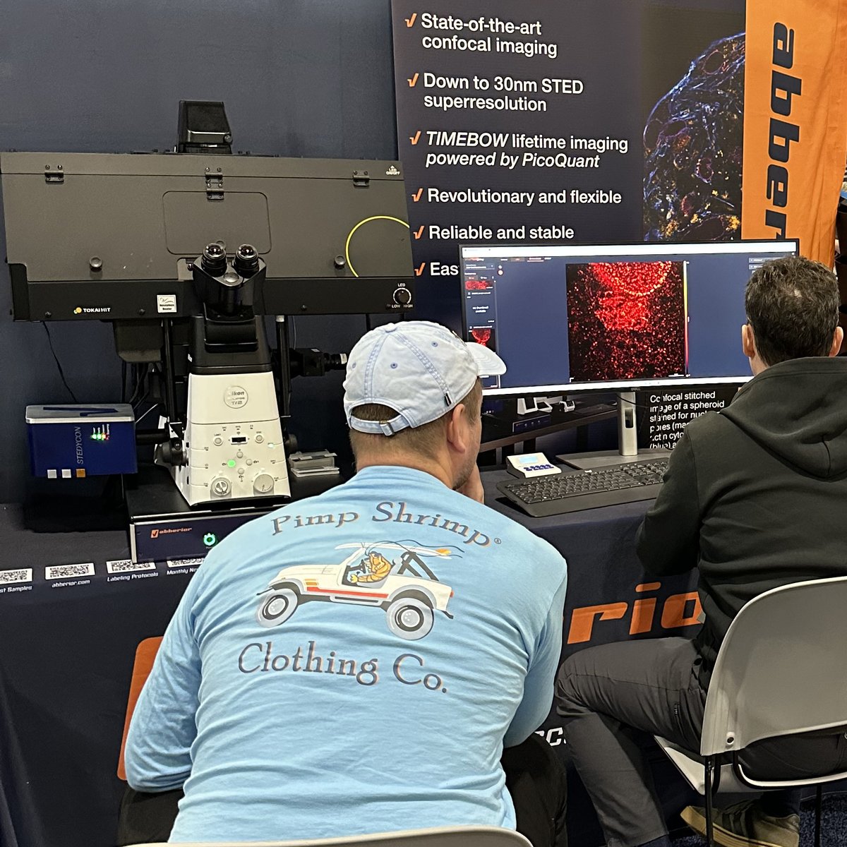
🔬 STEDYCON2 in action in Milan!
First STEDYCON2 demo in Europe is currently running at #UnitechNOLIMITS (https://t.co/Xg9EVakKeZ) at the Università degli Studi di Milano together with Alessandro Rossi from our italian partner Crisel Instruments. Scientists test the #STEDYCON2 on a Nikon body with their own samples and experience how easy STED imaging can be. In addition to super-resolution imaging, the system also enables fluorescence lifetime measurements (FLIM), opening new possibilities for multi-color imaging and studying biological and physical correlations.
Many thanks to Alex Costa and Miriam Ascagni for hosting us!
#STEDYCON2 #italy #FLIM #STED #confocal
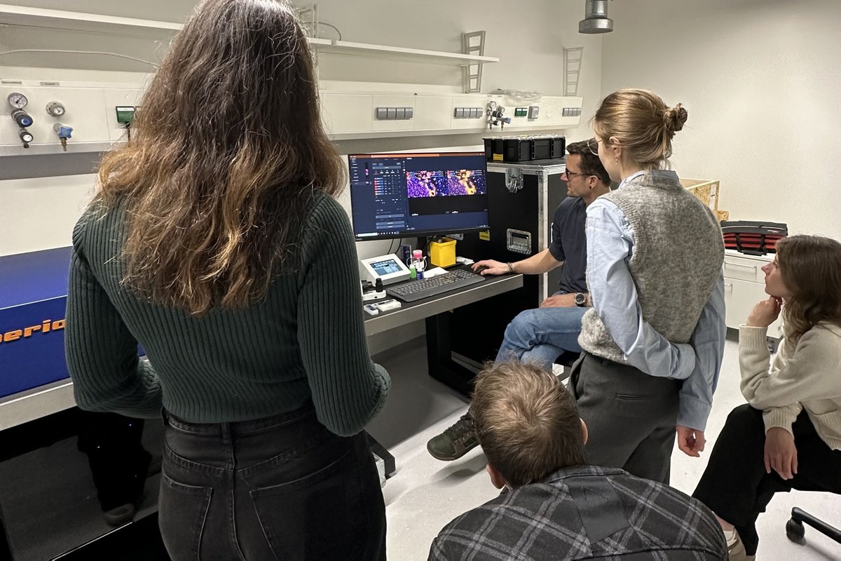
During last week's workshop @UKEHamburg scientists experienced first-hand how our #MIRAVAPOLYSCOPE facilitates imaging across scales, from millimeter samples all the way to single-digit nanometer localization precision.
Many thanks to Virgilio Failla for having us!
Superresolution microscopy has never been easier than with our STEDYCON.
Switch to STED with a single click to reveal subcellular details like the exact localization of these two proteins within the golgi.
https://t.co/W9MfYKXkwo
#STED#STEDYCON2#superresolution
Superresolution microscopy has never been easier than with our STEDYCON.
Switch to STED with a single click to reveal subcellular details like the exact localization of these two proteins within the golgi.
https://t.co/W9MfYKXkwo
#STED#STEDYCON2#superresolution
STEDYCON’s software is the definition of ease-of-use:
🔸Hardly any training required
🔸From system start to stunning images in no time
🔸Switch between confocal, STED, FLIM with only a few clicks
🔸Browser-based: monitor your session from anywhere
https://t.co/X9CI4RpWBb
#STEDYCON2
At #cellbio2025, our new #STEDYCON2 again did what it does best: impressing everyone with a rock-solid, high-quality performance in confocal, STED, and lifetime imaging 🤩
STEDYCON transforms your microscope into a full-fledged confocal and STED machine!
🔸independent of the manufacturer
🔸for inverted and upright microscopes
🔸for old and new frames
https://t.co/W9MfYKXSlW
#STEDYCON2
We will exhibit at #cellbio2025 in Philadelphia, PA! Get to know our brand-new STEDYCON 2 live and in action!
Bring your own samples and see for yourself how this compact and flexible microscope performs as it combines confocal, STED, and lifetime imaging in one flexible system.
Our colleagues will be happy to answer your questions. Hope to see you there!
#STEDYCON2 is engineered for ultimate robustness:
🔸All lasers aligned by design, including STED laser👉no need for beam calibration, ever
🔸Hardware-based STEADYFOCUS 👉focus position is permanently stable
🔸Enhanced optomechanics 👉extended remote serviceability
https://t.co/SpapqisjIY
#FLIM is now available for STEDYCON! 🌈
As #TIMEBOW lifetime imaging, it’s fully integrated into the STEDYCON smart control software and powered by @PicoQuant’s MultiHarp 150 hardware.
STEDYCON is your fast track to lifetime imaging.
https://t.co/S8h0gIWs6g
#STEDYCON2
Presenting the new #STEDYCON2 at #SfN25, booth no. 812! Bring your own sample and see for yourself how this highly compact and flexible confocal and STED microscope performs.
Our new #STEDYCON2 and crew is waiting for you at #Sfn25 booth 812! Bring your own samples and see for yourself how this flexible and easy-to-use system performs, be it in confocal, superresolution STED, or lifetime mode.
3D cell staining & imaging in action! Check out this confocal image of a NIH-3T3 spheroid stained with abberior dyes!
Spheroids are 3D cell cultures that better mimic tissue architecture than traditional 2D cultures, providing a more physiologically relevant model for studying cellular behavior, drug response, or developmental processes. abberior dyes offer highest brightness, excellent photostability, and are validated for a broad range of imaging techniques, making them ideal for capturing complex structures like spheroids.
Immunofluorescence staining:
• abberior STAR RED for nuclear pore complex (magenta)
• abberior STAR ORANGE for mitochondrial outer membrane protein TOM20 (yellow)
• abberior LIVE 510 actin (cyan)
The spheroids were grown in the µ-Slide VI 0.4 µ-Pattern ibiTreat (cir200, pit600, hex) and fixed with 4% PFA. Imaging was performed using the abberior #STEDYCON.
A special thank you to @ibidiCells for the cooperation!
#abberior #ibidi #spheroids #3Dcellculture #fluorescencefriday
Hey #imagingfacility managers:
Do you want to
👉speed up training sessions
👉increase productivity
👉make your users happy?
Our new STEDYCON 2 can do all that, combining confocal, STED, and lifetime imaging in one flexible system.
Join our webinar:
📆Nov 13 | 15:30 CET
Register: https://t.co/naur6hg4Y0
#FLIM #STED #confocal #webinar #stedycon2