Senior Medical Scientist, Electron Microscopy Unit, SA Pathology, Adelaide, Australia. Ultrastructural pathology of kidney, muscle, nerve, liver, tumours, etc.
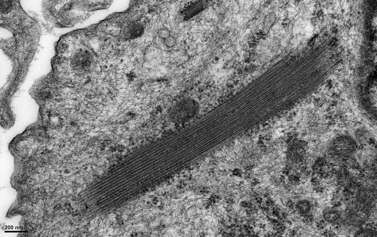
Another example of ribosome-lamella complexes within a podocyte from a case of transplant glomerulopathy. The significance of these structures is uncertain but possibly reflects aberrant protein sysnthesis within the cell.
Renal tx insertion bx. @electronmicroscopy shows a rare finding of ribosome-lamella complexes (arrows) in podocytes. Does anyone know if they are significant? I think it's a non-specific change reflecting cell stress. Not to be confused with crystals that can occur in podocytes.
@Zhou1217001 Any follow up on this case. The inclusions have an appearance similar to myelin - although I can't see if there is any lamellar substructure (need higher magnification for that).
Renal biopsy of a patient with T1DM. #electronmicroscopy of medullary tubules. Severe accumulation of glycogen in the epithelium. The nuclei (easily misinterpreted as vacuoles) contain so much glycogen that the chromatin and nucleoli have been pushed to the nuclear periphery.
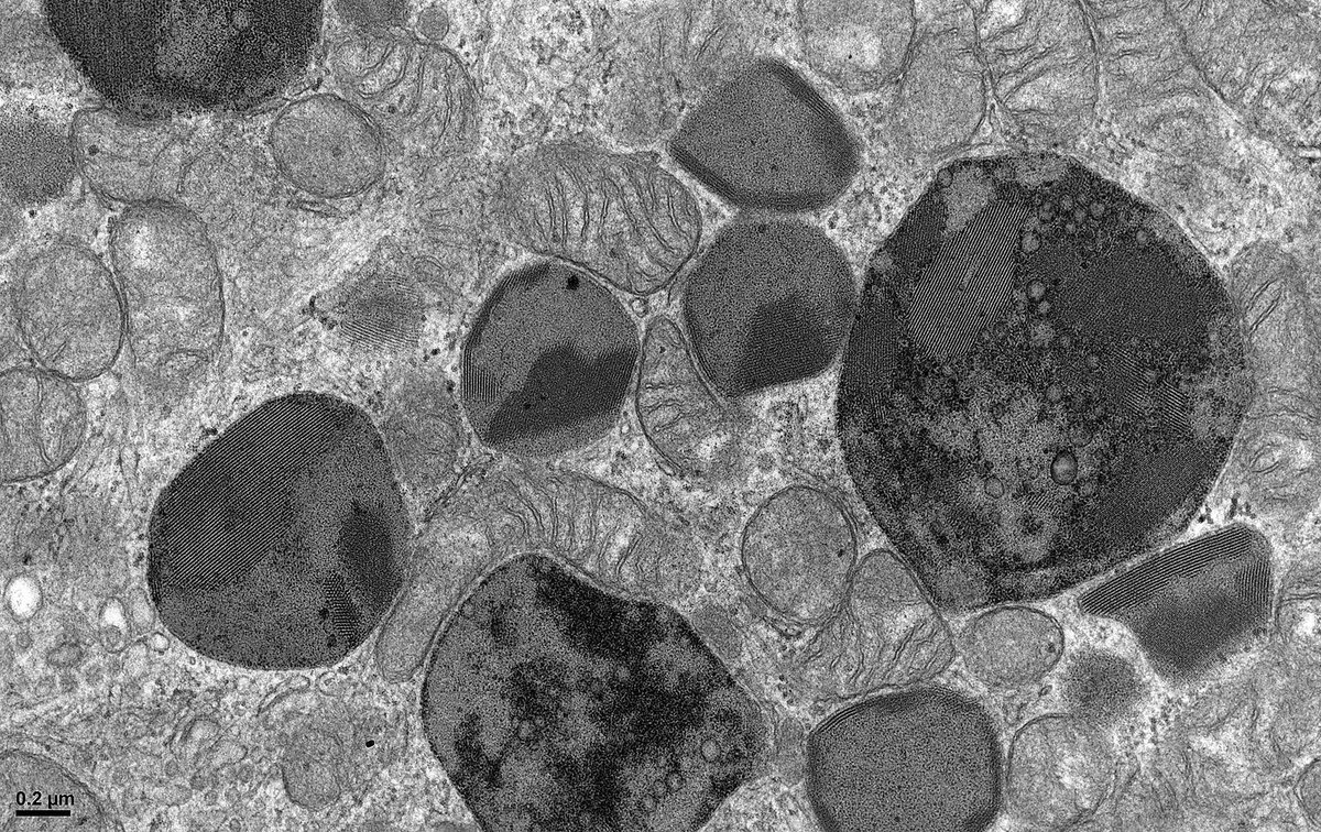
@GibsonIgibson That's a great question, but I don't know. Interestingly, image 2 shows a renin vesicle (with rhomboid crystal) in close apposition with the lipofuscin-like material. These images were taken from a patient with hypertension, so maybe chronic RAAS activation is a factor.
These structures are seen by @electronmicroscopy in smooth muscle cells of kidney arteriolar walls and juxtaglomerular cells. These 'lipofuscin-like granules' should not be misinterpreted as inclusions of the type found in some forms of neuronal ceroid lipofuscinosis.
@GibsonIgibson@SethiRenalPath@ChangUCanSpare@JZRenalPath The case is unresolved as there is an underlying suspicion of infection or malignancy in the patient. It's unclear to me how these conditions could relate to the EM findings. Any thoughts welcome.
@DrGeetikaSingh1@JZRenalPath I've seen lupus patients on HCQ w myelin figures in podocytes (but agree the lysosomes in endothelial cells are typically smaller). Geetika, your paper uses the term "HCQ toxicity". Is there proof the myelin figures indicate toxicity - or is it merely a sign the patient is on HCQ
@sam_albadri I've seen similar material by EM in the interstitium, possibly representing the remnants of tubules that have been destroyed. Do you think your material is in a tubule or glomerulus - I can't tell? Did it stain by Immunofluorescence (or was it too focal for IF to stain)?
#askrenal Can a plasmacytoma produce anti-LCAT autoantibodies?
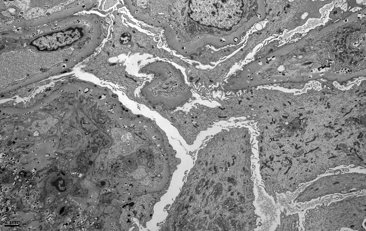
We have a case of a patient with a plasmacytoma. However, by #electronmicroscopy, the glomerular ultrastructure suggests LCAT deficiency.
#electronmicroscopy Renal tx bx from a child w renal dysplasia as native disease. Any thoughts on these structures in tubules - ? lipofuscin, ? peroxisomes, ? a drug reaction. It’s the structures w periodicity that interest me. Human peroxisomes typically lack a crystalloid core
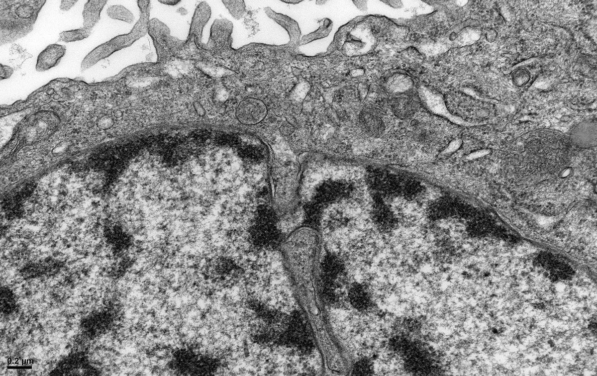
#electronmicroscopy Kidney bx shows a membranous nephropathy. #podocytes, typically post-mitotic, appear to be trying to divide. Nuclei are bi-lobed with narrow chromatin bridges. Cells are rounded with blebs + detachment from the GBM. Possibly an example of mitotic catastrophe.