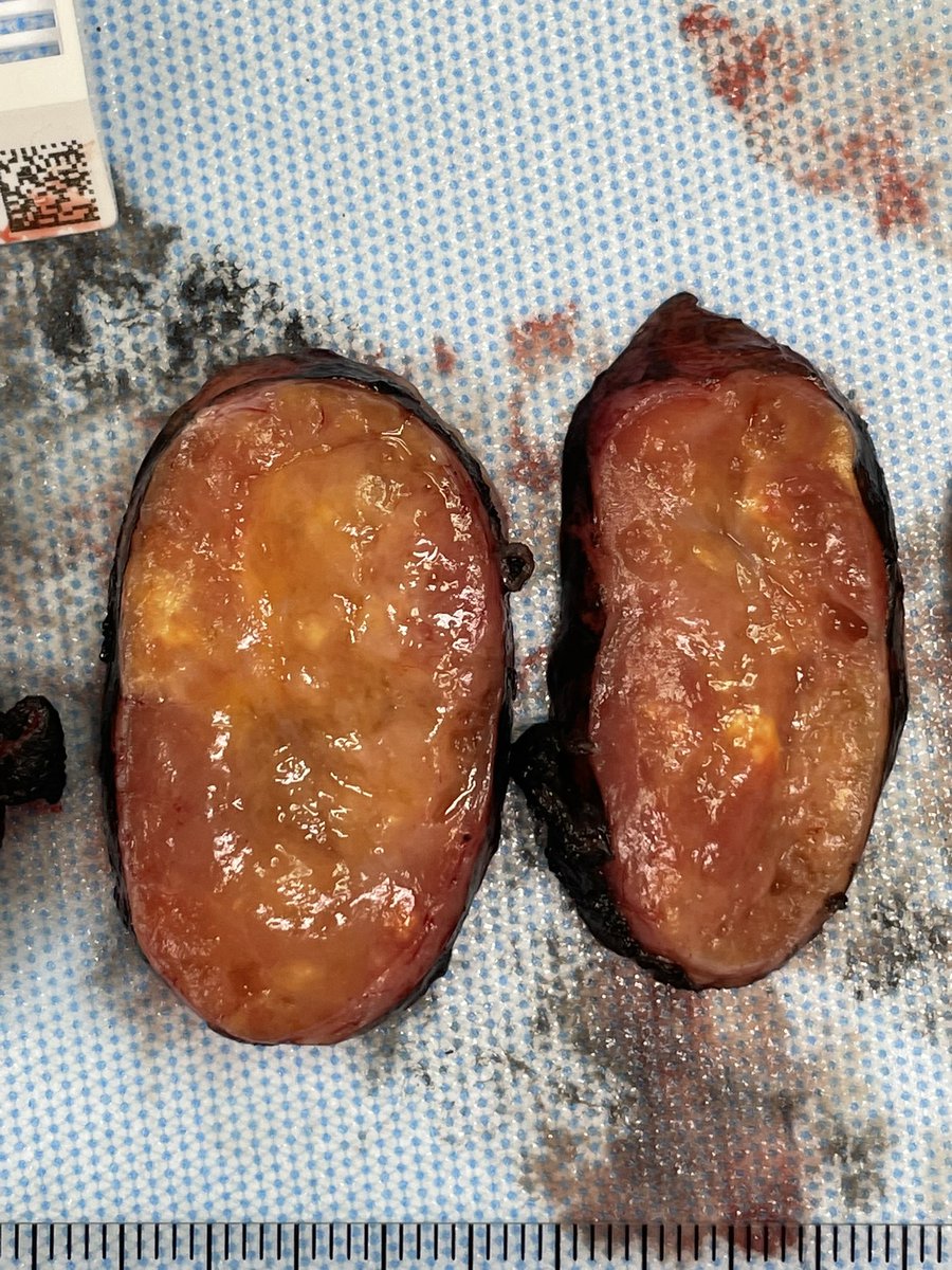
I was thinking of possible filler material, or fat necrosis. But looked really weird to me?? CD163 positive and keratin negative. Does this ring a bell for anyone ?
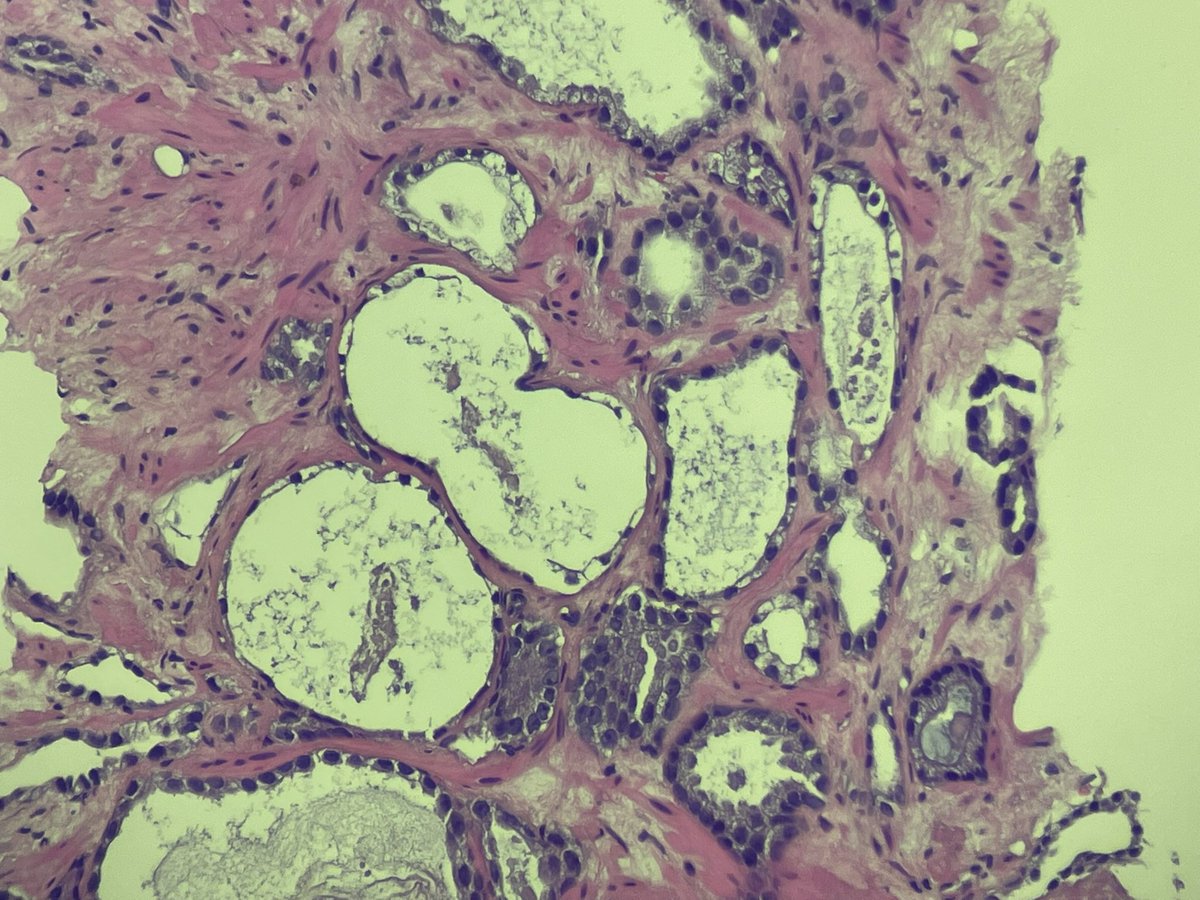
I have a strange case, was wondering if anybody has seen something like this… 37 F with gyn right labial mass. Cystic lesion with fat. I think shows papillary-like structures lined by histiocytes. And abundant degenerative material
@slusagar My very NEXT case after your post— atrophic cancer 😂. There are surrounding usual cancer glands so no problem, but out of context, a few these glands at the edge of a core… really tricky. And I think ihc may not help (thank you for all your great posts!)
@slusagar Great great great :) thank you very much for the links and references. I really appreciate it. This one focus looks angry for NA to me. But I believe that is what this must be. Thank You 👏
@slusagar Here’s CK7 and some PAX8 looking positive! Very few tubules and some stromal cells. So I think case closed fibromyxoid NA. (NKX3.1, GATA-3, CDX-2 negative)
@slusagar Thank you so much for the suggestion! History of brachytherapy would make sense too. Areas with benign-looking gland structures, and more atypical stromal looking cells. I saw the mucin and 😱. Added the Pax8 will keep you posted!
@CPremalata I didn’t do the EMA up front (perhaps I should have). Yes very richly vascular. I don’t think location makes sense for ependymoma (anterior pubic/abdominal mass) I am going to send the case out, so didn’t want to hold up any longer without knowing what it is
@kis_lorand Erg highlights the vessels, very faint staining in tumor cells (looks neg)
I can throw an inhibin or calretinin
There is some tumor necrosis and mitotic activity… so im not sure this is benign.