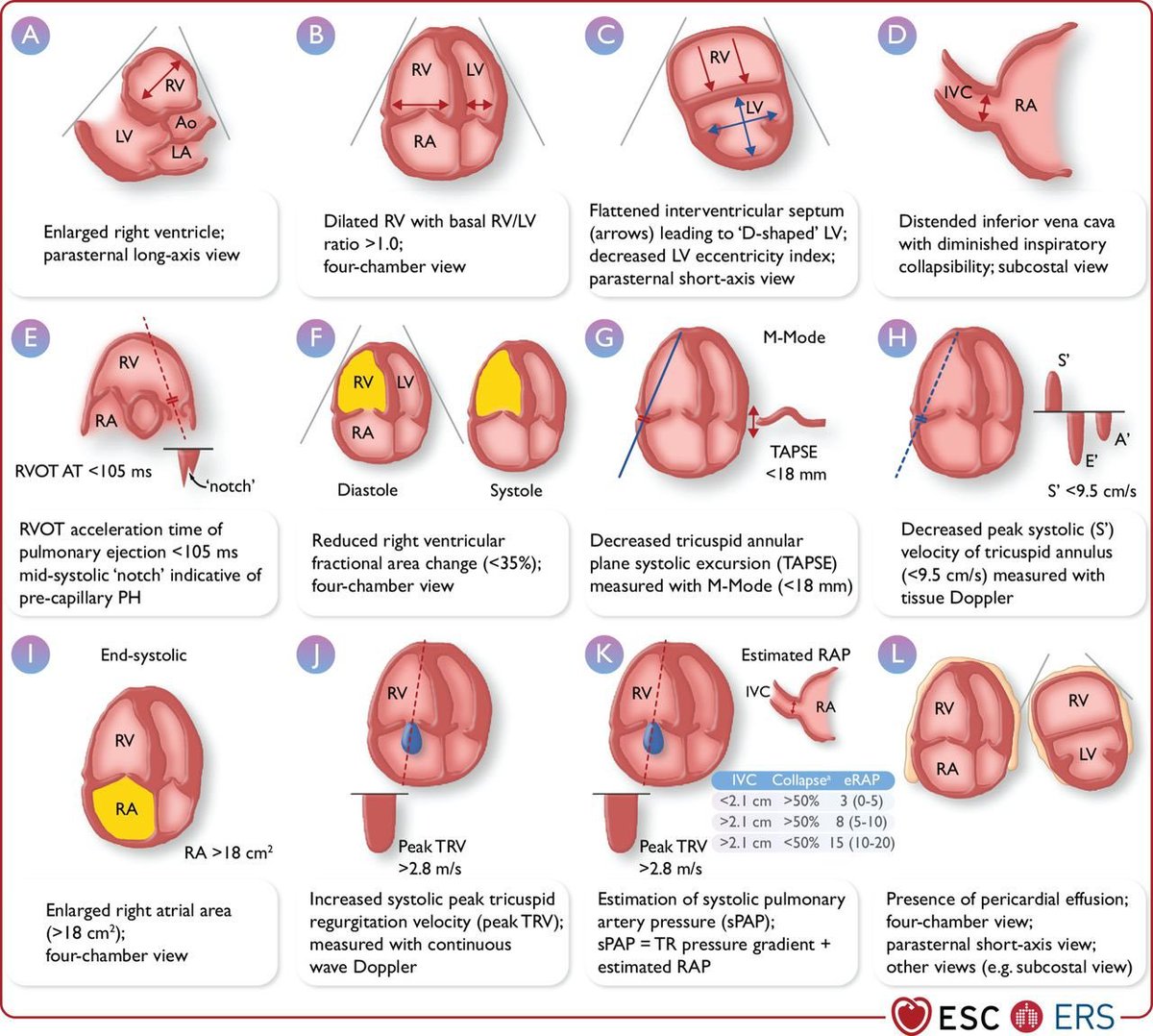
Echocardiographic parameters suggesting pulmonary hypertension
How do you recognize PH on echocardiography?
The 2022 ESC/ERS Guidelines recommend assessing multiple parameters rather than relying on a single measurement.
Key findings include:
1️⃣ Peak TRV >2.8 m/s
2️⃣ RV/LV basal diameter or area ratio >1.0
3️⃣ Flattening of the interventricular septum
4️⃣ RVOT acceleration time <105 ms or mid-systolic notching
5️⃣ TAPSE/sPAP ratio <0.55 mm/mmHg
6️⃣ RA area >18 cm²
7️⃣ IVC >21 mm with reduced inspiratory collapse
8️⃣ PA diameter >25 mm
9️⃣ Early diastolic pulmonary regurgitation velocity >2.2 m/s
Importantly, TRV should not be interpreted in isolation. Additional signs from at least 2 different categories are required to modify the echocardiographic probability of PH.
Echocardiography estimates the probability of PH; right-heart catheterization is required for definitive haemodynamic diagnosis.
Source: 2022 ESC/ERS Guidelines for the diagnosis and treatment of pulmonary hypertension.
一番くじ ドラゴンボール SPIRIT OF THE SAIYAN
10月17日発売予定
1回790円
ラストワン賞 孫悟空&孫悟飯 MASTERLISE PLUS
ラストワン賞には孫悟空と孫悟飯が筋斗雲に乗ってMASTERLISE PLUSシリーズに登場!
筋斗雲のボリューム感に加え、親子の絆が感じられるポージングで立体化され、空を駆けるような躍動感のある唯一無二のフィギュア!!
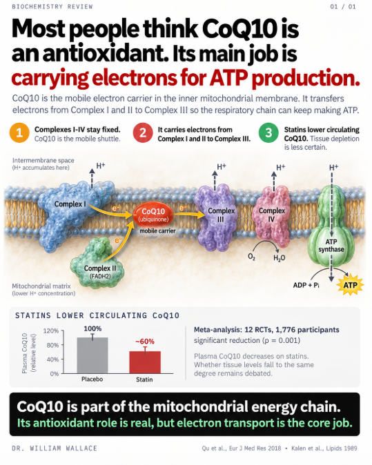
Most people who take CoQ10 think of it as an antioxidant. It is one. But that is not the most important thing it does.
CoQ10 is the only mobile electron carrier in the inner mitochondrial membrane. The electron transport chain has four protein complexes fixed in the membrane. Complex I accepts electrons from NADH. Complex II accepts them from FADH2. But neither can pass those electrons directly to Complex III. They hand them to CoQ10, which physically shuttles across the lipid bilayer to deliver them. Complex III passes them to Complex IV, which reduces oxygen to water and drives the proton gradient that ATP synthase uses to produce ATP.
Without CoQ10, the chain breaks between Complex I/II and Complex III. Electrons have nowhere to go. The proton gradient collapses. ATP production stalls. This is not an antioxidant function. This is the core mechanism of aerobic energy production.
CoQ10 is predominantly synthesized endogenously through the mevalonate pathway, the same pathway that produces cholesterol. HMG-CoA reductase is the rate-limiting enzyme. Statins inhibit HMG-CoA reductase. That is how they lower cholesterol. It is also how they lower CoQ10.
An updated meta-analysis by Qu et al. (2018) pooled 12 RCTs with 1,776 participants and found statins significantly reduced circulating CoQ10. The reduction was independent of statin type, intensity, or treatment duration. Both lipophilic and hydrophilic statins produced the same effect. This is consistent with what the biochemistry predicts: the pathway is shared.
On top of statin-induced depletion, CoQ10 in human heart tissue declines naturally with age. Kalén et al. (1989) measured CoQ10 concentrations in myocardial tissue and found levels peak around age 20, decline by more than 30% by age 40, and drop approximately 50% by age 80. The organ with the highest energy demand loses half its electron carrier over a lifetime.
A 2025 meta-analysis by Kovacic et al. (Journal of Nutritional Science, 7 RCTs, 389 patients) found CoQ10 supplementation significantly reduced statin-associated muscle symptoms measured by pain intensity. This is the most current pooled data on clinical outcomes.
One important nuance: while plasma CoQ10 depletion from statins is well established, whether intramuscular CoQ10 drops proportionally is inconsistent. Some studies found no change or even increases in muscle tissue CoQ10 during statin treatment. The plasma reduction may partly reflect reduced LDL particles, which are the primary carriers of CoQ10 in blood. The clinical significance of depletion beyond muscle symptoms remains debated.
Roughly 200 million people worldwide take statins. The mevalonate pathway that produces their target also produces the electron carrier their mitochondria depend on. The mechanism is not controversial. The clinical implications are still being defined.
Kalén et al., Lipids, 1989.
Qu et al., Eur J Med Res, 2018.
Kovacic et al., J Nutr Sci, 2025.
Most people take their full magnesium dose in one sitting. The absorption data says that strategy may not “maximize efficiency.”
Fine et al. gave healthy subjects a standard meal supplemented with increasing amounts of magnesium. At the lowest dose (36 mg), 65% was absorbed. At the highest dose (1,009 mg), only 11%. The curve was not linear. It dropped steeply at first, then flattened. Their model explained it as two simultaneous processes: an active transport channel that saturates, plus a passive route that absorbs a fixed ~7% of whatever is present.
The active channel is TRPM6. It sits in the intestinal epithelium and actively pulls magnesium ions across the membrane. It works well at low concentrations but has a ceiling. Once it is saturated, additional magnesium can only cross passively between cells (paracellular transport), driven by the concentration gradient. That passive route never saturates, but it only captures about 7% of the dose regardless of how much is present.
This is why splitting a 400 mg dose into two 200 mg doses absorbs more total magnesium. Each dose stays closer to the steep part of the curve where TRPM6 is still contributing. One large dose overwhelms the active channel, and most of the magnesium passes through unabsorbed. The unabsorbed fraction is osmotically active, pulls water into the colon, and causes the loose stools people commonly experience.
A question that comes up: does the form of magnesium change this? The absorption curve from Fine et al. used magnesium acetate, which is highly soluble. The form determines how completely and quickly the magnesium salt dissolves and releases free Mg2+ ions in the gut. Oxide dissolves poorly at intestinal pH, so much of it never becomes available. Citrate, glycinate, and acetate dissolve more readily. But once the ion is free, it faces the same TRPM6 and paracellular bottleneck regardless of what delivered it. Form determines how much Mg2+ reaches the membrane. The curve determines how much of that gets through. A poorly soluble form at a high dose is the worst combination. A highly soluble form split across meals is the best.
The RDA for magnesium is 310-420 mg per day. NHANES data consistently shows about half of US adults fall short. Splitting the dose is free, requires no product change, and the physiology is clear.
Fine et al., J Clin Invest, 1991.
Schuchardt & Hahn, Curr Nutr Food Sci, 2017.