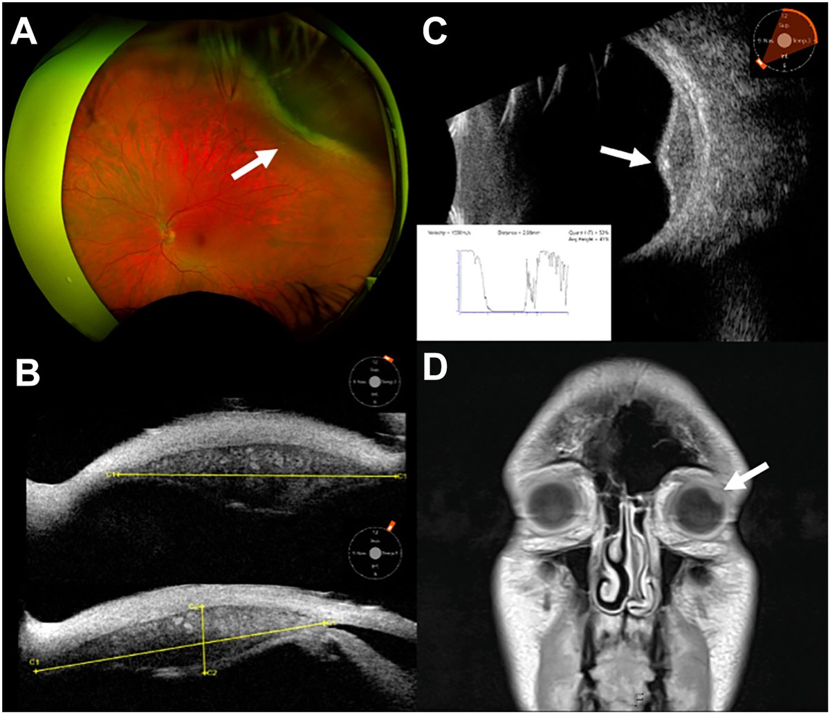
A 51-year-old man underwent brain magnetic resonance imaging that disclosed a left globe mass. Fundus imaging showed an amelanotic lesion with posterior exudative deposits, without orange pigment or subretinal fluid (A). Ultrasound biomicroscopy (B) and B-scan (C) revealed a solid ciliary body mass with marked intralesional vascularity, hyperechoic calcific deposits, and A-scan (C, subset) demonstrated low internal reflectivity. Magnetic resonance imaging showed a T1 hyperintense ciliary body mass, but was unchanged compared with magnetic resonance imaging from 10 years prior (D, 3.4 mm height). The constellation of a ciliary body mass, intrinsic vascularity, low reflectivity, increased transillumination, calcific deposits, and decade-long stability favored leiomyoma. Although definitive diagnosis would require histopathologic examination with surgical biopsy, the lesion’s indolent course supported continued surveillance.
https://t.co/CcEjynviZn
#ophthalmology #retina
@SkySportsNews That will be simpler. 2 tournaments of 32 teams (just like the old format). 64 games in South America, 64 games in Europe. 1 FINAL Boss Battle game in the middle of the Atlantic! 😁😁😁