Arachnoid cyst
It’s a fluid-filled sac located within the arachnoid membrane, one of the protective layers surrounding the brain and spinal cord. These cysts are typically congenital, meaning they develop before birth.
The brain’s most common sites for arachnoid cysts include: Middle cranial fossa, cerebellopontine angle, suprasellar region, parietal or frontal lobes.
The management of an arachnoid cyst depends on factors like size, location, and whether or not it is causing symptoms. Options include:
•Observation: If the cyst is small and asymptomatic, regular monitoring with periodic imaging may be sufficient.
•Surgical Intervention: If symptoms are present, surgery may be recommended. Procedures include:
•Cyst fenestration: Creating an opening in the cyst to allow the fluid to drain into the brain’s natural fluid spaces.
•Shunting: Inserting a shunt to drain fluid from the cyst into another part of the body, such as the abdomen.
•Endoscopic procedures: Minimally invasive techniques to drain the cyst.
Each case is individualized, and treatment decisions are made based on symptom severity, cyst location, and overall patient health.
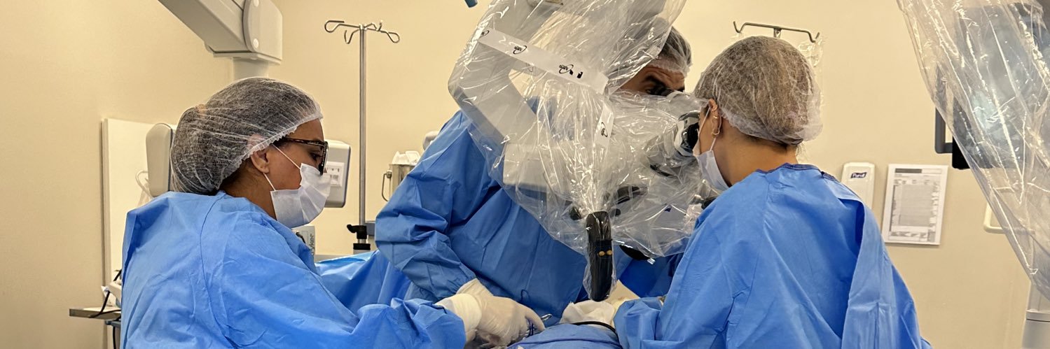
🏴🇬🇧Being involved in neurosurgery services abroad is an incredibly enriching experience. It offers a unique opportunity to learn new techniques, explore different clinical and surgical approaches, and become familiar with cutting-edge technologies that might not yet be available in one's home country.
🇧🇷 Estar envolvido em serviços de neurocirurgia no exterior é uma experiência incrivelmente enriquecedora. Ela oferece uma oportunidade única de aprender novas técnicas, explorar diferentes abordagens clínicas e cirúrgicas e se familiarizar com tecnologias de ponta que talvez ainda não estejam disponíveis em nosso país.
As shown below, Evan's index is used to support the diagnosis of normal pressure hydrocephalus.
NPH triad: cognitive decline, gait abnormality, and urinary incontinence.
#neurotip#dementia#neuroradiology
“Among the tales of sorrow and of ruin that come down to us from the darkness of those days there are yet some in which amid weeping there is joy and under the shadow of death light that endures.”
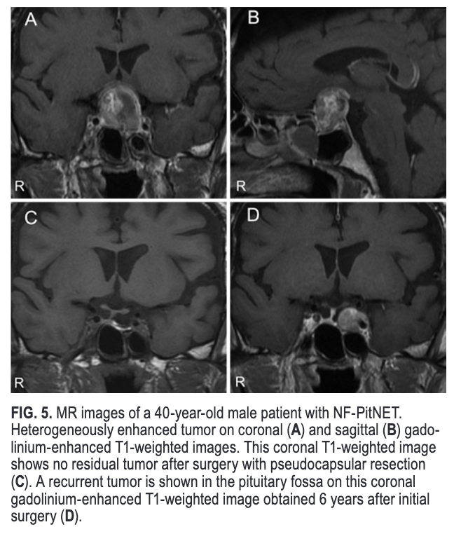
#OnlineFirst: Pseudocapsular resection to prevent recurrence in nonfunctioning pituitary neuroendocrine tumors: a retrospective, single-center study with more than 5 years of follow-up.
https://t.co/qwSOoVXqh3.
In this operative video, Dr. Cohen demonstrates anastomosis of the STA and MCA for low flow revascularization. The distal end of the donor STA is prepared by removing the surrounding soft tissue. Learn more here | https://t.co/RN7A4Bc3Nw. #MedTwitter#Neurosurgery#NSGY#surgery
Most of what we do in neurosurgery is deconstructive – taking things down and removing them. Bypass surgery is constructive – building something beneficial that did not previously exist. That’s why these moyamoya patients are among my favorite. Tips: handle intima carefully; use continuous suture technique for speed and rhythm; maximize the microscope zoom; and don’t over-snug the suture line…
#OnlineFirst: Microsurgical anatomy and approaches to thalamic gliomas. Part 1: A cartography guide for navigating to the thalamus. Integrating 3D model rendering with anatomical dissections.
https://t.co/28MobWVk1D.
Cervical spondylotic myelopathy (CSM) is a pathological condition resulting from chronic compression of the cervical spinal cord due to degenerative changes in the cervical spine. These changes include the formation of osteophytes, hypertrophy of the ligamentum flavum, spinal canal stenosis, and disc herniation, which can contribute to the compression of the spinal cord and/or nerve roots.
The clinical presentation of CSM can vary from mild symptoms to severe disability, including limb weakness, gait disturbance, loss of manual dexterity, and sphincter dysfunction. Clinical signs may include hyperreflexia, a positive Babinski sign, and sensory disturbances.
The diagnosis of CSM is based on the correlation of medical history, physical examination, and radiological findings. Magnetic resonance imaging (MRI) is the preferred imaging modality to assess spinal cord compression and soft tissue pathology. MRI can show direct spinal cord compression, intramedullary signal changes suggesting myelomalacia, and other degenerative changes in the cervical spine.
Management of CSM can be conservative or surgical. Conservative treatment includes medications, physical therapy, and activity modifications. However, in cases of progressive or severe symptoms, or when there is evidence of myelopathy on MRI, surgical intervention may be considered.
Surgical options for CSM include:
1. Anterior decompression, such as anterior cervical discectomy and fusion (ACDF) or corpectomy.
2. Posterior decompression, such as cervical laminoplasty or laminectomy, with or without fusion.
3. Combined decompression and fusion, which may be necessary in cases of significant deformity or instability.
The choice of surgical approach depends on several factors, including the location and extent of compression, the presence of spinal deformity or instability, and the surgeon's preference based on experience and clinical outcomes.
Surgery aims to decompress the spinal cord and stabilize the cervical spine to prevent the progression of myelopathy and improve symptoms. Postoperative recovery can vary, and some patients may require intensive rehabilitation to maximize functional recovery.
Long-term follow-up is important to monitor spinal stability, the progression of adjacent segment degenerative disease, and neurological function. CSM is a complex condition that requires a multidisciplinary approach for its management.
Intra‐arterial Selective Bevacizumab Administration in the Middle Meningeal Artery for Chronic Subdural Hematoma: An Early Experience in 12 Hemispheres | Stroke: Vascular and Interventional Neurology https://t.co/d4Rr5gJoce @SVINJournal@StrokeAHA_ASA@svinsociety
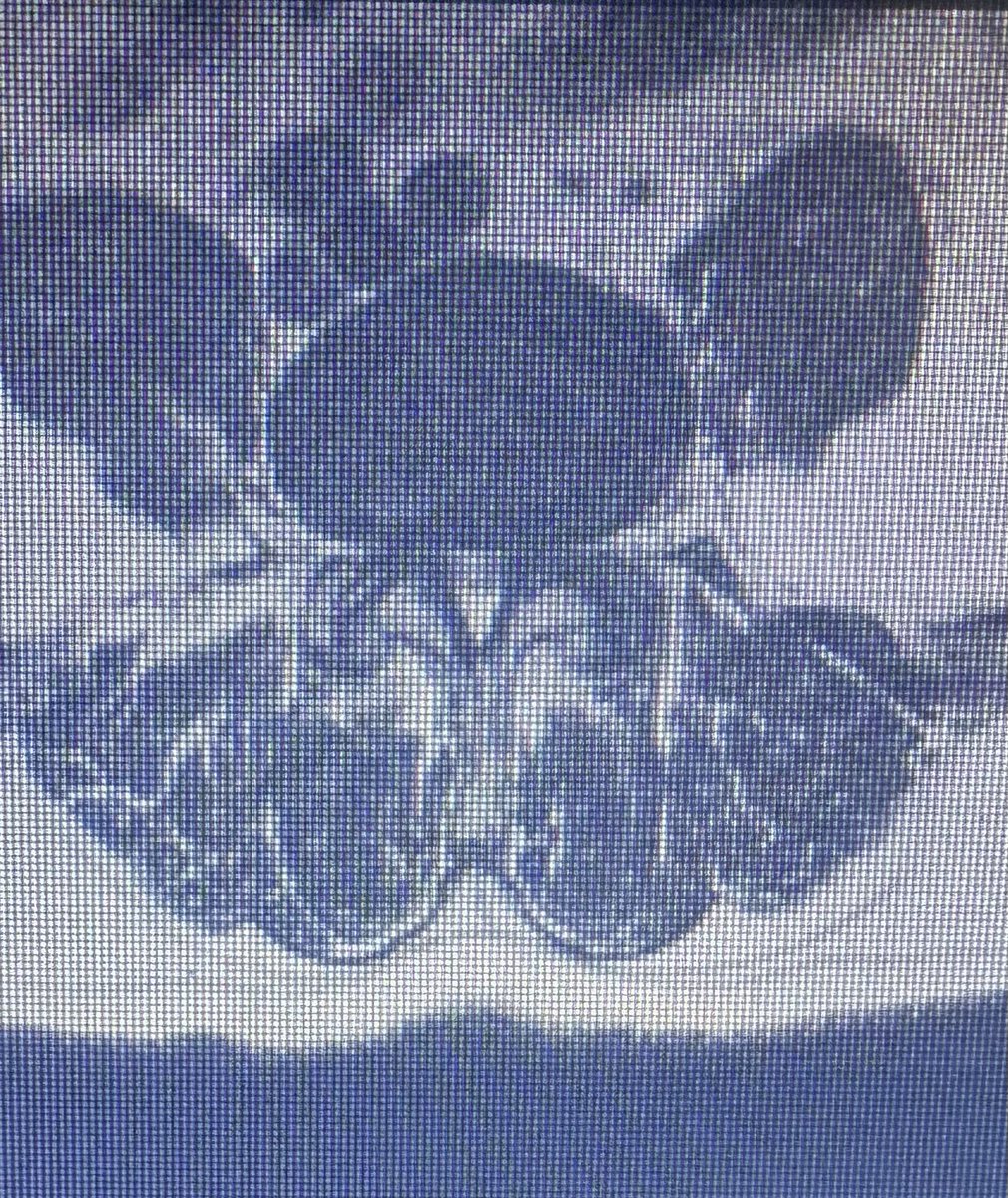
When the sagittal view don’t show us how severe is the stenosis. It’s necessary to evaluate the axial view that is indispensable for an accurate and complete diagnosis of conditions affecting the lumbar spine, providing crucial information that complements sagittal and coronal views in MRI.
The importance of this view includes:
1. Visualization of Neural Structures: It allows a clear visualization of the spinal cord, nerve roots, and dural sac, essential for assessing compressions, disc herniations, and stenosis.
2. Assessment of Facet Joints: It facilitates the observation of facet joints, enabling the identification of facet joint arthropathies, which can be a common cause of low back pain.
3. Identification of Disc Herniations: It helps to identify disc protrusions or extrusions that may not be as evident in sagittal views, as well as to evaluate the direction and extent of herniations.
4. Study of Bone Anatomy: It provides a cross-sectional view of the vertebrae, helping to identify fractures, bone lesions, and degenerative changes.
5. Detection of Pathological Lesions: It aids in the identification of tumors, infections, and other pathological lesions that may affect the lumbar spine.