Clinical 7T gradients can go fast — but peripheral nerve stimulation (PNS) stops you at high-res fMRI. ⚡
Meet POPE 🌍: we reshape EPI pulses to dodge PNS exactly when it spikes → robust 0.3mm fMRI, seeing intracortical veins. 🧠
w/ @SiemensHealth
👉 https://t.co/a4QexD81KH
Last chance to apply for the 2026 In Vivo Magnetic Resonance Gordon Research Conference! Deadline June 14. https://t.co/mo2xo0205D Join us July 12–17, NH, USA for frontier science, discussion and a friendly community. Theme: Advancing Beyond Limits.
Join us for the 7T Impulse Symposium at the Martinos Center, Nov. 5th, 2026.
Program & Registration: https://t.co/r4mh55DtSi
Location (in person): 114 16th street at Martinos Center
Virtual access will be provided.
Join the Connectome 2.0 Dissemination Workshop on June 3, 2026, 9 AM-4 PM EST at the MGH Martinos Center. Learn about ultra-high gradient MRI, diffusion MRI, connectivity, and microstructural imaging using the Connectome 2.0 scanner. Program details: https://t.co/Yw6oa8rSxz
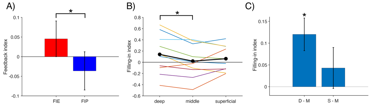
My first PhD project has finally taken shape and is now available as a preprint after 3 years! We found a feedback signature in V1 during Troxler perceptual filling-in using 7T layer-fMRI. Please check it out ;))
https://t.co/pmdjbOE7wz
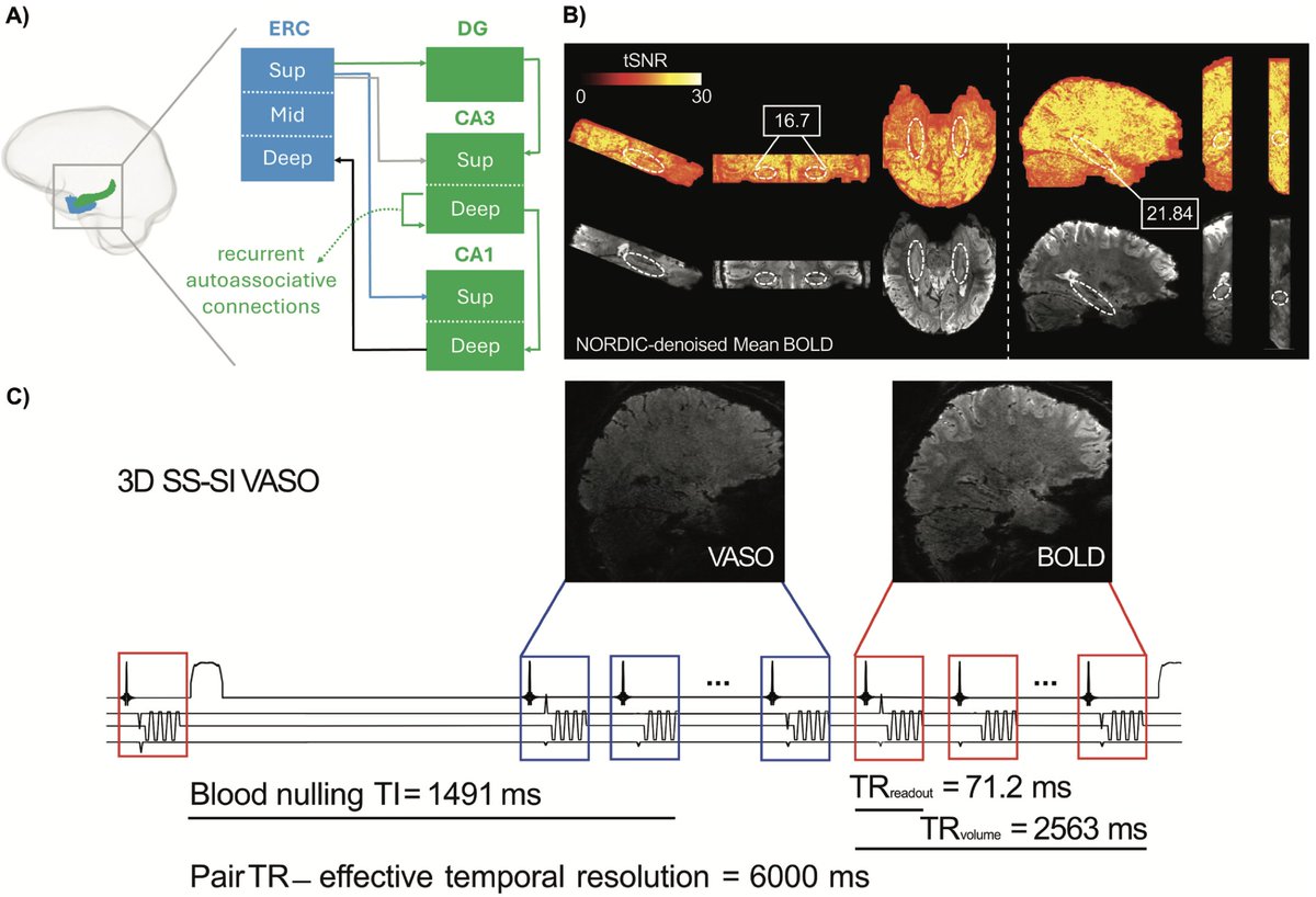
New paper in Imaging Neuroscience by Khazar Ahmadi, Nikolai Axmacher, et al:
Blood volume-sensitive laminar fMRI with VASO in human hippocampus: Capabilities and biophysical challenges at clinical 7T scanners
https://t.co/LZXbhYACxp
If you are setting up the meso-veins protocol for the first time at your 7T (https://t.co/FjE3J3vsE6), you should wath this video where I am providing the most up to date pointers. As well as the tips on quality controlling your images: https://t.co/Guplqdk76r
Interested in understanding your high-res data better? @ofgulban enhanced LayNii IDA with some very useful QA and interactive viewing tools. In the last LayNii release.
https://t.co/NafICaCMg6
New LayNii v2.10.0 is out!
https://t.co/2rExyxjxfF
🚀 LN2_FRISGO: This program mitigates the Fuzzy Ripple artifacts in dual polarity 3D-EPI fMRI data.
💾 Big data ready: Now handling >5GB NIFTIs to support the mesoscopic shift toward high-res, whole-brain images.
@layerfMRI
A new MRI protocol can scan the human #brain and its venous network in under 7 minutes, marking it as a potential tool for diagnosing cerebrovascular diseases and monitoring neurodegeneration. @ofgulban https://t.co/Ok30agpYxq
In a satirical commentary, @ofgulban examines the widening gap between traditional publication norms and emerging channels of science communication: https://t.co/8VqvrkLZqy
@OHBM#FutureofPublishing#SpecialIssue
Yesterday, @ofgulban gave a great talk about efficient imaging of small veins.
It was inspiring. Impressive how much information is there waiting to be harvested.
https://t.co/qFWabcCZPc
Happy to share that our new preprint is now available!
Digging into our previous dataset, we found a feedforward signature in FFA during passive face image presentations, suggesting face information is structured prior to FFA.
Please find more details in the manuscript 😁
New layer-fMRI preprint using deep sampling to track face information across layers in FFA.
50 times 15 min scanning with whole brain VASO and BOLD fMRI.
By Kenshu Koiso et al.
https://t.co/E6PltTV8Up