Mitral valve anatomy on TEE
The mitral valve has two leaflets:
⭕ Anterior leaflet: A1, A2, A3
⭕ Posterior leaflet: P1, P2, P3
The posterior leaflet has three anatomical scallops, while the anterior segments are named according to the opposing posterior scallops.
Think of it as:
A1 ↔ P1
A2 ↔ P2
A3 ↔ P3
The mitral apparatus is more than just the leaflets.
It includes:
◻️Mitral annulus
◻️Anterior and posterior leaflets
◻️ Commissures
◻️ Chordae tendineae
◻️ Anterolateral and posteromedial papillary muscles
◻️ Left atrium and left ventricle
The annulus has a characteristic saddle-shaped, nonplanar geometry, rather than being a simple flat circle.
Why does segmental anatomy matter?
When assessing mitral regurgitation, identifying the precise diseased segment helps define the mechanism and guides surgical or transcatheter treatment.
For example, a prolapsing P2 segment can be described much more precisely than simply saying (posterior leaflet prolapse).
2D TEE uses different multiplane angles and probe positions to interrogate the valve.
3D TEE provides an en-face view of the mitral valve from the left atrial perspective, closely resembling the surgeon’s view. This makes it particularly valuable for localizing leaflet pathology and planning structural interventions.
A practical memory aid:
P1 = lateral
P2 = middle
P3 = medial
And the corresponding anterior segments are:
A1 = lateral
A2 = middle
A3 = medial
Mastering A1–A3 and P1–P3 is fundamental for interpreting mitral valve pathology on echocardiography.
🫀🚨 𝗜𝗻𝘀𝘂𝗳𝗶𝗰𝗶𝗲𝗻𝗰𝗶𝗮 𝗰𝗮𝗿𝗱𝗶𝗮𝗰𝗮 𝗮𝗴𝘂𝗱𝗮 ‼️
@JACCJournals@ACCinTouch
👇🏼👇🏼👇🏼👇🏼
📑🔗🔑🔓
https://t.co/89ht2mBRSA
⬇️⬇️⬇️⬇️
🧵👇
La revisión State-of-the-Art de 𝙅𝘼𝘾𝘾 𝟮𝟬𝟮𝟲 propone un cambio de paradigma:
💧 aliviar congestión rápidamente
🔍 identificar y tratar el precipitante
🧬 definir la cardiopatía subyacente
💊 iniciar precozmente terapia modificadora de enfermedad
📈 planear la trayectoria después del alta
El acrónimo 𝙎𝙋𝙇𝘼𝙎𝙃 resume la evaluación inicial:
𝙎ymptoms
𝙋ast history
𝙇ife signs
𝘼ssess congestion
𝙎TEMI/equivalente
𝙃ypoperfusion
🔥 La hospitalización es una 𝙫𝙚𝙣𝙩𝙖𝙣𝙖 𝙩𝙚𝙧𝙖𝙥𝙚́𝙪𝙩𝙞𝙘𝙖 𝙥𝙖𝙧𝙖 𝙢𝙤𝙙𝙞𝙛𝙞𝙘𝙖𝙧 𝙚𝙡 𝙥𝙧𝙤𝙣𝙤́𝙨𝙩𝙞𝙘𝙤, no simplemente para hacer desaparecer la disnea.
💉💧 𝙁𝙪𝙧𝙤𝙨𝙚𝙢𝙞𝙙𝙖: 𝙣𝙤 𝙗𝙖𝙨𝙩𝙖 𝙘𝙤𝙣 𝙖𝙙𝙢𝙞𝙣𝙞𝙨𝙩𝙧𝙖𝙧𝙡𝙖. 𝙃𝙖𝙮 𝙦𝙪𝙚 𝙨𝙖𝙗𝙚𝙧 𝙚𝙣 𝟮 𝙝𝙤𝙧𝙖𝙨 𝙨𝙞 𝙛𝙪𝙣𝙘𝙞𝙤𝙣𝙤́
Si existe alta sospecha de IC aguda con congestión, el artículo recomienda iniciar el diurético IV 𝙨𝙞𝙣 𝙚𝙨𝙥𝙚𝙧𝙖𝙧 𝙖 𝙘𝙤𝙢𝙥𝙡𝙚𝙩𝙖𝙧 𝙩𝙤𝙙𝙖𝙨 𝙡𝙖𝙨 𝙥𝙧𝙪𝙚𝙗𝙖𝙨 𝙙𝙞𝙖𝙜𝙣𝙤́𝙨𝙩𝙞𝙘𝙖𝙨.
💊 Sin tratamiento previo o dosis baja: 𝙛𝙪𝙧𝙤𝙨𝙚𝙢𝙞𝙙𝙖 𝟰𝟬 𝙢𝙜 𝙄𝙑
💊 Dosis oral crónica alta: ≥𝟴𝟬 𝙢𝙜 𝙄𝙑 𝙤 ≈𝟮× 𝙡𝙖 𝙙𝙤𝙨𝙞𝙨 𝙤𝙧𝙖𝙡
A las 𝟮 𝙝𝙤𝙧𝙖𝙨, evaluar:
✅ 𝘿𝙞𝙪𝙧𝙚𝙨𝙞𝙨 ≥𝟯𝟬𝟬 𝙢𝙇
o
✅ 𝙉𝙖 𝙪𝙧𝙞𝙣𝙖𝙧𝙞𝙤 ≥𝟳𝟬–𝟴𝟬 𝙢𝙢𝙤𝙡/𝙇
Si no se alcanza:
⬆️ duplicar la dosis de asa
➕ considerar 𝙖𝙘𝙚𝙩𝙖𝙯𝙤𝙡𝙖𝙢𝙞𝙙𝙖 𝙤 𝙩𝙞𝙖𝙯𝙞𝙙𝙖
🔍 excluir causas intrarrenales/postrenales
🔥 Esperar 24 horas para descubrir que el diurético no funcionó es perder tiempo de descongestión.
🫘📈 𝘾𝙧𝙚𝙖𝙩𝙞𝙣𝙞𝙣𝙖 𝙦𝙪𝙚 𝙖𝙪𝙢𝙚𝙣𝙩𝙖 𝙙𝙪𝙧𝙖𝙣𝙩𝙚 𝙪𝙣𝙖 𝙙𝙚𝙨𝙘𝙤𝙣𝙜𝙚𝙨𝙩𝙞𝙤́𝙣 𝙚𝙛𝙞𝙘𝙖𝙯 ≠ 𝙛𝙧𝙖𝙘𝙖𝙨𝙤 𝙩𝙚𝙧𝙖𝙥𝙚́𝙪𝙩𝙞𝙘𝙤
Uno de los errores más frecuentes es reducir o suspender diuréticos automáticamente ante una elevación moderada de creatinina.
La revisión enfatiza:
🫘 en ERC pueden necesitarse 𝙙𝙤𝙨𝙞𝙨 𝙢𝙖𝙮𝙤𝙧𝙚𝙨 de diuréticos
📈 la creatinina puede aumentar transitoriamente durante una descongestión efectiva
🚫 esto no obliga por sí solo a detener el tratamiento
🎯 el contexto de 𝙘𝙤𝙣𝙜𝙚𝙨𝙩𝙞𝙤́𝙣 𝙧𝙚𝙨𝙞𝙙𝙪𝙖𝙡 𝙮 𝙧𝙚𝙨𝙥𝙪𝙚𝙨𝙩𝙖 𝙙𝙞𝙪𝙧𝙚́𝙩𝙞𝙘𝙖 importa más que una cifra aislada
Además, la congestión persistente al alta se asocia con peor pronóstico.
📌 Una reducción de aproximadamente 𝟯𝟬% 𝙙𝙚𝙡 𝙉𝙏-𝙥𝙧𝙤𝘽𝙉𝙋 desde el ingreso al alta se considera compatible con descongestión adecuada dentro de una valoración multimodal.
🔥 En IC aguda, proteger la creatinina a costa de dejar al paciente congestionado puede ser una falsa victoria.
💊⚡ 𝙀𝙡 𝙜𝙧𝙖𝙣 𝙘𝙖𝙢𝙗𝙞𝙤: 𝙡𝙖 𝙂𝘿𝙈𝙏 𝙙𝙚𝙗𝙚 𝙘𝙤𝙢𝙚𝙣𝙯𝙖𝙧 𝙙𝙪𝙧𝙖𝙣𝙩𝙚 𝙡𝙖 𝙝𝙤𝙨𝙥𝙞𝙩𝙖𝙡𝙞𝙯𝙖𝙘𝙞𝙤́𝙣, 𝙣𝙤 “𝙖𝙡𝙜𝙪́𝙣 𝙙𝙞́𝙖” 𝙚𝙣 𝙘𝙤𝙣𝙨𝙪𝙡𝙩𝙖 𝙚𝙭𝙩𝙚𝙧𝙣𝙖
Secuencia rápida después de la estabilización:
🔹 𝙞𝙎𝙂𝙇𝙏𝟮 → desde los primeros días si eGFR ≥20 mL/min/1,73 m²
🔹 𝘼𝙍𝙈 → tempranamente si presión, función renal y K⁺ lo permiten
🔹 𝘼𝙍𝙉𝙄/𝙍𝘼𝙎𝙄 → tras estabilización, preferentemente con congestión leve o euvolemia
🔹 β-𝙗𝙡𝙤𝙦𝙪𝙚𝙖𝙙𝙤𝙧 → mantener si estable; iniciar en HFrEF cuando mejore la congestión
📊 La evidencia citada es potente:
⚡ 𝙋𝙄𝙊𝙉𝙀𝙀𝙍-𝙃𝙁: sacubitrilo/valsartán → ↓66% del endpoint clínico compuesto exploratorio
⚡ 𝙀𝙈𝙋𝙐𝙇𝙎𝙀: empagliflozina → ↓31% de muerte CV/eventos de IC
⚡ 𝙎𝙊𝙇𝙊𝙄𝙎𝙏-𝙒𝙃𝙁: sotagliflozina → ↓33%
⚡ 𝙎𝙏𝙍𝙊𝙉𝙂-𝙃𝙁: estrategia intensiva → ↓34% de muerte o rehospitalización por IC
🔥 El predictor más fuerte de que un paciente NO reciba GDMT después del alta es que 𝙣𝙤 𝙨𝙚 𝙡𝙖 𝙝𝙖𝙮𝙖𝙣 𝙞𝙣𝙞𝙘𝙞𝙖𝙙𝙤 𝙙𝙪𝙧𝙖𝙣𝙩𝙚 𝙡𝙖 𝙝𝙤𝙨𝙥𝙞𝙩𝙖𝙡𝙞𝙯𝙖𝙘𝙞𝙤́𝙣.
🎯 𝙀𝙡 𝙖𝙡𝙩𝙖 𝙣𝙤 𝙩𝙚𝙧𝙢𝙞𝙣𝙖 𝙚𝙡 𝙩𝙧𝙖𝙩𝙖𝙢𝙞𝙚𝙣𝙩𝙤: 𝙘𝙤𝙢𝙞𝙚𝙣𝙯𝙖 𝙡𝙖 “𝙛𝙖𝙨𝙚 𝙫𝙪𝙡𝙣𝙚𝙧𝙖𝙗𝙡𝙚”
La estrategia contemporánea exige salir del hospital con:
✅ 𝙚𝙪𝙫𝙤𝙡𝙚𝙢𝙞𝙖 𝙤 𝙙𝙚𝙨𝙘𝙤𝙣𝙜𝙚𝙨𝙩𝙞𝙤́𝙣 𝙤́𝙥𝙩𝙞𝙢𝙖
✅ GDMT iniciada y plan de titulación
✅ educación del paciente y cuidadores
✅ evaluación de hierro, función renal y comorbilidades
✅ plan para dispositivos cuando corresponda
✅ seguimiento 𝙨𝙚𝙢𝙖𝙣𝙖𝙡 𝙤 𝙘𝙖𝙙𝙖 𝟮 𝙨𝙚𝙢𝙖𝙣𝙖𝙨 𝙙𝙪𝙧𝙖𝙣𝙩𝙚 𝙚𝙡 𝙥𝙧𝙞𝙢𝙚𝙧 𝙢𝙚𝙨
Objetivo:
🚀 alcanzar la implementación completa y dosis toleradas de las terapias indicadas en 𝙨𝙚𝙢𝙖𝙣𝙖𝙨, idealmente antes de 𝟯 𝙢𝙚𝙨𝙚𝙨.
❌ paradigma antiguo:
𝙘𝙤𝙣𝙜𝙚𝙨𝙩𝙞𝙤́𝙣 → 𝙙𝙞𝙪𝙧𝙚́𝙩𝙞𝙘𝙤 → 𝙖𝙡𝙩𝙖 → 𝙣𝙪𝙚𝙫𝙖 𝙘𝙤𝙣𝙜𝙚𝙨𝙩𝙞𝙤́𝙣
✅ paradigma moderno:
𝙙𝙚𝙨𝙘𝙤𝙣𝙜𝙚𝙨𝙩𝙞𝙤́𝙣 → 𝙂𝘿𝙈𝙏 𝙧𝙖́𝙥𝙞𝙙𝙖 → 𝙧𝙚𝙢𝙤𝙙𝙚𝙡𝙖𝙙𝙤 𝙧𝙚𝙫𝙚𝙧𝙨𝙤 → 𝙢𝙚𝙣𝙤𝙧 𝙧𝙞𝙚𝙨𝙜𝙤 𝙙𝙚 𝙣𝙪𝙚𝙫𝙖 𝙄𝘾 → 𝙥𝙤𝙨𝙞𝙗𝙡𝙚 𝙧𝙚𝙢𝙞𝙨𝙞𝙤́𝙣
🔥 𝙈𝙚𝙣𝙨𝙖𝙟𝙚 𝙛𝙞𝙣𝙖𝙡: el tratamiento contemporáneo de la insuficiencia cardiaca aguda ya no termina cuando el paciente deja de tener edema. El verdadero objetivo es utilizar la hospitalización para 𝙩𝙧𝙖𝙣𝙨𝙛𝙤𝙧𝙢𝙖𝙧 𝙪𝙣𝙖 𝙙𝙚𝙨𝙘𝙤𝙢𝙥𝙚𝙣𝙨𝙖𝙘𝙞𝙤́𝙣 𝙖𝙜𝙪𝙙𝙖 𝙚𝙣 𝙚𝙡 𝙞𝙣𝙞𝙘𝙞𝙤 𝙙𝙚 𝙪𝙣𝙖 𝙚𝙨𝙩𝙧𝙖𝙩𝙚𝙜𝙞𝙖 𝙙𝙚 𝙧𝙚𝙢𝙞𝙨𝙞𝙤́𝙣 𝙙𝙚 𝙡𝙖 𝙚𝙣𝙛𝙚𝙧𝙢𝙚𝙙𝙖𝙙
📚📖 Más en 𝕏 @MarlonVFZR y en el blog 👉 [https://t.co/wbCJFKHCXL]
‼️Si te sirve: ❤️ Me gusta | 🔁 Repost | ➕ Follow para más👇🏼👇🏼👇🏼👇🏼
📚📖#ClubCrit👨🏻⚕️👨🏻🏫🧠🫶
#InsuficienciaCardiaca #AcuteHeartFailure #Cardiología
#FOAMed #FOAMcc #CriticalCare #CriticalCare #CuidadoCritico #MedTwitter #MedX #IntensiveCare #MedXCommunity #MedED
@fsmcruz Entendo não gostarem de Neto. Mas fingir que o PT tem feito um bom trabalho na Bahia, não dá. Pior ainda é tentar forçar que Jerônimo lacrou nesse debate.
🟥Acoplamiento Ventrículo-Arterial izquierdo [VAC]. 🔎🫀💥
🔴El VAC describe que tan bien están coordinados el ventrículo izquierdo y el sistema arterial durante la eyección. Esta relación se expresa como: VAC = Ea/Ees.
🔺️Ea (elastancia arterial): representa la carga arterial global o poscarga que enfrente el VI. Integra la resistencia vascular, distensibilidad arterial y FC. Una Ea ⤴️ sugiere mayor carga para el VI.
🔺️Ees (elastancia telesistólica): representa la capacidad contractil del VI para generar presión. Una Ees ⤵️ sugiere menor capacidad contractil.
📐Actualmente puede estimarse de forma no invasiva mediante ecocardiografía, utilizando el método se "un sólo latido", descrito por Chen et al.
📊Valores normales:
🔺️Ea: 2.2 ± 0.8 mmHg/mL.
🔺️Ees: 2.3 ± 1.0 mmHg/mL.
🔺️Ea/Ees: 1.0 ± 0.36
*En sujetos sanos, el sistema tiende fisiológicamente hacia un VAC ~0.5, asociado con una eficiencia metabólica óptima y una FEVI ~66%.
✅️VAC ~0.5 = máxima eficiencia energética. 👌🏻💓
⚠️VAC ~1.0 = máximo trabajo sistólico.
🚫VAC >1.3-1.4 = desacoplamiento.
https://t.co/rvQd0x1XtY
@jonasdiandrade@brunabragaxx O povo acha que 40 mil reais pagando aluguel e prestação de carro, plano de saúde, vive ostentando, é muito? Conheço gente que ganha muito mais e tá endividado.
The #BLUEProtocol is a rapid bedside #lung ultrasound technique for patients with acute respiratory failure. Using 6 standardized scanning points, this protocol helps identify common causes of acute dyspnea, such as #pneumonia, pulmonary edema, and pneumothorax, in less than 5 minutes.
📖 Read the full article to learn more: https://t.co/S7YvgiGeMs
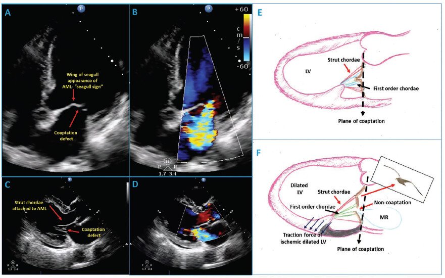
The Seagull Sign of Anterior Mitral Leaflet
🩺 What is the Seagull Sign?
In echo, the mitral valve seagull sign refers to a distinctive wing-like distortion or "kink" in the middle of the anterior mitral valve leaflet during systole, mimicking the wings of a flying seagull.
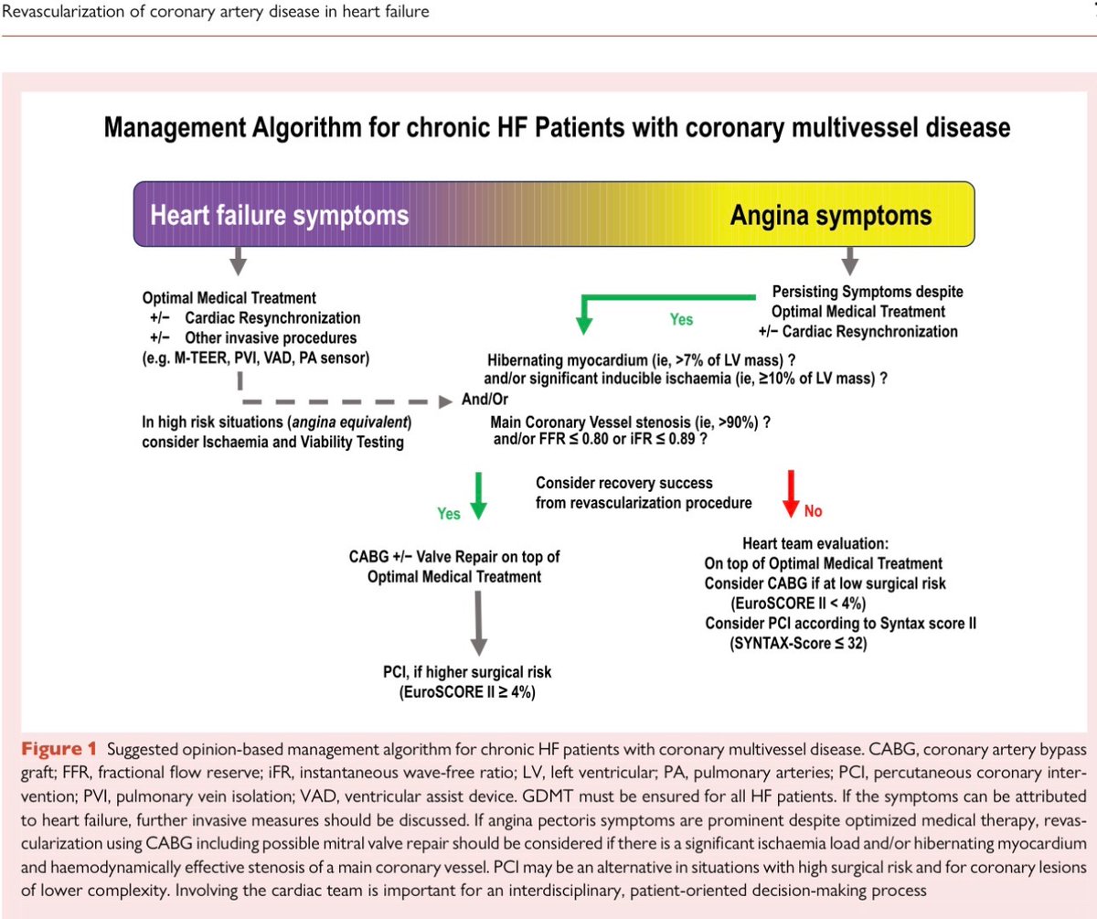
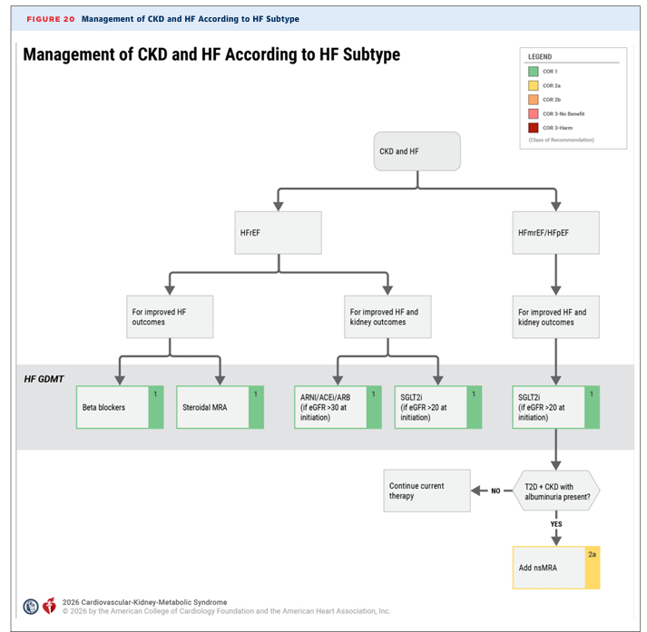
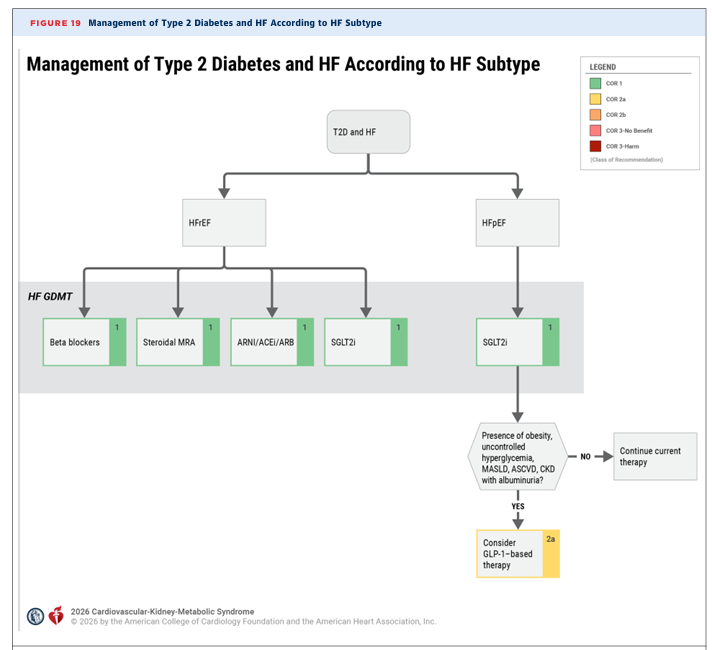
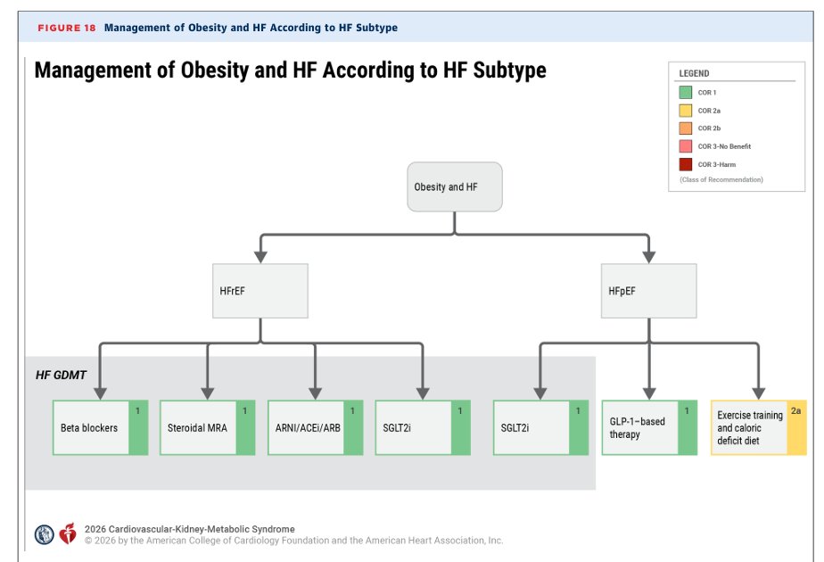
New HFpEF consensus pathway lays out clear approach:
💠SGLT2i + (ns)MRA, unless contraindicated
💠+GLP1, if obese
💠Consider ARNI
💠AVOID BB as able
CKM GL give framework in context of comorbidities.
🚨Need to make sure we don't delay effective therapies down the decision tree!🚨
Precisión diagnóstica de la ecocardiografía transesofágica mediante aproximación bicomisural en la evaluación de la anatomía valvular mitral
🗂️ https://t.co/INQVIKsKT5 ◀️
Repaso del comentario crítico publicado en "Lo mejor de la literatura en..."
For SAVR, the current scheme works pretty well.
PR cannot be captured by Doppler but increased likelihood in (a) TAVI (and Doppler often higher than invasive in TAVi for other reasons than PR) (b) at the center of bileaflet AVR (mostly not clin relevant) and (c) when small aorta < 3 cm. PR is less an issue in true severe stenosis, much more in true moderate AS.
El ECO puede AYUDAR a elegir candidatos a Terapia de Resincronizacion Cardiaca
No es solo el retraso eléctrico (QRS)…
Es la dissinconía mecánica la que importa
Qué buscar en el eco:
✅ Flash septal (SF) → contracción septal temprana + rebote
✅ Balanceo apical (ApRock) → movimiento bifásico del ápice
✅ Juntos = SFoAR = dissinconía mecánica
1️⃣ Adquisición multiviendo de alta calidad
2️⃣ Detectar flash septal
3️⃣ Detectar balanceo apical
4️⃣ Integrar → diagnosticar dissinconía
Por qué importa?
❗ 20–45% de los pacientes con CRT son no respondedores
Presencia de SF/ApRock = mayor probabilidad de respuesta
Evaluación visual = herramienta simple, reproducible, al lado de la cama
Dos conceptos: Eléctrico ≠ mecánico
(No es lo mismo)
El eco puede refinar la selección de pacientes más allá de las guías


![MarlonVFZR's tweet photo. 🫀🚨 𝗜𝗻𝘀𝘂𝗳𝗶𝗰𝗶𝗲𝗻𝗰𝗶𝗮 𝗰𝗮𝗿𝗱𝗶𝗮𝗰𝗮 𝗮𝗴𝘂𝗱𝗮 ‼️
@JACCJournals @ACCinTouch
👇🏼👇🏼👇🏼👇🏼
📑🔗🔑🔓
https://t.co/89ht2mBRSA
⬇️⬇️⬇️⬇️
🧵👇
La revisión State-of-the-Art de 𝙅𝘼𝘾𝘾 𝟮𝟬𝟮𝟲 propone un cambio de paradigma:
💧 aliviar congestión rápidamente
🔍 identificar y tratar el precipitante
🧬 definir la cardiopatía subyacente
💊 iniciar precozmente terapia modificadora de enfermedad
📈 planear la trayectoria después del alta
El acrónimo 𝙎𝙋𝙇𝘼𝙎𝙃 resume la evaluación inicial:
𝙎ymptoms
𝙋ast history
𝙇ife signs
𝘼ssess congestion
𝙎TEMI/equivalente
𝙃ypoperfusion
🔥 La hospitalización es una 𝙫𝙚𝙣𝙩𝙖𝙣𝙖 𝙩𝙚𝙧𝙖𝙥𝙚́𝙪𝙩𝙞𝙘𝙖 𝙥𝙖𝙧𝙖 𝙢𝙤𝙙𝙞𝙛𝙞𝙘𝙖𝙧 𝙚𝙡 𝙥𝙧𝙤𝙣𝙤́𝙨𝙩𝙞𝙘𝙤, no simplemente para hacer desaparecer la disnea.
💉💧 𝙁𝙪𝙧𝙤𝙨𝙚𝙢𝙞𝙙𝙖: 𝙣𝙤 𝙗𝙖𝙨𝙩𝙖 𝙘𝙤𝙣 𝙖𝙙𝙢𝙞𝙣𝙞𝙨𝙩𝙧𝙖𝙧𝙡𝙖. 𝙃𝙖𝙮 𝙦𝙪𝙚 𝙨𝙖𝙗𝙚𝙧 𝙚𝙣 𝟮 𝙝𝙤𝙧𝙖𝙨 𝙨𝙞 𝙛𝙪𝙣𝙘𝙞𝙤𝙣𝙤́
Si existe alta sospecha de IC aguda con congestión, el artículo recomienda iniciar el diurético IV 𝙨𝙞𝙣 𝙚𝙨𝙥𝙚𝙧𝙖𝙧 𝙖 𝙘𝙤𝙢𝙥𝙡𝙚𝙩𝙖𝙧 𝙩𝙤𝙙𝙖𝙨 𝙡𝙖𝙨 𝙥𝙧𝙪𝙚𝙗𝙖𝙨 𝙙𝙞𝙖𝙜𝙣𝙤́𝙨𝙩𝙞𝙘𝙖𝙨.
💊 Sin tratamiento previo o dosis baja: 𝙛𝙪𝙧𝙤𝙨𝙚𝙢𝙞𝙙𝙖 𝟰𝟬 𝙢𝙜 𝙄𝙑
💊 Dosis oral crónica alta: ≥𝟴𝟬 𝙢𝙜 𝙄𝙑 𝙤 ≈𝟮× 𝙡𝙖 𝙙𝙤𝙨𝙞𝙨 𝙤𝙧𝙖𝙡
A las 𝟮 𝙝𝙤𝙧𝙖𝙨, evaluar:
✅ 𝘿𝙞𝙪𝙧𝙚𝙨𝙞𝙨 ≥𝟯𝟬𝟬 𝙢𝙇
o
✅ 𝙉𝙖 𝙪𝙧𝙞𝙣𝙖𝙧𝙞𝙤 ≥𝟳𝟬–𝟴𝟬 𝙢𝙢𝙤𝙡/𝙇
Si no se alcanza:
⬆️ duplicar la dosis de asa
➕ considerar 𝙖𝙘𝙚𝙩𝙖𝙯𝙤𝙡𝙖𝙢𝙞𝙙𝙖 𝙤 𝙩𝙞𝙖𝙯𝙞𝙙𝙖
🔍 excluir causas intrarrenales/postrenales
🔥 Esperar 24 horas para descubrir que el diurético no funcionó es perder tiempo de descongestión.
🫘📈 𝘾𝙧𝙚𝙖𝙩𝙞𝙣𝙞𝙣𝙖 𝙦𝙪𝙚 𝙖𝙪𝙢𝙚𝙣𝙩𝙖 𝙙𝙪𝙧𝙖𝙣𝙩𝙚 𝙪𝙣𝙖 𝙙𝙚𝙨𝙘𝙤𝙣𝙜𝙚𝙨𝙩𝙞𝙤́𝙣 𝙚𝙛𝙞𝙘𝙖𝙯 ≠ 𝙛𝙧𝙖𝙘𝙖𝙨𝙤 𝙩𝙚𝙧𝙖𝙥𝙚́𝙪𝙩𝙞𝙘𝙤
Uno de los errores más frecuentes es reducir o suspender diuréticos automáticamente ante una elevación moderada de creatinina.
La revisión enfatiza:
🫘 en ERC pueden necesitarse 𝙙𝙤𝙨𝙞𝙨 𝙢𝙖𝙮𝙤𝙧𝙚𝙨 de diuréticos
📈 la creatinina puede aumentar transitoriamente durante una descongestión efectiva
🚫 esto no obliga por sí solo a detener el tratamiento
🎯 el contexto de 𝙘𝙤𝙣𝙜𝙚𝙨𝙩𝙞𝙤́𝙣 𝙧𝙚𝙨𝙞𝙙𝙪𝙖𝙡 𝙮 𝙧𝙚𝙨𝙥𝙪𝙚𝙨𝙩𝙖 𝙙𝙞𝙪𝙧𝙚́𝙩𝙞𝙘𝙖 importa más que una cifra aislada
Además, la congestión persistente al alta se asocia con peor pronóstico.
📌 Una reducción de aproximadamente 𝟯𝟬% 𝙙𝙚𝙡 𝙉𝙏-𝙥𝙧𝙤𝘽𝙉𝙋 desde el ingreso al alta se considera compatible con descongestión adecuada dentro de una valoración multimodal.
🔥 En IC aguda, proteger la creatinina a costa de dejar al paciente congestionado puede ser una falsa victoria.
💊⚡ 𝙀𝙡 𝙜𝙧𝙖𝙣 𝙘𝙖𝙢𝙗𝙞𝙤: 𝙡𝙖 𝙂𝘿𝙈𝙏 𝙙𝙚𝙗𝙚 𝙘𝙤𝙢𝙚𝙣𝙯𝙖𝙧 𝙙𝙪𝙧𝙖𝙣𝙩𝙚 𝙡𝙖 𝙝𝙤𝙨𝙥𝙞𝙩𝙖𝙡𝙞𝙯𝙖𝙘𝙞𝙤́𝙣, 𝙣𝙤 “𝙖𝙡𝙜𝙪́𝙣 𝙙𝙞́𝙖” 𝙚𝙣 𝙘𝙤𝙣𝙨𝙪𝙡𝙩𝙖 𝙚𝙭𝙩𝙚𝙧𝙣𝙖
Secuencia rápida después de la estabilización:
🔹 𝙞𝙎𝙂𝙇𝙏𝟮 → desde los primeros días si eGFR ≥20 mL/min/1,73 m²
🔹 𝘼𝙍𝙈 → tempranamente si presión, función renal y K⁺ lo permiten
🔹 𝘼𝙍𝙉𝙄/𝙍𝘼𝙎𝙄 → tras estabilización, preferentemente con congestión leve o euvolemia
🔹 β-𝙗𝙡𝙤𝙦𝙪𝙚𝙖𝙙𝙤𝙧 → mantener si estable; iniciar en HFrEF cuando mejore la congestión
📊 La evidencia citada es potente:
⚡ 𝙋𝙄𝙊𝙉𝙀𝙀𝙍-𝙃𝙁: sacubitrilo/valsartán → ↓66% del endpoint clínico compuesto exploratorio
⚡ 𝙀𝙈𝙋𝙐𝙇𝙎𝙀: empagliflozina → ↓31% de muerte CV/eventos de IC
⚡ 𝙎𝙊𝙇𝙊𝙄𝙎𝙏-𝙒𝙃𝙁: sotagliflozina → ↓33%
⚡ 𝙎𝙏𝙍𝙊𝙉𝙂-𝙃𝙁: estrategia intensiva → ↓34% de muerte o rehospitalización por IC
🔥 El predictor más fuerte de que un paciente NO reciba GDMT después del alta es que 𝙣𝙤 𝙨𝙚 𝙡𝙖 𝙝𝙖𝙮𝙖𝙣 𝙞𝙣𝙞𝙘𝙞𝙖𝙙𝙤 𝙙𝙪𝙧𝙖𝙣𝙩𝙚 𝙡𝙖 𝙝𝙤𝙨𝙥𝙞𝙩𝙖𝙡𝙞𝙯𝙖𝙘𝙞𝙤́𝙣.
🎯 𝙀𝙡 𝙖𝙡𝙩𝙖 𝙣𝙤 𝙩𝙚𝙧𝙢𝙞𝙣𝙖 𝙚𝙡 𝙩𝙧𝙖𝙩𝙖𝙢𝙞𝙚𝙣𝙩𝙤: 𝙘𝙤𝙢𝙞𝙚𝙣𝙯𝙖 𝙡𝙖 “𝙛𝙖𝙨𝙚 𝙫𝙪𝙡𝙣𝙚𝙧𝙖𝙗𝙡𝙚”
La estrategia contemporánea exige salir del hospital con:
✅ 𝙚𝙪𝙫𝙤𝙡𝙚𝙢𝙞𝙖 𝙤 𝙙𝙚𝙨𝙘𝙤𝙣𝙜𝙚𝙨𝙩𝙞𝙤́𝙣 𝙤́𝙥𝙩𝙞𝙢𝙖
✅ GDMT iniciada y plan de titulación
✅ educación del paciente y cuidadores
✅ evaluación de hierro, función renal y comorbilidades
✅ plan para dispositivos cuando corresponda
✅ seguimiento 𝙨𝙚𝙢𝙖𝙣𝙖𝙡 𝙤 𝙘𝙖𝙙𝙖 𝟮 𝙨𝙚𝙢𝙖𝙣𝙖𝙨 𝙙𝙪𝙧𝙖𝙣𝙩𝙚 𝙚𝙡 𝙥𝙧𝙞𝙢𝙚𝙧 𝙢𝙚𝙨
Objetivo:
🚀 alcanzar la implementación completa y dosis toleradas de las terapias indicadas en 𝙨𝙚𝙢𝙖𝙣𝙖𝙨, idealmente antes de 𝟯 𝙢𝙚𝙨𝙚𝙨.
❌ paradigma antiguo:
𝙘𝙤𝙣𝙜𝙚𝙨𝙩𝙞𝙤́𝙣 → 𝙙𝙞𝙪𝙧𝙚́𝙩𝙞𝙘𝙤 → 𝙖𝙡𝙩𝙖 → 𝙣𝙪𝙚𝙫𝙖 𝙘𝙤𝙣𝙜𝙚𝙨𝙩𝙞𝙤́𝙣
✅ paradigma moderno:
𝙙𝙚𝙨𝙘𝙤𝙣𝙜𝙚𝙨𝙩𝙞𝙤́𝙣 → 𝙂𝘿𝙈𝙏 𝙧𝙖́𝙥𝙞𝙙𝙖 → 𝙧𝙚𝙢𝙤𝙙𝙚𝙡𝙖𝙙𝙤 𝙧𝙚𝙫𝙚𝙧𝙨𝙤 → 𝙢𝙚𝙣𝙤𝙧 𝙧𝙞𝙚𝙨𝙜𝙤 𝙙𝙚 𝙣𝙪𝙚𝙫𝙖 𝙄𝘾 → 𝙥𝙤𝙨𝙞𝙗𝙡𝙚 𝙧𝙚𝙢𝙞𝙨𝙞𝙤́𝙣
🔥 𝙈𝙚𝙣𝙨𝙖𝙟𝙚 𝙛𝙞𝙣𝙖𝙡: el tratamiento contemporáneo de la insuficiencia cardiaca aguda ya no termina cuando el paciente deja de tener edema. El verdadero objetivo es utilizar la hospitalización para 𝙩𝙧𝙖𝙣𝙨𝙛𝙤𝙧𝙢𝙖𝙧 𝙪𝙣𝙖 𝙙𝙚𝙨𝙘𝙤𝙢𝙥𝙚𝙣𝙨𝙖𝙘𝙞𝙤́𝙣 𝙖𝙜𝙪𝙙𝙖 𝙚𝙣 𝙚𝙡 𝙞𝙣𝙞𝙘𝙞𝙤 𝙙𝙚 𝙪𝙣𝙖 𝙚𝙨𝙩𝙧𝙖𝙩𝙚𝙜𝙞𝙖 𝙙𝙚 𝙧𝙚𝙢𝙞𝙨𝙞𝙤́𝙣 𝙙𝙚 𝙡𝙖 𝙚𝙣𝙛𝙚𝙧𝙢𝙚𝙙𝙖𝙙
📚📖 Más en 𝕏 @MarlonVFZR y en el blog 👉 [https://t.co/wbCJFKHCXL]
‼️Si te sirve: ❤️ Me gusta | 🔁 Repost | ➕ Follow para más👇🏼👇🏼👇🏼👇🏼
📚📖#ClubCrit👨🏻⚕️👨🏻🏫🧠🫶
#InsuficienciaCardiaca #AcuteHeartFailure #Cardiología
#FOAMed #FOAMcc #CriticalCare #CriticalCare #CuidadoCritico #MedTwitter #MedX #IntensiveCare #MedXCommunity #MedED](https://pbs.twimg.com/media/HPrz5WEXIAAJNs-.jpg)
![MarlonVFZR's tweet photo. 🫀🚨 𝗜𝗻𝘀𝘂𝗳𝗶𝗰𝗶𝗲𝗻𝗰𝗶𝗮 𝗰𝗮𝗿𝗱𝗶𝗮𝗰𝗮 𝗮𝗴𝘂𝗱𝗮 ‼️
@JACCJournals @ACCinTouch
👇🏼👇🏼👇🏼👇🏼
📑🔗🔑🔓
https://t.co/89ht2mBRSA
⬇️⬇️⬇️⬇️
🧵👇
La revisión State-of-the-Art de 𝙅𝘼𝘾𝘾 𝟮𝟬𝟮𝟲 propone un cambio de paradigma:
💧 aliviar congestión rápidamente
🔍 identificar y tratar el precipitante
🧬 definir la cardiopatía subyacente
💊 iniciar precozmente terapia modificadora de enfermedad
📈 planear la trayectoria después del alta
El acrónimo 𝙎𝙋𝙇𝘼𝙎𝙃 resume la evaluación inicial:
𝙎ymptoms
𝙋ast history
𝙇ife signs
𝘼ssess congestion
𝙎TEMI/equivalente
𝙃ypoperfusion
🔥 La hospitalización es una 𝙫𝙚𝙣𝙩𝙖𝙣𝙖 𝙩𝙚𝙧𝙖𝙥𝙚́𝙪𝙩𝙞𝙘𝙖 𝙥𝙖𝙧𝙖 𝙢𝙤𝙙𝙞𝙛𝙞𝙘𝙖𝙧 𝙚𝙡 𝙥𝙧𝙤𝙣𝙤́𝙨𝙩𝙞𝙘𝙤, no simplemente para hacer desaparecer la disnea.
💉💧 𝙁𝙪𝙧𝙤𝙨𝙚𝙢𝙞𝙙𝙖: 𝙣𝙤 𝙗𝙖𝙨𝙩𝙖 𝙘𝙤𝙣 𝙖𝙙𝙢𝙞𝙣𝙞𝙨𝙩𝙧𝙖𝙧𝙡𝙖. 𝙃𝙖𝙮 𝙦𝙪𝙚 𝙨𝙖𝙗𝙚𝙧 𝙚𝙣 𝟮 𝙝𝙤𝙧𝙖𝙨 𝙨𝙞 𝙛𝙪𝙣𝙘𝙞𝙤𝙣𝙤́
Si existe alta sospecha de IC aguda con congestión, el artículo recomienda iniciar el diurético IV 𝙨𝙞𝙣 𝙚𝙨𝙥𝙚𝙧𝙖𝙧 𝙖 𝙘𝙤𝙢𝙥𝙡𝙚𝙩𝙖𝙧 𝙩𝙤𝙙𝙖𝙨 𝙡𝙖𝙨 𝙥𝙧𝙪𝙚𝙗𝙖𝙨 𝙙𝙞𝙖𝙜𝙣𝙤́𝙨𝙩𝙞𝙘𝙖𝙨.
💊 Sin tratamiento previo o dosis baja: 𝙛𝙪𝙧𝙤𝙨𝙚𝙢𝙞𝙙𝙖 𝟰𝟬 𝙢𝙜 𝙄𝙑
💊 Dosis oral crónica alta: ≥𝟴𝟬 𝙢𝙜 𝙄𝙑 𝙤 ≈𝟮× 𝙡𝙖 𝙙𝙤𝙨𝙞𝙨 𝙤𝙧𝙖𝙡
A las 𝟮 𝙝𝙤𝙧𝙖𝙨, evaluar:
✅ 𝘿𝙞𝙪𝙧𝙚𝙨𝙞𝙨 ≥𝟯𝟬𝟬 𝙢𝙇
o
✅ 𝙉𝙖 𝙪𝙧𝙞𝙣𝙖𝙧𝙞𝙤 ≥𝟳𝟬–𝟴𝟬 𝙢𝙢𝙤𝙡/𝙇
Si no se alcanza:
⬆️ duplicar la dosis de asa
➕ considerar 𝙖𝙘𝙚𝙩𝙖𝙯𝙤𝙡𝙖𝙢𝙞𝙙𝙖 𝙤 𝙩𝙞𝙖𝙯𝙞𝙙𝙖
🔍 excluir causas intrarrenales/postrenales
🔥 Esperar 24 horas para descubrir que el diurético no funcionó es perder tiempo de descongestión.
🫘📈 𝘾𝙧𝙚𝙖𝙩𝙞𝙣𝙞𝙣𝙖 𝙦𝙪𝙚 𝙖𝙪𝙢𝙚𝙣𝙩𝙖 𝙙𝙪𝙧𝙖𝙣𝙩𝙚 𝙪𝙣𝙖 𝙙𝙚𝙨𝙘𝙤𝙣𝙜𝙚𝙨𝙩𝙞𝙤́𝙣 𝙚𝙛𝙞𝙘𝙖𝙯 ≠ 𝙛𝙧𝙖𝙘𝙖𝙨𝙤 𝙩𝙚𝙧𝙖𝙥𝙚́𝙪𝙩𝙞𝙘𝙤
Uno de los errores más frecuentes es reducir o suspender diuréticos automáticamente ante una elevación moderada de creatinina.
La revisión enfatiza:
🫘 en ERC pueden necesitarse 𝙙𝙤𝙨𝙞𝙨 𝙢𝙖𝙮𝙤𝙧𝙚𝙨 de diuréticos
📈 la creatinina puede aumentar transitoriamente durante una descongestión efectiva
🚫 esto no obliga por sí solo a detener el tratamiento
🎯 el contexto de 𝙘𝙤𝙣𝙜𝙚𝙨𝙩𝙞𝙤́𝙣 𝙧𝙚𝙨𝙞𝙙𝙪𝙖𝙡 𝙮 𝙧𝙚𝙨𝙥𝙪𝙚𝙨𝙩𝙖 𝙙𝙞𝙪𝙧𝙚́𝙩𝙞𝙘𝙖 importa más que una cifra aislada
Además, la congestión persistente al alta se asocia con peor pronóstico.
📌 Una reducción de aproximadamente 𝟯𝟬% 𝙙𝙚𝙡 𝙉𝙏-𝙥𝙧𝙤𝘽𝙉𝙋 desde el ingreso al alta se considera compatible con descongestión adecuada dentro de una valoración multimodal.
🔥 En IC aguda, proteger la creatinina a costa de dejar al paciente congestionado puede ser una falsa victoria.
💊⚡ 𝙀𝙡 𝙜𝙧𝙖𝙣 𝙘𝙖𝙢𝙗𝙞𝙤: 𝙡𝙖 𝙂𝘿𝙈𝙏 𝙙𝙚𝙗𝙚 𝙘𝙤𝙢𝙚𝙣𝙯𝙖𝙧 𝙙𝙪𝙧𝙖𝙣𝙩𝙚 𝙡𝙖 𝙝𝙤𝙨𝙥𝙞𝙩𝙖𝙡𝙞𝙯𝙖𝙘𝙞𝙤́𝙣, 𝙣𝙤 “𝙖𝙡𝙜𝙪́𝙣 𝙙𝙞́𝙖” 𝙚𝙣 𝙘𝙤𝙣𝙨𝙪𝙡𝙩𝙖 𝙚𝙭𝙩𝙚𝙧𝙣𝙖
Secuencia rápida después de la estabilización:
🔹 𝙞𝙎𝙂𝙇𝙏𝟮 → desde los primeros días si eGFR ≥20 mL/min/1,73 m²
🔹 𝘼𝙍𝙈 → tempranamente si presión, función renal y K⁺ lo permiten
🔹 𝘼𝙍𝙉𝙄/𝙍𝘼𝙎𝙄 → tras estabilización, preferentemente con congestión leve o euvolemia
🔹 β-𝙗𝙡𝙤𝙦𝙪𝙚𝙖𝙙𝙤𝙧 → mantener si estable; iniciar en HFrEF cuando mejore la congestión
📊 La evidencia citada es potente:
⚡ 𝙋𝙄𝙊𝙉𝙀𝙀𝙍-𝙃𝙁: sacubitrilo/valsartán → ↓66% del endpoint clínico compuesto exploratorio
⚡ 𝙀𝙈𝙋𝙐𝙇𝙎𝙀: empagliflozina → ↓31% de muerte CV/eventos de IC
⚡ 𝙎𝙊𝙇𝙊𝙄𝙎𝙏-𝙒𝙃𝙁: sotagliflozina → ↓33%
⚡ 𝙎𝙏𝙍𝙊𝙉𝙂-𝙃𝙁: estrategia intensiva → ↓34% de muerte o rehospitalización por IC
🔥 El predictor más fuerte de que un paciente NO reciba GDMT después del alta es que 𝙣𝙤 𝙨𝙚 𝙡𝙖 𝙝𝙖𝙮𝙖𝙣 𝙞𝙣𝙞𝙘𝙞𝙖𝙙𝙤 𝙙𝙪𝙧𝙖𝙣𝙩𝙚 𝙡𝙖 𝙝𝙤𝙨𝙥𝙞𝙩𝙖𝙡𝙞𝙯𝙖𝙘𝙞𝙤́𝙣.
🎯 𝙀𝙡 𝙖𝙡𝙩𝙖 𝙣𝙤 𝙩𝙚𝙧𝙢𝙞𝙣𝙖 𝙚𝙡 𝙩𝙧𝙖𝙩𝙖𝙢𝙞𝙚𝙣𝙩𝙤: 𝙘𝙤𝙢𝙞𝙚𝙣𝙯𝙖 𝙡𝙖 “𝙛𝙖𝙨𝙚 𝙫𝙪𝙡𝙣𝙚𝙧𝙖𝙗𝙡𝙚”
La estrategia contemporánea exige salir del hospital con:
✅ 𝙚𝙪𝙫𝙤𝙡𝙚𝙢𝙞𝙖 𝙤 𝙙𝙚𝙨𝙘𝙤𝙣𝙜𝙚𝙨𝙩𝙞𝙤́𝙣 𝙤́𝙥𝙩𝙞𝙢𝙖
✅ GDMT iniciada y plan de titulación
✅ educación del paciente y cuidadores
✅ evaluación de hierro, función renal y comorbilidades
✅ plan para dispositivos cuando corresponda
✅ seguimiento 𝙨𝙚𝙢𝙖𝙣𝙖𝙡 𝙤 𝙘𝙖𝙙𝙖 𝟮 𝙨𝙚𝙢𝙖𝙣𝙖𝙨 𝙙𝙪𝙧𝙖𝙣𝙩𝙚 𝙚𝙡 𝙥𝙧𝙞𝙢𝙚𝙧 𝙢𝙚𝙨
Objetivo:
🚀 alcanzar la implementación completa y dosis toleradas de las terapias indicadas en 𝙨𝙚𝙢𝙖𝙣𝙖𝙨, idealmente antes de 𝟯 𝙢𝙚𝙨𝙚𝙨.
❌ paradigma antiguo:
𝙘𝙤𝙣𝙜𝙚𝙨𝙩𝙞𝙤́𝙣 → 𝙙𝙞𝙪𝙧𝙚́𝙩𝙞𝙘𝙤 → 𝙖𝙡𝙩𝙖 → 𝙣𝙪𝙚𝙫𝙖 𝙘𝙤𝙣𝙜𝙚𝙨𝙩𝙞𝙤́𝙣
✅ paradigma moderno:
𝙙𝙚𝙨𝙘𝙤𝙣𝙜𝙚𝙨𝙩𝙞𝙤́𝙣 → 𝙂𝘿𝙈𝙏 𝙧𝙖́𝙥𝙞𝙙𝙖 → 𝙧𝙚𝙢𝙤𝙙𝙚𝙡𝙖𝙙𝙤 𝙧𝙚𝙫𝙚𝙧𝙨𝙤 → 𝙢𝙚𝙣𝙤𝙧 𝙧𝙞𝙚𝙨𝙜𝙤 𝙙𝙚 𝙣𝙪𝙚𝙫𝙖 𝙄𝘾 → 𝙥𝙤𝙨𝙞𝙗𝙡𝙚 𝙧𝙚𝙢𝙞𝙨𝙞𝙤́𝙣
🔥 𝙈𝙚𝙣𝙨𝙖𝙟𝙚 𝙛𝙞𝙣𝙖𝙡: el tratamiento contemporáneo de la insuficiencia cardiaca aguda ya no termina cuando el paciente deja de tener edema. El verdadero objetivo es utilizar la hospitalización para 𝙩𝙧𝙖𝙣𝙨𝙛𝙤𝙧𝙢𝙖𝙧 𝙪𝙣𝙖 𝙙𝙚𝙨𝙘𝙤𝙢𝙥𝙚𝙣𝙨𝙖𝙘𝙞𝙤́𝙣 𝙖𝙜𝙪𝙙𝙖 𝙚𝙣 𝙚𝙡 𝙞𝙣𝙞𝙘𝙞𝙤 𝙙𝙚 𝙪𝙣𝙖 𝙚𝙨𝙩𝙧𝙖𝙩𝙚𝙜𝙞𝙖 𝙙𝙚 𝙧𝙚𝙢𝙞𝙨𝙞𝙤́𝙣 𝙙𝙚 𝙡𝙖 𝙚𝙣𝙛𝙚𝙧𝙢𝙚𝙙𝙖𝙙
📚📖 Más en 𝕏 @MarlonVFZR y en el blog 👉 [https://t.co/wbCJFKHCXL]
‼️Si te sirve: ❤️ Me gusta | 🔁 Repost | ➕ Follow para más👇🏼👇🏼👇🏼👇🏼
📚📖#ClubCrit👨🏻⚕️👨🏻🏫🧠🫶
#InsuficienciaCardiaca #AcuteHeartFailure #Cardiología
#FOAMed #FOAMcc #CriticalCare #CriticalCare #CuidadoCritico #MedTwitter #MedX #IntensiveCare #MedXCommunity #MedED](https://pbs.twimg.com/media/HPrz5TIWUAEAHUa.jpg)
![MarlonVFZR's tweet photo. 🫀🚨 𝗜𝗻𝘀𝘂𝗳𝗶𝗰𝗶𝗲𝗻𝗰𝗶𝗮 𝗰𝗮𝗿𝗱𝗶𝗮𝗰𝗮 𝗮𝗴𝘂𝗱𝗮 ‼️
@JACCJournals @ACCinTouch
👇🏼👇🏼👇🏼👇🏼
📑🔗🔑🔓
https://t.co/89ht2mBRSA
⬇️⬇️⬇️⬇️
🧵👇
La revisión State-of-the-Art de 𝙅𝘼𝘾𝘾 𝟮𝟬𝟮𝟲 propone un cambio de paradigma:
💧 aliviar congestión rápidamente
🔍 identificar y tratar el precipitante
🧬 definir la cardiopatía subyacente
💊 iniciar precozmente terapia modificadora de enfermedad
📈 planear la trayectoria después del alta
El acrónimo 𝙎𝙋𝙇𝘼𝙎𝙃 resume la evaluación inicial:
𝙎ymptoms
𝙋ast history
𝙇ife signs
𝘼ssess congestion
𝙎TEMI/equivalente
𝙃ypoperfusion
🔥 La hospitalización es una 𝙫𝙚𝙣𝙩𝙖𝙣𝙖 𝙩𝙚𝙧𝙖𝙥𝙚́𝙪𝙩𝙞𝙘𝙖 𝙥𝙖𝙧𝙖 𝙢𝙤𝙙𝙞𝙛𝙞𝙘𝙖𝙧 𝙚𝙡 𝙥𝙧𝙤𝙣𝙤́𝙨𝙩𝙞𝙘𝙤, no simplemente para hacer desaparecer la disnea.
💉💧 𝙁𝙪𝙧𝙤𝙨𝙚𝙢𝙞𝙙𝙖: 𝙣𝙤 𝙗𝙖𝙨𝙩𝙖 𝙘𝙤𝙣 𝙖𝙙𝙢𝙞𝙣𝙞𝙨𝙩𝙧𝙖𝙧𝙡𝙖. 𝙃𝙖𝙮 𝙦𝙪𝙚 𝙨𝙖𝙗𝙚𝙧 𝙚𝙣 𝟮 𝙝𝙤𝙧𝙖𝙨 𝙨𝙞 𝙛𝙪𝙣𝙘𝙞𝙤𝙣𝙤́
Si existe alta sospecha de IC aguda con congestión, el artículo recomienda iniciar el diurético IV 𝙨𝙞𝙣 𝙚𝙨𝙥𝙚𝙧𝙖𝙧 𝙖 𝙘𝙤𝙢𝙥𝙡𝙚𝙩𝙖𝙧 𝙩𝙤𝙙𝙖𝙨 𝙡𝙖𝙨 𝙥𝙧𝙪𝙚𝙗𝙖𝙨 𝙙𝙞𝙖𝙜𝙣𝙤́𝙨𝙩𝙞𝙘𝙖𝙨.
💊 Sin tratamiento previo o dosis baja: 𝙛𝙪𝙧𝙤𝙨𝙚𝙢𝙞𝙙𝙖 𝟰𝟬 𝙢𝙜 𝙄𝙑
💊 Dosis oral crónica alta: ≥𝟴𝟬 𝙢𝙜 𝙄𝙑 𝙤 ≈𝟮× 𝙡𝙖 𝙙𝙤𝙨𝙞𝙨 𝙤𝙧𝙖𝙡
A las 𝟮 𝙝𝙤𝙧𝙖𝙨, evaluar:
✅ 𝘿𝙞𝙪𝙧𝙚𝙨𝙞𝙨 ≥𝟯𝟬𝟬 𝙢𝙇
o
✅ 𝙉𝙖 𝙪𝙧𝙞𝙣𝙖𝙧𝙞𝙤 ≥𝟳𝟬–𝟴𝟬 𝙢𝙢𝙤𝙡/𝙇
Si no se alcanza:
⬆️ duplicar la dosis de asa
➕ considerar 𝙖𝙘𝙚𝙩𝙖𝙯𝙤𝙡𝙖𝙢𝙞𝙙𝙖 𝙤 𝙩𝙞𝙖𝙯𝙞𝙙𝙖
🔍 excluir causas intrarrenales/postrenales
🔥 Esperar 24 horas para descubrir que el diurético no funcionó es perder tiempo de descongestión.
🫘📈 𝘾𝙧𝙚𝙖𝙩𝙞𝙣𝙞𝙣𝙖 𝙦𝙪𝙚 𝙖𝙪𝙢𝙚𝙣𝙩𝙖 𝙙𝙪𝙧𝙖𝙣𝙩𝙚 𝙪𝙣𝙖 𝙙𝙚𝙨𝙘𝙤𝙣𝙜𝙚𝙨𝙩𝙞𝙤́𝙣 𝙚𝙛𝙞𝙘𝙖𝙯 ≠ 𝙛𝙧𝙖𝙘𝙖𝙨𝙤 𝙩𝙚𝙧𝙖𝙥𝙚́𝙪𝙩𝙞𝙘𝙤
Uno de los errores más frecuentes es reducir o suspender diuréticos automáticamente ante una elevación moderada de creatinina.
La revisión enfatiza:
🫘 en ERC pueden necesitarse 𝙙𝙤𝙨𝙞𝙨 𝙢𝙖𝙮𝙤𝙧𝙚𝙨 de diuréticos
📈 la creatinina puede aumentar transitoriamente durante una descongestión efectiva
🚫 esto no obliga por sí solo a detener el tratamiento
🎯 el contexto de 𝙘𝙤𝙣𝙜𝙚𝙨𝙩𝙞𝙤́𝙣 𝙧𝙚𝙨𝙞𝙙𝙪𝙖𝙡 𝙮 𝙧𝙚𝙨𝙥𝙪𝙚𝙨𝙩𝙖 𝙙𝙞𝙪𝙧𝙚́𝙩𝙞𝙘𝙖 importa más que una cifra aislada
Además, la congestión persistente al alta se asocia con peor pronóstico.
📌 Una reducción de aproximadamente 𝟯𝟬% 𝙙𝙚𝙡 𝙉𝙏-𝙥𝙧𝙤𝘽𝙉𝙋 desde el ingreso al alta se considera compatible con descongestión adecuada dentro de una valoración multimodal.
🔥 En IC aguda, proteger la creatinina a costa de dejar al paciente congestionado puede ser una falsa victoria.
💊⚡ 𝙀𝙡 𝙜𝙧𝙖𝙣 𝙘𝙖𝙢𝙗𝙞𝙤: 𝙡𝙖 𝙂𝘿𝙈𝙏 𝙙𝙚𝙗𝙚 𝙘𝙤𝙢𝙚𝙣𝙯𝙖𝙧 𝙙𝙪𝙧𝙖𝙣𝙩𝙚 𝙡𝙖 𝙝𝙤𝙨𝙥𝙞𝙩𝙖𝙡𝙞𝙯𝙖𝙘𝙞𝙤́𝙣, 𝙣𝙤 “𝙖𝙡𝙜𝙪́𝙣 𝙙𝙞́𝙖” 𝙚𝙣 𝙘𝙤𝙣𝙨𝙪𝙡𝙩𝙖 𝙚𝙭𝙩𝙚𝙧𝙣𝙖
Secuencia rápida después de la estabilización:
🔹 𝙞𝙎𝙂𝙇𝙏𝟮 → desde los primeros días si eGFR ≥20 mL/min/1,73 m²
🔹 𝘼𝙍𝙈 → tempranamente si presión, función renal y K⁺ lo permiten
🔹 𝘼𝙍𝙉𝙄/𝙍𝘼𝙎𝙄 → tras estabilización, preferentemente con congestión leve o euvolemia
🔹 β-𝙗𝙡𝙤𝙦𝙪𝙚𝙖𝙙𝙤𝙧 → mantener si estable; iniciar en HFrEF cuando mejore la congestión
📊 La evidencia citada es potente:
⚡ 𝙋𝙄𝙊𝙉𝙀𝙀𝙍-𝙃𝙁: sacubitrilo/valsartán → ↓66% del endpoint clínico compuesto exploratorio
⚡ 𝙀𝙈𝙋𝙐𝙇𝙎𝙀: empagliflozina → ↓31% de muerte CV/eventos de IC
⚡ 𝙎𝙊𝙇𝙊𝙄𝙎𝙏-𝙒𝙃𝙁: sotagliflozina → ↓33%
⚡ 𝙎𝙏𝙍𝙊𝙉𝙂-𝙃𝙁: estrategia intensiva → ↓34% de muerte o rehospitalización por IC
🔥 El predictor más fuerte de que un paciente NO reciba GDMT después del alta es que 𝙣𝙤 𝙨𝙚 𝙡𝙖 𝙝𝙖𝙮𝙖𝙣 𝙞𝙣𝙞𝙘𝙞𝙖𝙙𝙤 𝙙𝙪𝙧𝙖𝙣𝙩𝙚 𝙡𝙖 𝙝𝙤𝙨𝙥𝙞𝙩𝙖𝙡𝙞𝙯𝙖𝙘𝙞𝙤́𝙣.
🎯 𝙀𝙡 𝙖𝙡𝙩𝙖 𝙣𝙤 𝙩𝙚𝙧𝙢𝙞𝙣𝙖 𝙚𝙡 𝙩𝙧𝙖𝙩𝙖𝙢𝙞𝙚𝙣𝙩𝙤: 𝙘𝙤𝙢𝙞𝙚𝙣𝙯𝙖 𝙡𝙖 “𝙛𝙖𝙨𝙚 𝙫𝙪𝙡𝙣𝙚𝙧𝙖𝙗𝙡𝙚”
La estrategia contemporánea exige salir del hospital con:
✅ 𝙚𝙪𝙫𝙤𝙡𝙚𝙢𝙞𝙖 𝙤 𝙙𝙚𝙨𝙘𝙤𝙣𝙜𝙚𝙨𝙩𝙞𝙤́𝙣 𝙤́𝙥𝙩𝙞𝙢𝙖
✅ GDMT iniciada y plan de titulación
✅ educación del paciente y cuidadores
✅ evaluación de hierro, función renal y comorbilidades
✅ plan para dispositivos cuando corresponda
✅ seguimiento 𝙨𝙚𝙢𝙖𝙣𝙖𝙡 𝙤 𝙘𝙖𝙙𝙖 𝟮 𝙨𝙚𝙢𝙖𝙣𝙖𝙨 𝙙𝙪𝙧𝙖𝙣𝙩𝙚 𝙚𝙡 𝙥𝙧𝙞𝙢𝙚𝙧 𝙢𝙚𝙨
Objetivo:
🚀 alcanzar la implementación completa y dosis toleradas de las terapias indicadas en 𝙨𝙚𝙢𝙖𝙣𝙖𝙨, idealmente antes de 𝟯 𝙢𝙚𝙨𝙚𝙨.
❌ paradigma antiguo:
𝙘𝙤𝙣𝙜𝙚𝙨𝙩𝙞𝙤́𝙣 → 𝙙𝙞𝙪𝙧𝙚́𝙩𝙞𝙘𝙤 → 𝙖𝙡𝙩𝙖 → 𝙣𝙪𝙚𝙫𝙖 𝙘𝙤𝙣𝙜𝙚𝙨𝙩𝙞𝙤́𝙣
✅ paradigma moderno:
𝙙𝙚𝙨𝙘𝙤𝙣𝙜𝙚𝙨𝙩𝙞𝙤́𝙣 → 𝙂𝘿𝙈𝙏 𝙧𝙖́𝙥𝙞𝙙𝙖 → 𝙧𝙚𝙢𝙤𝙙𝙚𝙡𝙖𝙙𝙤 𝙧𝙚𝙫𝙚𝙧𝙨𝙤 → 𝙢𝙚𝙣𝙤𝙧 𝙧𝙞𝙚𝙨𝙜𝙤 𝙙𝙚 𝙣𝙪𝙚𝙫𝙖 𝙄𝘾 → 𝙥𝙤𝙨𝙞𝙗𝙡𝙚 𝙧𝙚𝙢𝙞𝙨𝙞𝙤́𝙣
🔥 𝙈𝙚𝙣𝙨𝙖𝙟𝙚 𝙛𝙞𝙣𝙖𝙡: el tratamiento contemporáneo de la insuficiencia cardiaca aguda ya no termina cuando el paciente deja de tener edema. El verdadero objetivo es utilizar la hospitalización para 𝙩𝙧𝙖𝙣𝙨𝙛𝙤𝙧𝙢𝙖𝙧 𝙪𝙣𝙖 𝙙𝙚𝙨𝙘𝙤𝙢𝙥𝙚𝙣𝙨𝙖𝙘𝙞𝙤́𝙣 𝙖𝙜𝙪𝙙𝙖 𝙚𝙣 𝙚𝙡 𝙞𝙣𝙞𝙘𝙞𝙤 𝙙𝙚 𝙪𝙣𝙖 𝙚𝙨𝙩𝙧𝙖𝙩𝙚𝙜𝙞𝙖 𝙙𝙚 𝙧𝙚𝙢𝙞𝙨𝙞𝙤́𝙣 𝙙𝙚 𝙡𝙖 𝙚𝙣𝙛𝙚𝙧𝙢𝙚𝙙𝙖𝙙
📚📖 Más en 𝕏 @MarlonVFZR y en el blog 👉 [https://t.co/wbCJFKHCXL]
‼️Si te sirve: ❤️ Me gusta | 🔁 Repost | ➕ Follow para más👇🏼👇🏼👇🏼👇🏼
📚📖#ClubCrit👨🏻⚕️👨🏻🏫🧠🫶
#InsuficienciaCardiaca #AcuteHeartFailure #Cardiología
#FOAMed #FOAMcc #CriticalCare #CriticalCare #CuidadoCritico #MedTwitter #MedX #IntensiveCare #MedXCommunity #MedED](https://pbs.twimg.com/media/HPrz5R0W8AAWBvQ.jpg)





![MarlonVFZR's tweet photo. 🫀🚨 𝗜𝗻𝘀𝘂𝗳𝗶𝗰𝗶𝗲𝗻𝗰𝗶𝗮 𝗰𝗮𝗿𝗱𝗶𝗮𝗰𝗮 𝗮𝗴𝘂𝗱𝗮 ‼️
@JACCJournals @ACCinTouch
👇🏼👇🏼👇🏼👇🏼
📑🔗🔑🔓
https://t.co/89ht2mBRSA
⬇️⬇️⬇️⬇️
🧵👇
La revisión State-of-the-Art de 𝙅𝘼𝘾𝘾 𝟮𝟬𝟮𝟲 propone un cambio de paradigma:
💧 aliviar congestión rápidamente
🔍 identificar y tratar el precipitante
🧬 definir la cardiopatía subyacente
💊 iniciar precozmente terapia modificadora de enfermedad
📈 planear la trayectoria después del alta
El acrónimo 𝙎𝙋𝙇𝘼𝙎𝙃 resume la evaluación inicial:
𝙎ymptoms
𝙋ast history
𝙇ife signs
𝘼ssess congestion
𝙎TEMI/equivalente
𝙃ypoperfusion
🔥 La hospitalización es una 𝙫𝙚𝙣𝙩𝙖𝙣𝙖 𝙩𝙚𝙧𝙖𝙥𝙚́𝙪𝙩𝙞𝙘𝙖 𝙥𝙖𝙧𝙖 𝙢𝙤𝙙𝙞𝙛𝙞𝙘𝙖𝙧 𝙚𝙡 𝙥𝙧𝙤𝙣𝙤́𝙨𝙩𝙞𝙘𝙤, no simplemente para hacer desaparecer la disnea.
💉💧 𝙁𝙪𝙧𝙤𝙨𝙚𝙢𝙞𝙙𝙖: 𝙣𝙤 𝙗𝙖𝙨𝙩𝙖 𝙘𝙤𝙣 𝙖𝙙𝙢𝙞𝙣𝙞𝙨𝙩𝙧𝙖𝙧𝙡𝙖. 𝙃𝙖𝙮 𝙦𝙪𝙚 𝙨𝙖𝙗𝙚𝙧 𝙚𝙣 𝟮 𝙝𝙤𝙧𝙖𝙨 𝙨𝙞 𝙛𝙪𝙣𝙘𝙞𝙤𝙣𝙤́
Si existe alta sospecha de IC aguda con congestión, el artículo recomienda iniciar el diurético IV 𝙨𝙞𝙣 𝙚𝙨𝙥𝙚𝙧𝙖𝙧 𝙖 𝙘𝙤𝙢𝙥𝙡𝙚𝙩𝙖𝙧 𝙩𝙤𝙙𝙖𝙨 𝙡𝙖𝙨 𝙥𝙧𝙪𝙚𝙗𝙖𝙨 𝙙𝙞𝙖𝙜𝙣𝙤́𝙨𝙩𝙞𝙘𝙖𝙨.
💊 Sin tratamiento previo o dosis baja: 𝙛𝙪𝙧𝙤𝙨𝙚𝙢𝙞𝙙𝙖 𝟰𝟬 𝙢𝙜 𝙄𝙑
💊 Dosis oral crónica alta: ≥𝟴𝟬 𝙢𝙜 𝙄𝙑 𝙤 ≈𝟮× 𝙡𝙖 𝙙𝙤𝙨𝙞𝙨 𝙤𝙧𝙖𝙡
A las 𝟮 𝙝𝙤𝙧𝙖𝙨, evaluar:
✅ 𝘿𝙞𝙪𝙧𝙚𝙨𝙞𝙨 ≥𝟯𝟬𝟬 𝙢𝙇
o
✅ 𝙉𝙖 𝙪𝙧𝙞𝙣𝙖𝙧𝙞𝙤 ≥𝟳𝟬–𝟴𝟬 𝙢𝙢𝙤𝙡/𝙇
Si no se alcanza:
⬆️ duplicar la dosis de asa
➕ considerar 𝙖𝙘𝙚𝙩𝙖𝙯𝙤𝙡𝙖𝙢𝙞𝙙𝙖 𝙤 𝙩𝙞𝙖𝙯𝙞𝙙𝙖
🔍 excluir causas intrarrenales/postrenales
🔥 Esperar 24 horas para descubrir que el diurético no funcionó es perder tiempo de descongestión.
🫘📈 𝘾𝙧𝙚𝙖𝙩𝙞𝙣𝙞𝙣𝙖 𝙦𝙪𝙚 𝙖𝙪𝙢𝙚𝙣𝙩𝙖 𝙙𝙪𝙧𝙖𝙣𝙩𝙚 𝙪𝙣𝙖 𝙙𝙚𝙨𝙘𝙤𝙣𝙜𝙚𝙨𝙩𝙞𝙤́𝙣 𝙚𝙛𝙞𝙘𝙖𝙯 ≠ 𝙛𝙧𝙖𝙘𝙖𝙨𝙤 𝙩𝙚𝙧𝙖𝙥𝙚́𝙪𝙩𝙞𝙘𝙤
Uno de los errores más frecuentes es reducir o suspender diuréticos automáticamente ante una elevación moderada de creatinina.
La revisión enfatiza:
🫘 en ERC pueden necesitarse 𝙙𝙤𝙨𝙞𝙨 𝙢𝙖𝙮𝙤𝙧𝙚𝙨 de diuréticos
📈 la creatinina puede aumentar transitoriamente durante una descongestión efectiva
🚫 esto no obliga por sí solo a detener el tratamiento
🎯 el contexto de 𝙘𝙤𝙣𝙜𝙚𝙨𝙩𝙞𝙤́𝙣 𝙧𝙚𝙨𝙞𝙙𝙪𝙖𝙡 𝙮 𝙧𝙚𝙨𝙥𝙪𝙚𝙨𝙩𝙖 𝙙𝙞𝙪𝙧𝙚́𝙩𝙞𝙘𝙖 importa más que una cifra aislada
Además, la congestión persistente al alta se asocia con peor pronóstico.
📌 Una reducción de aproximadamente 𝟯𝟬% 𝙙𝙚𝙡 𝙉𝙏-𝙥𝙧𝙤𝘽𝙉𝙋 desde el ingreso al alta se considera compatible con descongestión adecuada dentro de una valoración multimodal.
🔥 En IC aguda, proteger la creatinina a costa de dejar al paciente congestionado puede ser una falsa victoria.
💊⚡ 𝙀𝙡 𝙜𝙧𝙖𝙣 𝙘𝙖𝙢𝙗𝙞𝙤: 𝙡𝙖 𝙂𝘿𝙈𝙏 𝙙𝙚𝙗𝙚 𝙘𝙤𝙢𝙚𝙣𝙯𝙖𝙧 𝙙𝙪𝙧𝙖𝙣𝙩𝙚 𝙡𝙖 𝙝𝙤𝙨𝙥𝙞𝙩𝙖𝙡𝙞𝙯𝙖𝙘𝙞𝙤́𝙣, 𝙣𝙤 “𝙖𝙡𝙜𝙪́𝙣 𝙙𝙞́𝙖” 𝙚𝙣 𝙘𝙤𝙣𝙨𝙪𝙡𝙩𝙖 𝙚𝙭𝙩𝙚𝙧𝙣𝙖
Secuencia rápida después de la estabilización:
🔹 𝙞𝙎𝙂𝙇𝙏𝟮 → desde los primeros días si eGFR ≥20 mL/min/1,73 m²
🔹 𝘼𝙍𝙈 → tempranamente si presión, función renal y K⁺ lo permiten
🔹 𝘼𝙍𝙉𝙄/𝙍𝘼𝙎𝙄 → tras estabilización, preferentemente con congestión leve o euvolemia
🔹 β-𝙗𝙡𝙤𝙦𝙪𝙚𝙖𝙙𝙤𝙧 → mantener si estable; iniciar en HFrEF cuando mejore la congestión
📊 La evidencia citada es potente:
⚡ 𝙋𝙄𝙊𝙉𝙀𝙀𝙍-𝙃𝙁: sacubitrilo/valsartán → ↓66% del endpoint clínico compuesto exploratorio
⚡ 𝙀𝙈𝙋𝙐𝙇𝙎𝙀: empagliflozina → ↓31% de muerte CV/eventos de IC
⚡ 𝙎𝙊𝙇𝙊𝙄𝙎𝙏-𝙒𝙃𝙁: sotagliflozina → ↓33%
⚡ 𝙎𝙏𝙍𝙊𝙉𝙂-𝙃𝙁: estrategia intensiva → ↓34% de muerte o rehospitalización por IC
🔥 El predictor más fuerte de que un paciente NO reciba GDMT después del alta es que 𝙣𝙤 𝙨𝙚 𝙡𝙖 𝙝𝙖𝙮𝙖𝙣 𝙞𝙣𝙞𝙘𝙞𝙖𝙙𝙤 𝙙𝙪𝙧𝙖𝙣𝙩𝙚 𝙡𝙖 𝙝𝙤𝙨𝙥𝙞𝙩𝙖𝙡𝙞𝙯𝙖𝙘𝙞𝙤́𝙣.
🎯 𝙀𝙡 𝙖𝙡𝙩𝙖 𝙣𝙤 𝙩𝙚𝙧𝙢𝙞𝙣𝙖 𝙚𝙡 𝙩𝙧𝙖𝙩𝙖𝙢𝙞𝙚𝙣𝙩𝙤: 𝙘𝙤𝙢𝙞𝙚𝙣𝙯𝙖 𝙡𝙖 “𝙛𝙖𝙨𝙚 𝙫𝙪𝙡𝙣𝙚𝙧𝙖𝙗𝙡𝙚”
La estrategia contemporánea exige salir del hospital con:
✅ 𝙚𝙪𝙫𝙤𝙡𝙚𝙢𝙞𝙖 𝙤 𝙙𝙚𝙨𝙘𝙤𝙣𝙜𝙚𝙨𝙩𝙞𝙤́𝙣 𝙤́𝙥𝙩𝙞𝙢𝙖
✅ GDMT iniciada y plan de titulación
✅ educación del paciente y cuidadores
✅ evaluación de hierro, función renal y comorbilidades
✅ plan para dispositivos cuando corresponda
✅ seguimiento 𝙨𝙚𝙢𝙖𝙣𝙖𝙡 𝙤 𝙘𝙖𝙙𝙖 𝟮 𝙨𝙚𝙢𝙖𝙣𝙖𝙨 𝙙𝙪𝙧𝙖𝙣𝙩𝙚 𝙚𝙡 𝙥𝙧𝙞𝙢𝙚𝙧 𝙢𝙚𝙨
Objetivo:
🚀 alcanzar la implementación completa y dosis toleradas de las terapias indicadas en 𝙨𝙚𝙢𝙖𝙣𝙖𝙨, idealmente antes de 𝟯 𝙢𝙚𝙨𝙚𝙨.
❌ paradigma antiguo:
𝙘𝙤𝙣𝙜𝙚𝙨𝙩𝙞𝙤́𝙣 → 𝙙𝙞𝙪𝙧𝙚́𝙩𝙞𝙘𝙤 → 𝙖𝙡𝙩𝙖 → 𝙣𝙪𝙚𝙫𝙖 𝙘𝙤𝙣𝙜𝙚𝙨𝙩𝙞𝙤́𝙣
✅ paradigma moderno:
𝙙𝙚𝙨𝙘𝙤𝙣𝙜𝙚𝙨𝙩𝙞𝙤́𝙣 → 𝙂𝘿𝙈𝙏 𝙧𝙖́𝙥𝙞𝙙𝙖 → 𝙧𝙚𝙢𝙤𝙙𝙚𝙡𝙖𝙙𝙤 𝙧𝙚𝙫𝙚𝙧𝙨𝙤 → 𝙢𝙚𝙣𝙤𝙧 𝙧𝙞𝙚𝙨𝙜𝙤 𝙙𝙚 𝙣𝙪𝙚𝙫𝙖 𝙄𝘾 → 𝙥𝙤𝙨𝙞𝙗𝙡𝙚 𝙧𝙚𝙢𝙞𝙨𝙞𝙤́𝙣
🔥 𝙈𝙚𝙣𝙨𝙖𝙟𝙚 𝙛𝙞𝙣𝙖𝙡: el tratamiento contemporáneo de la insuficiencia cardiaca aguda ya no termina cuando el paciente deja de tener edema. El verdadero objetivo es utilizar la hospitalización para 𝙩𝙧𝙖𝙣𝙨𝙛𝙤𝙧𝙢𝙖𝙧 𝙪𝙣𝙖 𝙙𝙚𝙨𝙘𝙤𝙢𝙥𝙚𝙣𝙨𝙖𝙘𝙞𝙤́𝙣 𝙖𝙜𝙪𝙙𝙖 𝙚𝙣 𝙚𝙡 𝙞𝙣𝙞𝙘𝙞𝙤 𝙙𝙚 𝙪𝙣𝙖 𝙚𝙨𝙩𝙧𝙖𝙩𝙚𝙜𝙞𝙖 𝙙𝙚 𝙧𝙚𝙢𝙞𝙨𝙞𝙤́𝙣 𝙙𝙚 𝙡𝙖 𝙚𝙣𝙛𝙚𝙧𝙢𝙚𝙙𝙖𝙙
📚📖 Más en 𝕏 @MarlonVFZR y en el blog 👉 [https://t.co/wbCJFKHCXL]
‼️Si te sirve: ❤️ Me gusta | 🔁 Repost | ➕ Follow para más👇🏼👇🏼👇🏼👇🏼
📚📖#ClubCrit👨🏻⚕️👨🏻🏫🧠🫶
#InsuficienciaCardiaca #AcuteHeartFailure #Cardiología
#FOAMed #FOAMcc #CriticalCare #CriticalCare #CuidadoCritico #MedTwitter #MedX #IntensiveCare #MedXCommunity #MedED](https://pbs.twimg.com/media/HPrz5WgW8AAXauO.jpg)
![javier20ch's tweet photo. 🟥Acoplamiento Ventrículo-Arterial izquierdo [VAC]. 🔎🫀💥
🔴El VAC describe que tan bien están coordinados el ventrículo izquierdo y el sistema arterial durante la eyección. Esta relación se expresa como: VAC = Ea/Ees.
🔺️Ea (elastancia arterial): representa la carga arterial global o poscarga que enfrente el VI. Integra la resistencia vascular, distensibilidad arterial y FC. Una Ea ⤴️ sugiere mayor carga para el VI.
🔺️Ees (elastancia telesistólica): representa la capacidad contractil del VI para generar presión. Una Ees ⤵️ sugiere menor capacidad contractil.
📐Actualmente puede estimarse de forma no invasiva mediante ecocardiografía, utilizando el método se "un sólo latido", descrito por Chen et al.
📊Valores normales:
🔺️Ea: 2.2 ± 0.8 mmHg/mL.
🔺️Ees: 2.3 ± 1.0 mmHg/mL.
🔺️Ea/Ees: 1.0 ± 0.36
*En sujetos sanos, el sistema tiende fisiológicamente hacia un VAC ~0.5, asociado con una eficiencia metabólica óptima y una FEVI ~66%.
✅️VAC ~0.5 = máxima eficiencia energética. 👌🏻💓
⚠️VAC ~1.0 = máximo trabajo sistólico.
🚫VAC >1.3-1.4 = desacoplamiento.
https://t.co/rvQd0x1XtY](https://pbs.twimg.com/media/HPTUpUiWcAEdJEw.jpg)