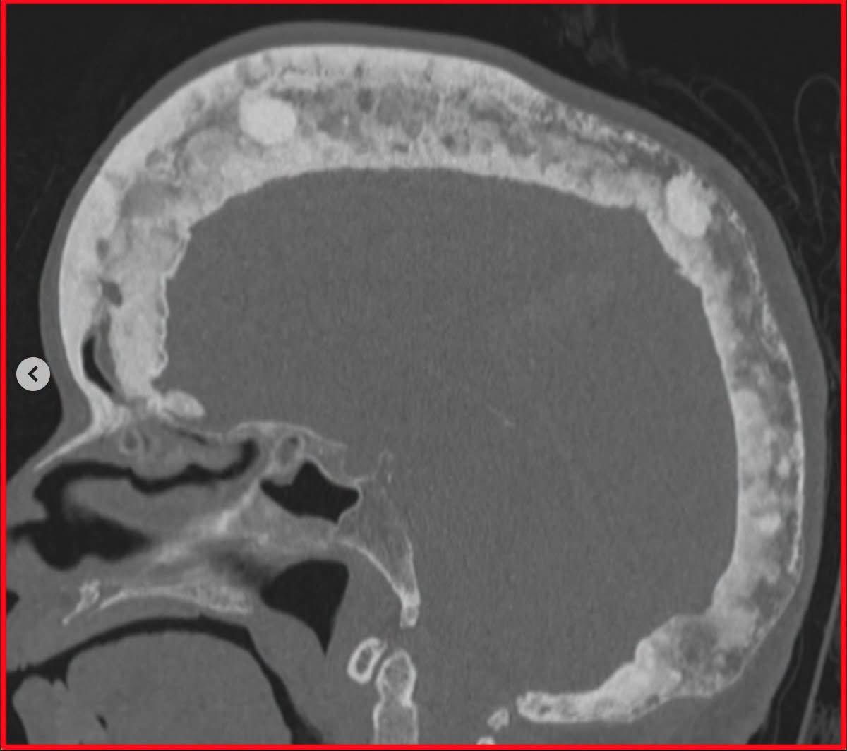
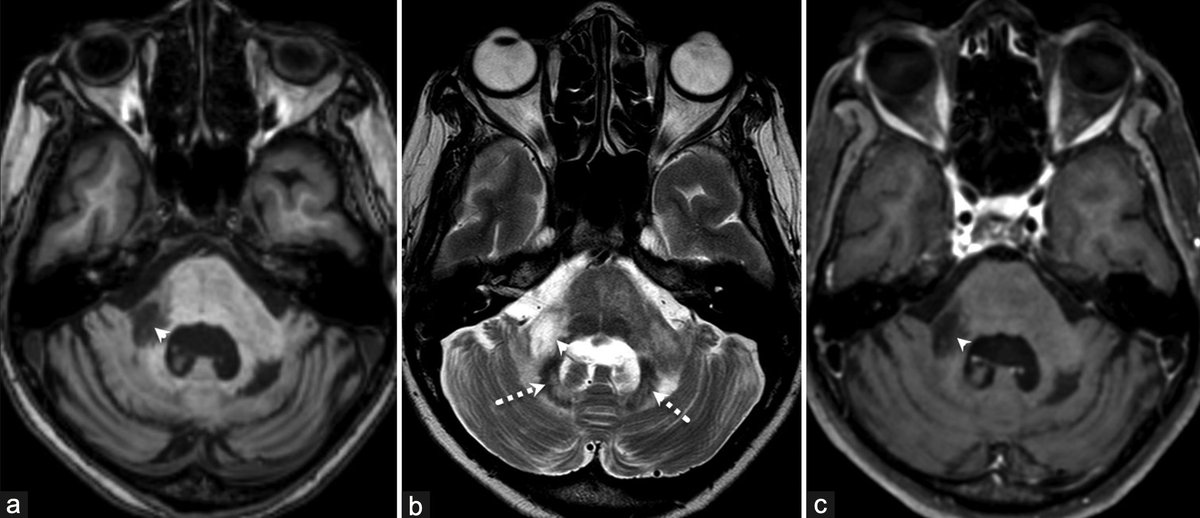
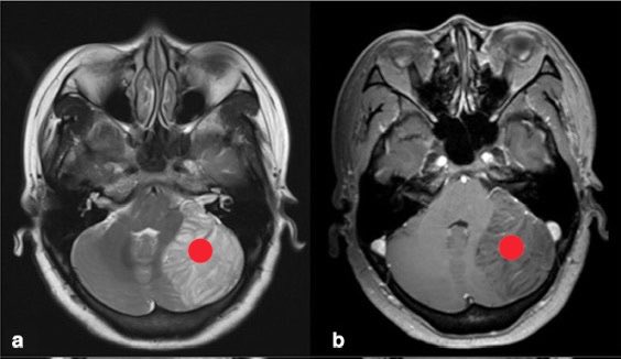
This teenaged boy of nonconsanguineous parents presented with abnormal body movements, gait difficulty, and slurring of speech for 2 years.
Findings?
Diagnosis?
CTX should be suspected in any patient
with childhood cataracts, tendon xanthomas & neurological findings of ataxia, spasticity, extrapyramidal features & dementia. MRI reveals B/L cerebellar atrophy and dentate hyperintensitites. CTX -> treatable
Link: 10.4103/aian.AIAN_126_18
Feel like waving the white flag over white matter disease?
Do you grade the degree of small vessel disease, or just mention & hope no one asks for more?
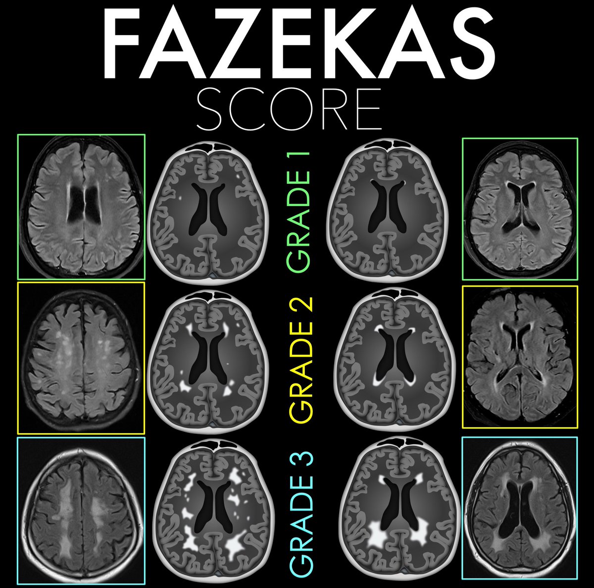
Degree of small vessel disease is important & the Fazekas scale is an easy way to semi-quantitate disease burden
Here’s how to remember it:
Think about how the number looks or feels — it grows in intensity!
Deep White Matter Fazekas:
1 → “1-hit wonder” Punctate foci — just tiny spots, like little dots.
2 → “2 = two-gether” Beginning confluence of foci — two dots coming together, mild merging.
3 → “3 is a crowd” Large, confluent areas — everything blending into a mess, too much white.
Periventricular Fazekas:
1 → “1 is a pencil” ✏️ Pencil looks a number 1 (pencil-thin “cap” or rim along ventricle).
2 → “2 curves into a halo” ⭕ Smooth halo of hyperintensity — picture the roundness of 2 encircling the ventricle.
3 → “3 branches out” 🌿 Irregular fingers of white matter reaching into the deep — the edge of the number 3 look like irregular finger
Now you know the Fazekas score for small vessel disease!
Now your knowledge of small vessel disease will be anything but small!
🧠 #SPINTweetorialWednesday
🧩 Case Challenge
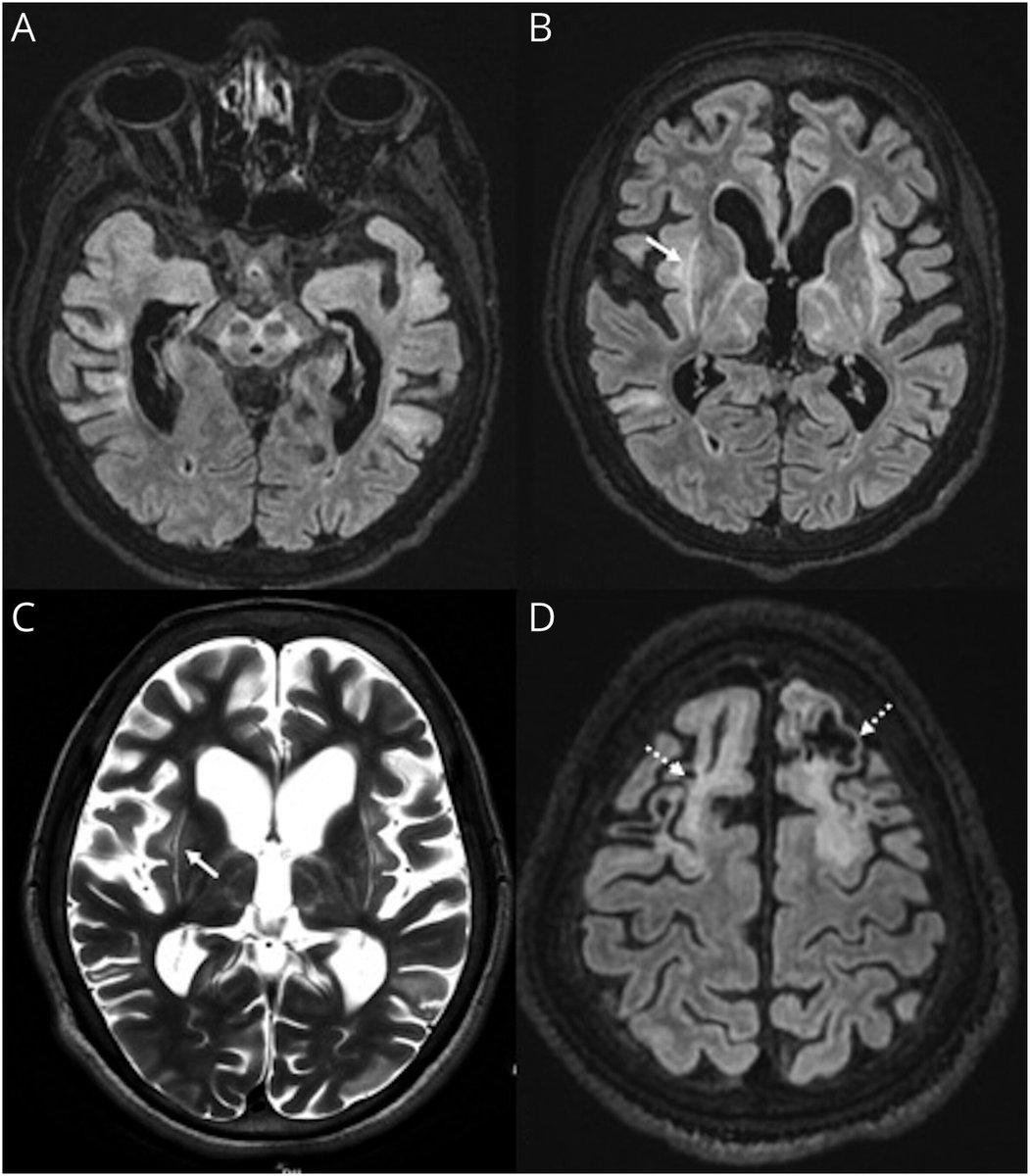
A 2½-year-old child develops rapid neurological deterioration after a viral respiratory illness.
What's your diagnosis?
🛑MCL bursa located along the middle 1/3 of the medial aspect of knee between superficial & deep portions of MCL.
🛑MCL bursitis is rare. Calcification of MCL is also rare but found either inside the bursa (calcific bursitis) or outside of it confused with meniscal tears,gout,..
A previously healthy man in his 50s presented with progressively worsening headaches and bluish nodular skin lesions.
Fast-field echo MRI image of the brain 👇
Likely diagnosis?
Shrimp sign has an 85% sensitivity, 100% specificity and 100% PPV for diagnosing PML. Asymmetry of the MCP lesions is paramount! Ajay Garg et al published one such case highlighting the "Shrimp sign" 🍤 in PML. @neuro_ian#PML Link: 10.4103/aian.aian_932_23
Polypoid lesions in the sinonasal region are mostly mucosal polyps but if you see a 'cerebriform' appearance (aqua arrows), especially one associated with a bony spur (chartreuse arrows), think Inverted papilloma! The bone spur must come out, to avoid recurrence.
Olivary hypertrophy is an exceptional consequence of brainstem insults. Usually occurs after a latency period of few months. @atabisneuro et al reported a case of bilateral olivary degeneration following Scrub typhus rhombencephalitis @neuro_ian Link: 10.4103/aian.aian_931_23
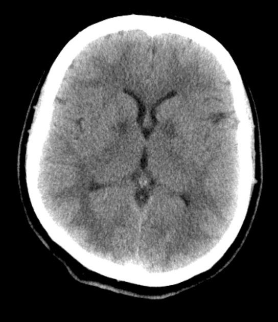
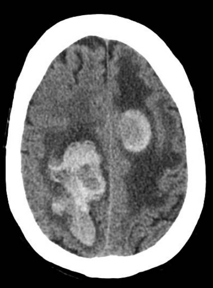
Top 3 most common causes of hemorrhagic metastatic lesions to the brain include?
How would you treat the edema caused by the tumor?
Drop your thought below
👇👇
⭐️ ~17% of patients with classic trigeminal neuralgia have aberrant nerve anatomy which may cause or contribute to their symptoms and is associated with worse surgical outcomes! ⚡️
⭐️ In patients with classic trigeminal neuralgia: Check the angle of the trigeminal nerve as it crosses through the porus trigeminus into Meckel's cave!
🔷Case example in 🧵
#Neurology #Neurosurgery #radres #futureradres #Medicine #MRI #FOAMed @ASNRographics@TheAJNR@Radiopaedia
#SPINTweetorialWednesday 🧵
Hypothalamic Hamartoma: The Little Lesion That Laughs ⚡🧠
A benign developmental malformation with a striking clinical signature:
😂 Gelastic seizures
⏩ Precocious puberty
🧠 Cognitive and behavioral disturbances
Let's dive in. 👇