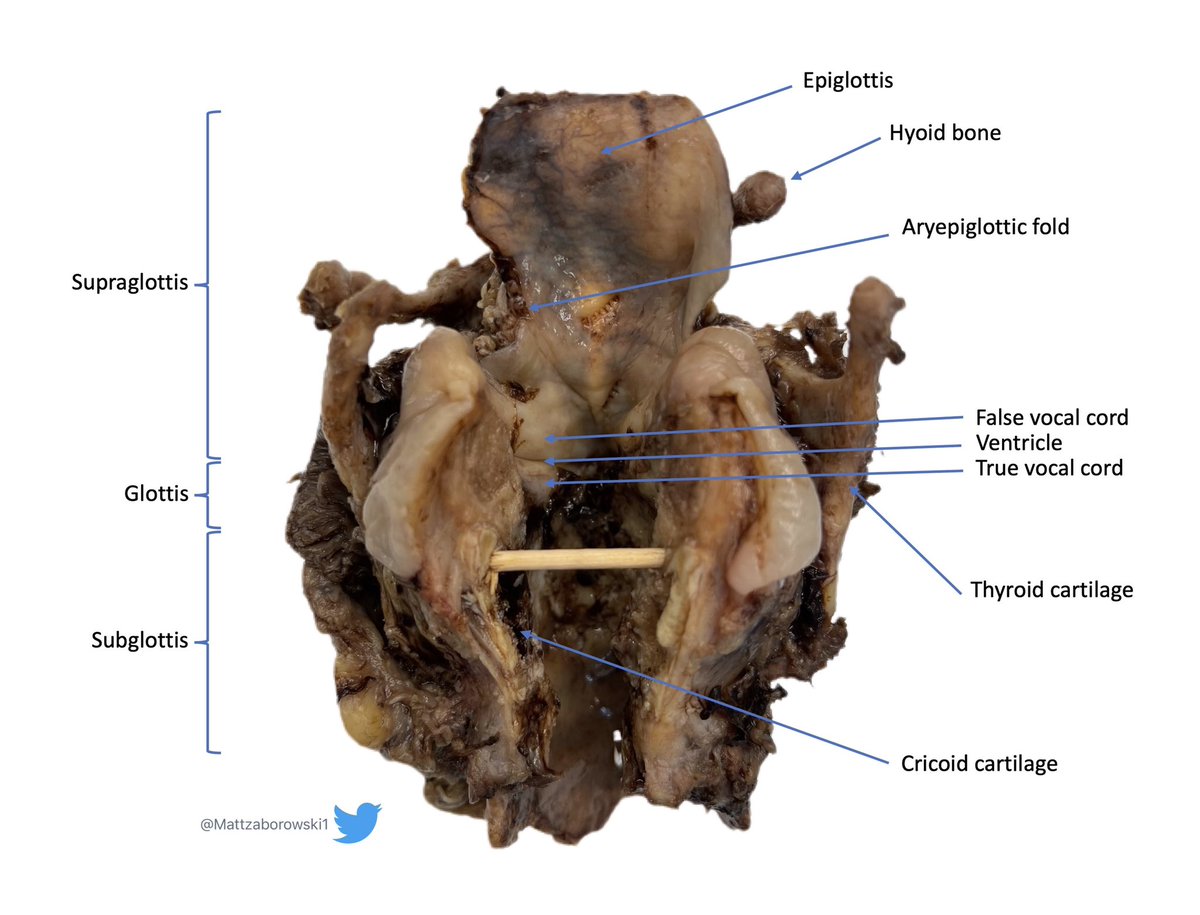
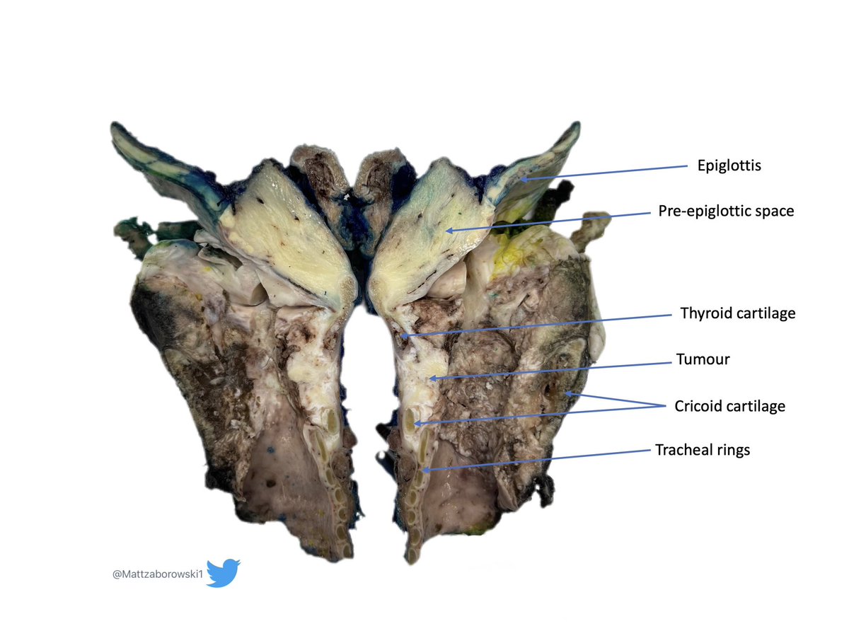
Larynx landmarks which might be useful (I find easier to correlate with compared to digital photos). #gross#larynx
Pathological stage classification is based on location: supraglottis, glottis and subglottis. https://t.co/d0ZPvL370s
✅SATB2 exhibits strong nuclear positivity in ~75–80% of Merkel cell carcinomas (MCC), with sensitivity comparable to CK20
✅other neuroendocrine carcinomas (e.g., pulmonary small cell carcinoma), which typically show absent or only weak/focal SATB2 staining
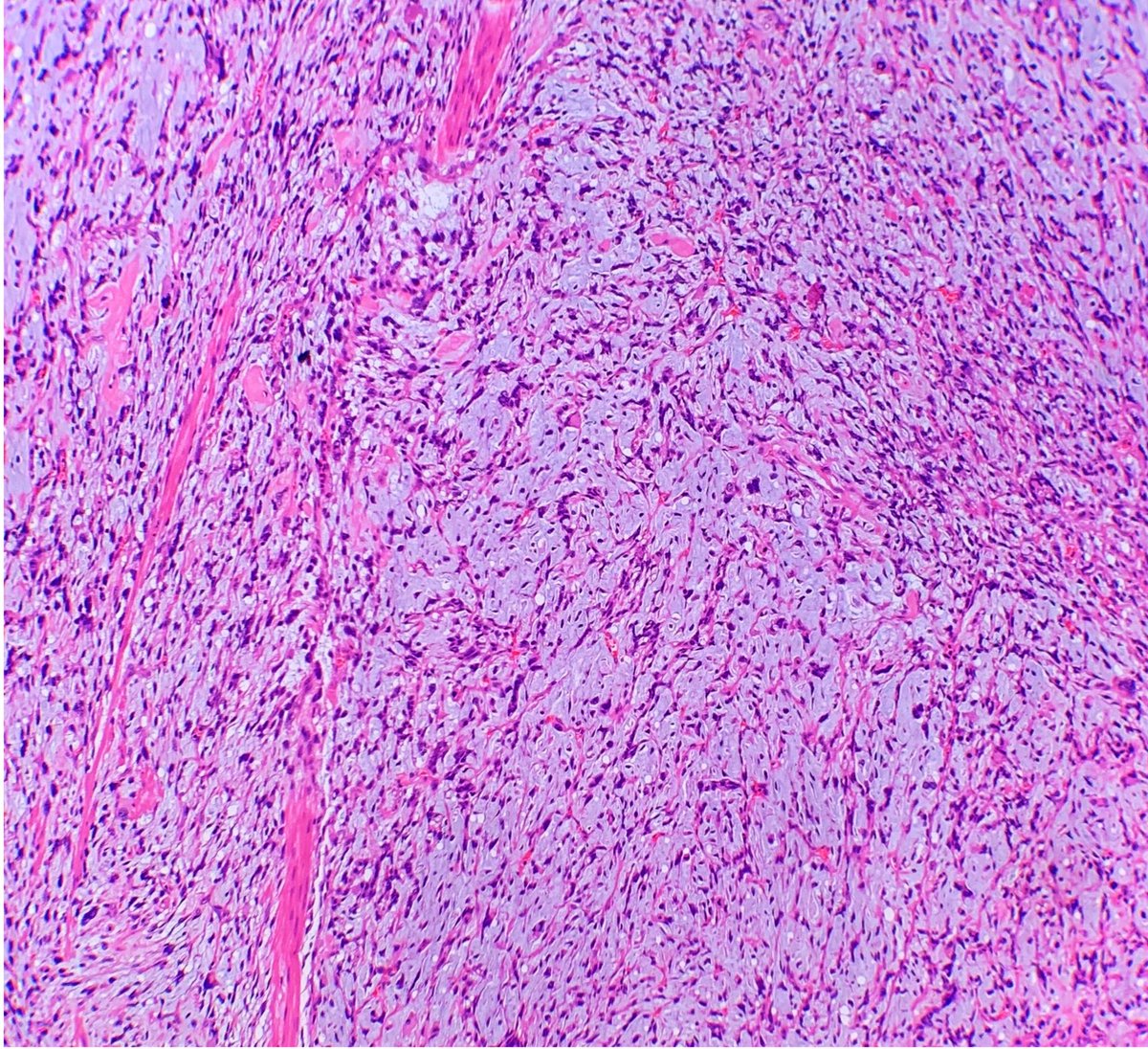
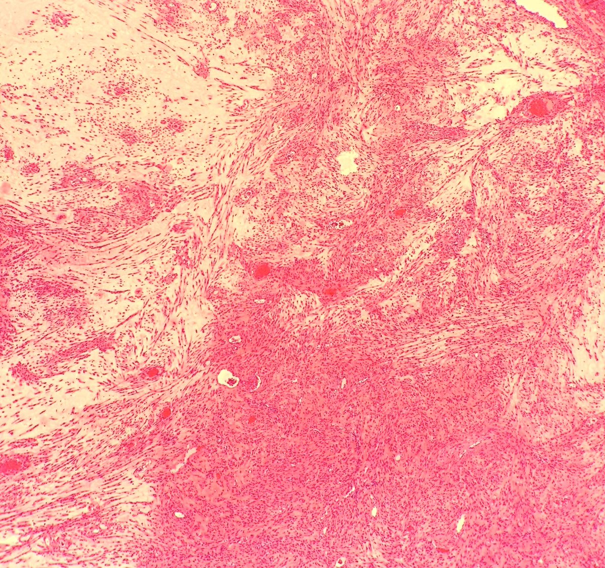
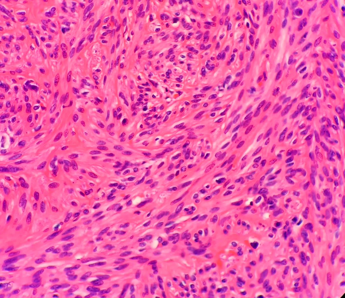
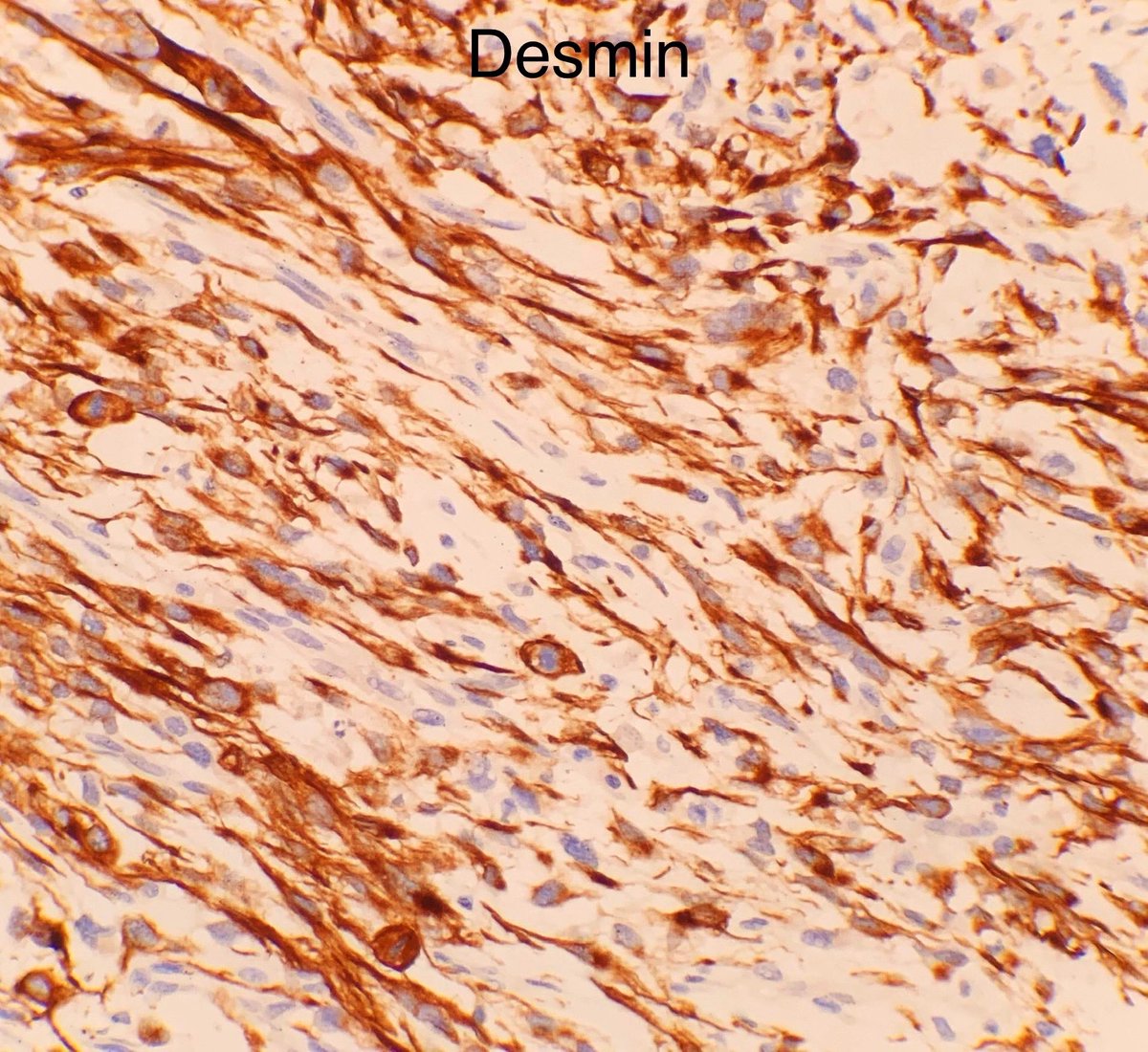
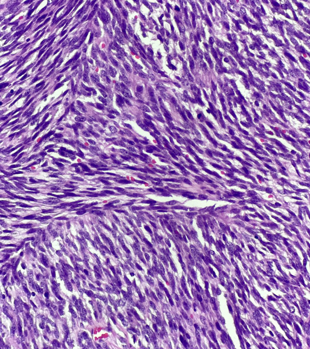
🟪Uterine myxoid leiomyosarcoma🟪
Macroscopic:
➡️Appearance is often gelatinous or glistening
Histopathology:
➡️Myxoid stroma
➡️Vague fascicular growth
Diagnostic criteria 🔬(1 or more of the following):
➡️Tumour necrosis
➡️Moderate to severe cytological atypia
➡️>1 mitosis per 10 HPF (0.55mm diameter field)
➡️Infiltrative margins
Immunohistochemistry:
➡️Desmin
➡️SMA
Take home point:
☑️Thorough sampling might be necessary to detect areas indicative of malignancy
Image
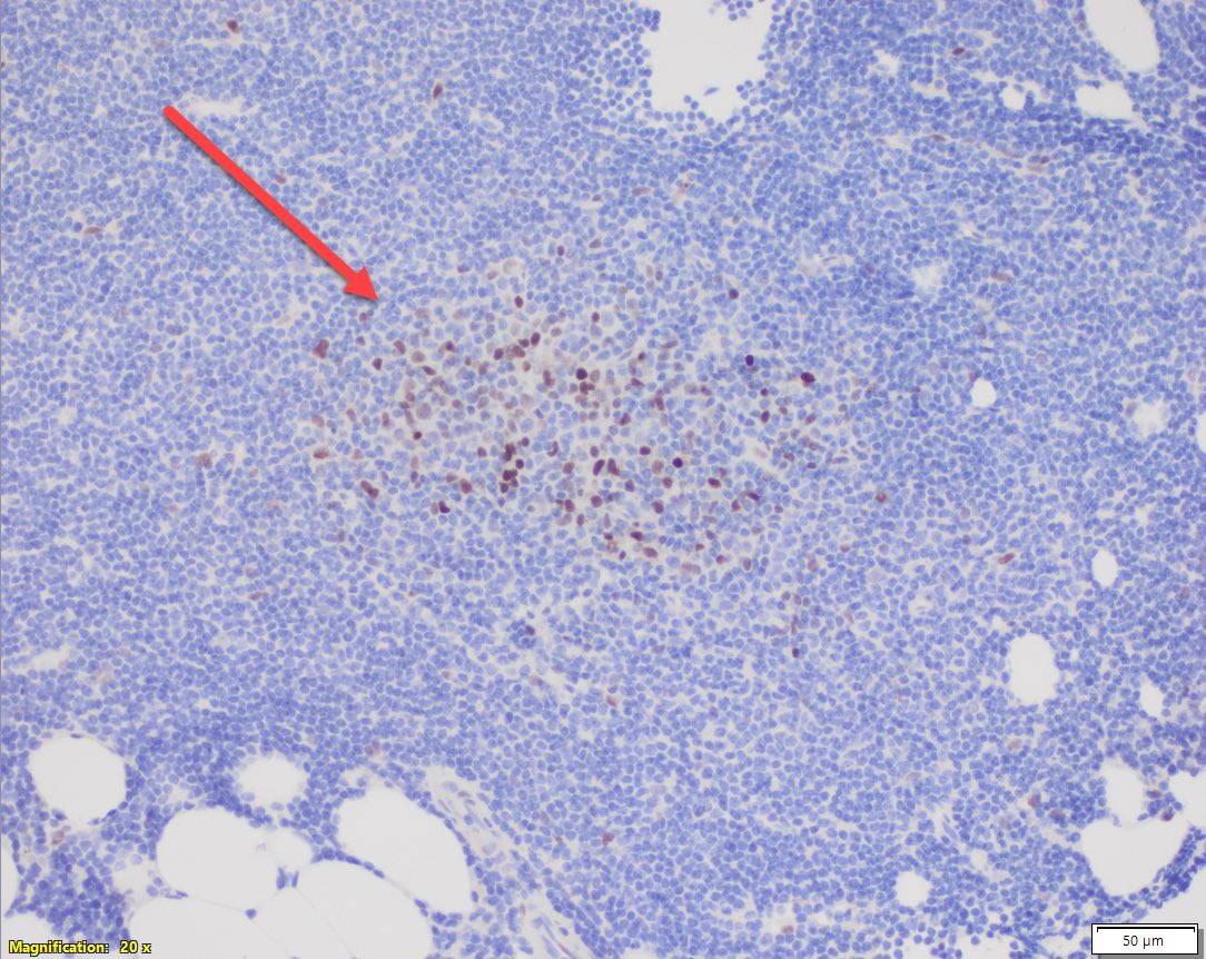
#hemepath#lymsm pitfall 🚨
CLL proliferation centers typically show uprgularion of cyclin D1. Don’t confuse this w/ mantle cell lymphoma.
There is no CCND1-r.
Read more about CLL w/ proliferation centers in the BM in our paper in @Human_Pathology
https://t.co/znIYZtOg9g
The serum free light chain FLC assay. Tutorial. #MedTwitter#MyelomaVR
Frequently ordered and frequently abnormal. This thread will reduce unnecessary headaches and referrals.
1/ Don’t order it unless you are suspecting myeloma, amyloid or related disorder
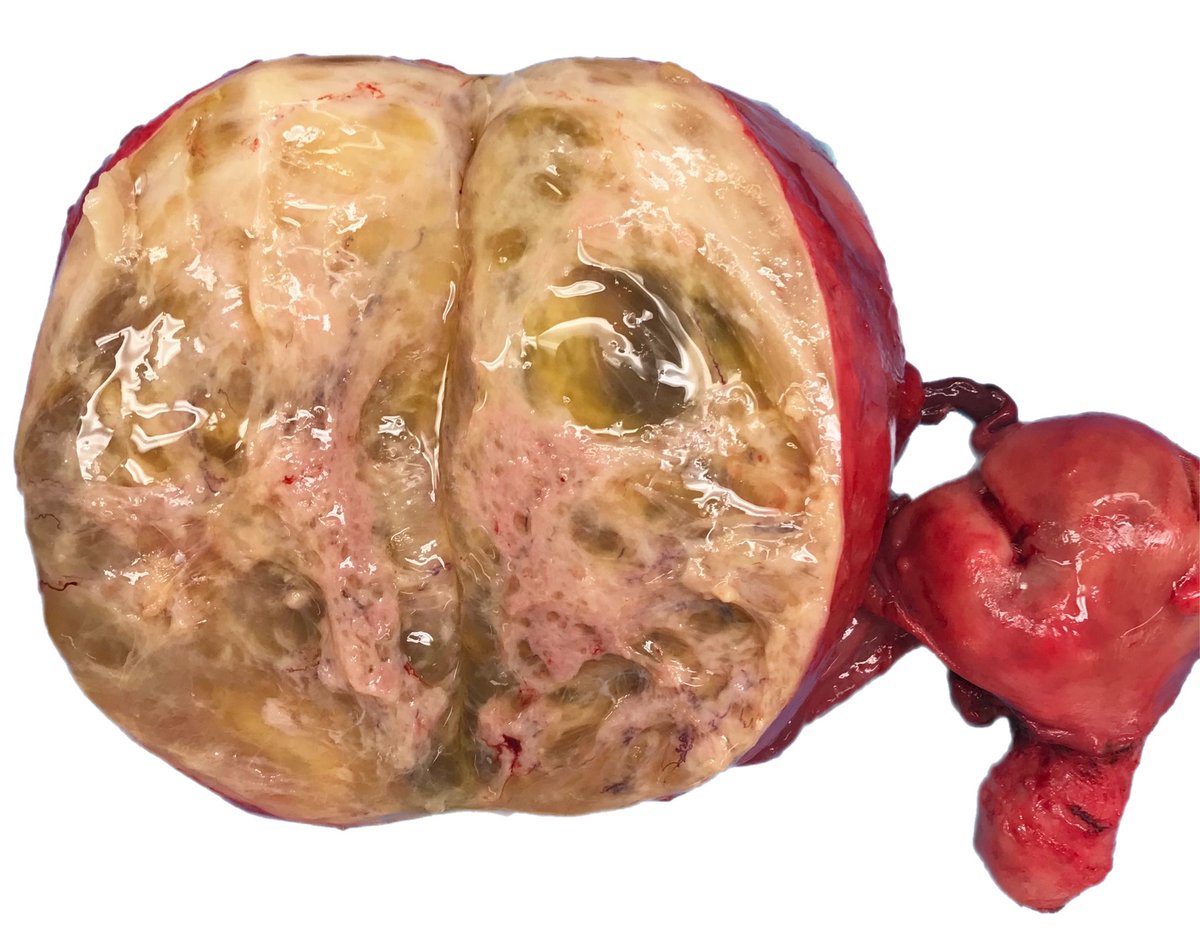
🟠Leiomyoma of the broad ligament🟠
☑️ circumscribed, firm mass
☑️ may develop prominent hydropic and degenerative cystic change
☑️ similar patterns and changes to its uterine counterpart, including criteria for malignancy