���� Take-home: In obese patients scanned on both systems, photon-counting CT gave better image quality (quantitative AND subjective) at ~30% lower dose, with the biggest gains in the largest patients.
https://t.co/tDUi8ArZCX
@VChernyakMD @SuhnyAbbara
🧵#RadInTraining#Tweetorial
CT in obese patients is a trade-off: ⬆️ X-ray attenuation usually means cranking up the dose just to keep images diagnostic.
New in @Radiology_RSNA: can photon-counting CT break that trade-off? Same patients, both scanners
@RITEditor@RadiologyEditor
🩻 Why it matters:
Obese patients are a group where CT quality often suffers and dose climbs. Here, PCCT improved both at once, and helped most in the largest patients.
A rare win-win in CT imaging.
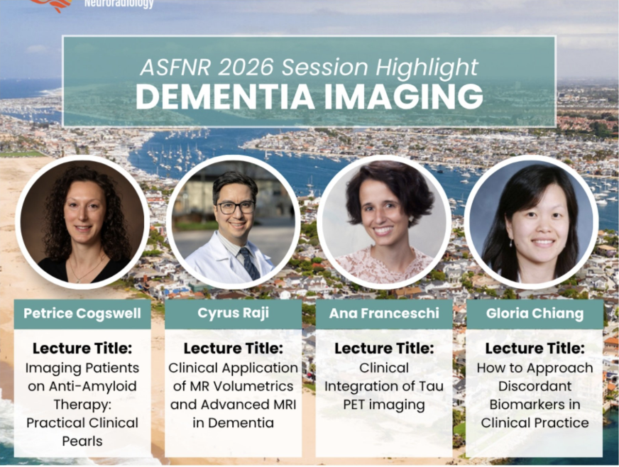
I'm very excited to give two talks today at the @theASFNR in Newport Beach, both on the same question: how do we translate advanced brain MRI from the research lab into everyday patient care?
10:42 AM | Clinical Application of MR Volumetrics and Advanced MRI in Dementia
Part of “Dementia Imaging: Navigating Real-World Cases with the Experts.Honored to share the session with Petrice Cogswell, MD, PhD of the Mayo Clinic @AFrances_MDPhD of Northwell Health and @GloriaChiangMD of Cornell University
12:30 PM | Lunch & Learn: Clinical Impact of MR Spectroscopy (sponsored by @Brain_Spec )
We are past due to apply MR spectroscopy to broader clinical neuroradiology practice. With FDA-cleared quantification and a Category I CPT code, that is changing. I’ll join Dr. Otto Rapalino and Dr. Alexander Lin of Mass General Brigham to walk through case studies where brain chemistry adds value to the team of advanced neuroimaging tools.
If you’re at ASFNR, come say hello, discuss the meeting topics and the #BrainHealthImaging mission I look forward to building at @IURadiology
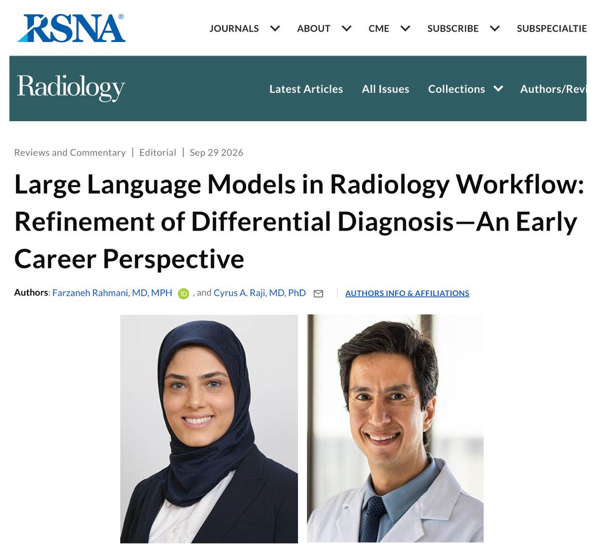
In other news, @cyrusraji & I published this editorial piece in @radiology_rsna
We weigh in on a growing body of papers including this latest by Li et al. on AI-assisted impression generation in radiology reports👇
https://t.co/t9VzTZBUZc
@cyrusraji@IURadiology Congratulations, Dr. Raji! It has been such a privilege to work with you and learn from you at WashU. I am truly grateful for your mentorship, guidance, and all that I have learned from you. Wishing you continued success at IU. They are very fortunate to have you!
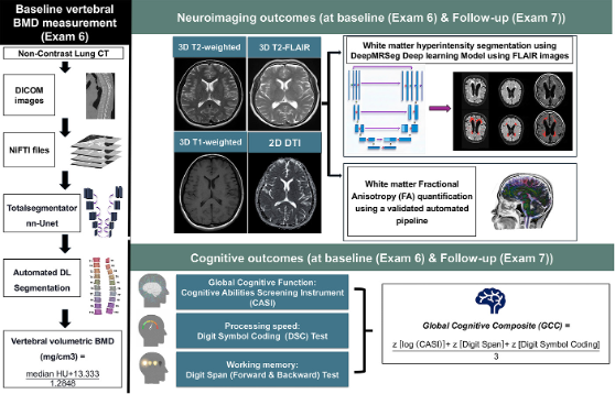
Hi #RadTwitter! I’m Mona Asghariahmadabad, MD, an international medical graduate and postdoctoral scholar at UCSF Radiology. I’m excited to be applying to radiology residency in #Match2027!
My research focuses on MRI in stroke and Alzheimer disease, and I’m eager to continue learning through clinical training.
Outside work, I’m usually looking for a new hiking trail, trying a new recipe, or working on a 3D printing project.
I’m looking forward to connecting with fellow applicants, residents, and faculty this season! @ProjectImg
AAMC ID: 16650132
#FutureRadRes
Hi #MedTwitter!
I'm Sara, a @TehUofMed grad and research fellow at @Hopkins_Rad. I'm so excited to apply to Diagnostic #Radiology in #Match2027. I'm passionate about AI-derived imaging markers that tell us more about a patient's health. Can't wait to connect and join you on this journey!
#FutureRadRes #RadRes #InsideTheMatch
🧵#Tweetorial
Conventional 3D DSA covers ~17 cm per spin, so head-and-neck angiography needs multiple selective injections. New in @Radiology_RSNA: spin the C-arm while sliding it along the patient, and ONE injection covers aortic arch to top of skull: https://t.co/R2Z3QWmiVg
⚠️ Limitations:
• n=1 human à need larger prospective studies
• Minor degradation at scan extremes (inexact reconstruction)
• ⬆️per-scan dose (50 vs 33 mGy); but 1 spin may replace 2–3 conventional ones à total dose still unquantified
• Descriptive statistics only