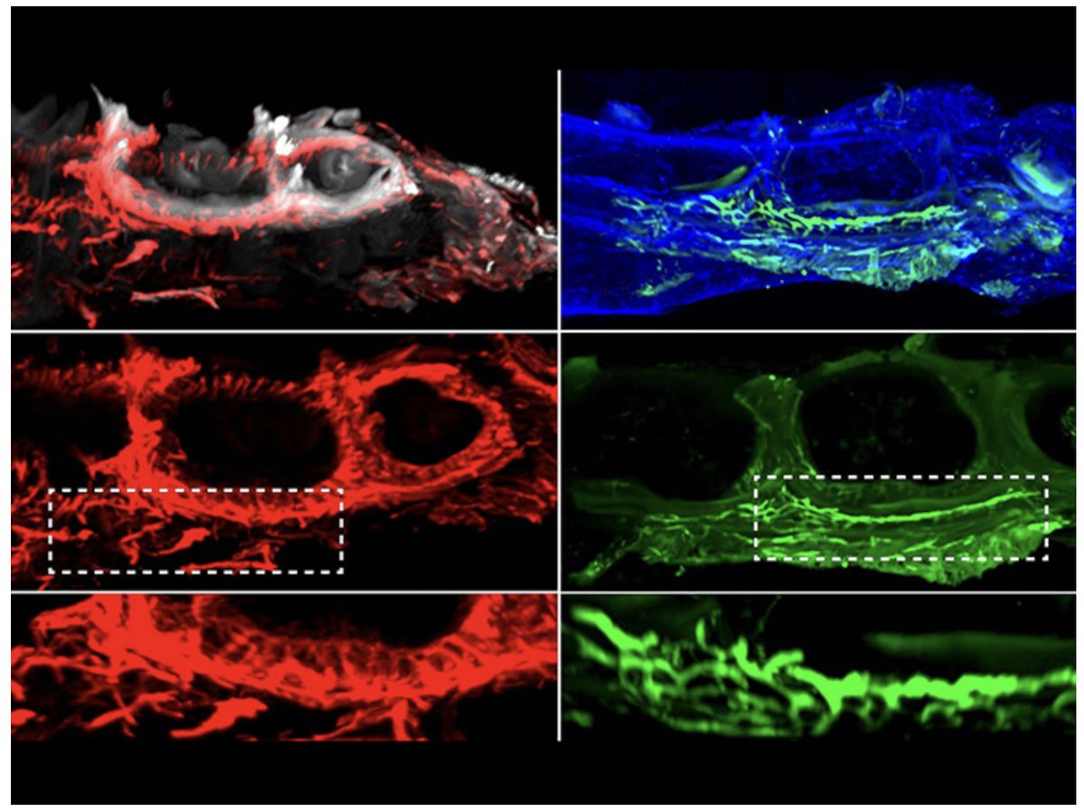
Excited to share our latest work in Nature Communications revealing vascular and lymphatic dysfunction as a defining hallmark of osteonecrosis of the jaw.
https://t.co/nELHDAKU0y
Pleased to see our ERC project featured by European Commission’s CORDIS
Our work reveals how ageing alters vascular niches to influence tumour cells, while vascular and lymphatic microenvironments shape skeletal and tissue aging and regeneration 👇
https://t.co/f6iPixjIdl
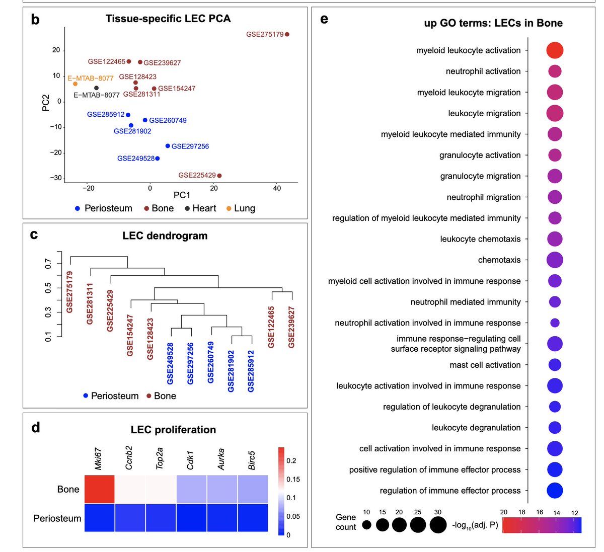
Finally online after a long journey!🎉 Multiple lines of evidence show that lymphatics are present in healthy bone and support bone regeneration. Huge respect for the persistence in validating and strengthening this finding! 🦴🔬@Anjali_Kusumbe@Anjali_Kusumbe
John Gurdon https://t.co/QXMjHLKlS3, beacon of a generation that tackled head on the deepest question of biology: the development of an organism. The best tribute to him and his peers should be to remember and follow the path they forged for how to do Science in form and content.
Proteomic profiling independently validates alternate transcriptional interpretation, revealing that old skull bone and bone marrow exhibit an impairment of niche-supportive programs.
Proteomic analysis here also proves skull bone is more like long bone and is not protected from aging but also that vertebrae are.
This work is definitive as science should be.
Dr. Adams’s PMID: 39537918; Extended Data Fig. 11a–c scRNA-seq data shows:
decline of aEC : arterial Endothelail Cells &
sEC : sinusoidal Endothelial Cells in old skulls - opposite to the paper’s conclusion of age-dependent vascular expansion 👇
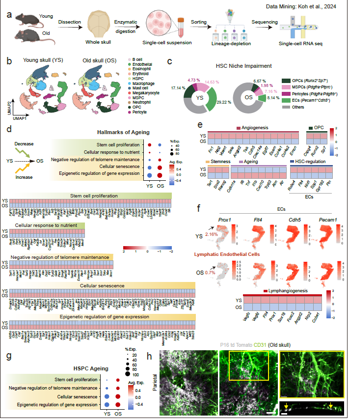
Reanalysis of Dr. Adams’s scRNA-seq dataset PMID 39537918 👇
While we show reanalysis & age-dependent changes, use of whole-skull precludes any conclusions about localization.
In PMID: 39537918 entire skulls were used but interpretations presented as specific to bone marrow.
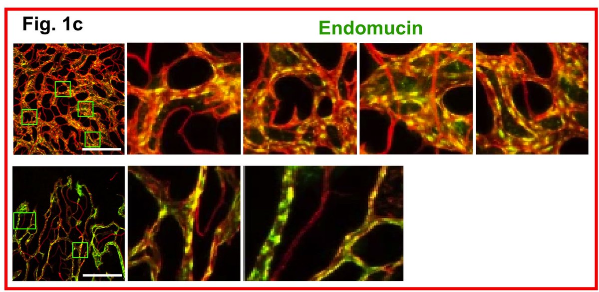
In Dr. Adams’s (PMID: 39537918; e.g. images below) confocal microscopy images of uncleared whole skull display aberrant nuclear localization of endothelial cell surface marker Endomucin, which contradicts its well-established membrane-restricted expression in previous studies
Very happy to share our multi-lab preprint:
Degeneration and Impaired Resilience of Skull Bone & Hematopoietic Bone Marrow 👇
https://t.co/qPRUChQqgJ
The skull shows pronounced age-related decline, while vertebrae are relatively spared
🧵
Very happy to share our multi-lab preprint:
Degeneration and Impaired Resilience of Skull Bone & Hematopoietic Bone Marrow 👇
https://t.co/qPRUChQqgJ
The skull shows pronounced age-related decline, while vertebrae are relatively spared
🧵
@E11BIO is excited to unveil PRISM technology for mapping brain wiring with simple light microscopes. Today, brain mapping in humans and other mammals is bottlenecked by accurate neuron tracing. PRISM uses molecular ID codes and AI to help neurons trace themselves. We discovered a new cell barcoding approach exceeding comparable methods by more than 750x. This is the heart of PRISM. We integrated this capability with microscopy and AI image analysis to automatically trace neurons at high resolution and annotate them with molecular features.
This is a key advance towards economically viable brain mapping - 95% of costs stem from neuron tracing. It is also an important step towards democratizing neuron tracing for everyday neuroscience. Solving these problems is critical for curing brain disorders, building safer and human-like AI, and even simulating brain function.
In our first pilot study, we acquired a unique dataset in mouse hippocampus. Barcodes improved the accuracy of tracing genetically labelled neurons by 8x – with a clear path to 100x or more. They also permit tracing across spatial gaps – essential for mitigating tissue section loss in whole-brain scaling. Using molecular annotation, we uncover an intriguing feature of synaptic organization, demonstrating how PRISM can be used for systematic discovery 🧵
Select your cell: I'm prototyping an interactive sc RNA-seq portal where the UMAP is dynamically linked to morphing 3D artistic renders of each cell type. You can play with the initial build below.
Our new review in Trends in Cell Biology : "The Wnt–NAD⁺ axis in cancer, aging, and tissue regeneration"
https://t.co/kHFrGcK3tn
👏 Kudos Lamprecht & Abu Sanad and 🙏 @KusumbeLab
Apologies to colleagues not cited (journal limits) + 🙏 to colleagues, reviewers & editor.
I am co-editing a Special Collection in npj Regenerative Medicine with Sharon Gerecht
"Repairing & Regenerating the Vascular Code Across Tissues & Time"
Vascular aging, regeneration to rejuvenation — submit your boldest science
Submit now
https://t.co/k0r7TtNzu1
Very grateful to all the fantastic speakers (including those not in the photo) at #AVIHM2025 for sharing exceptional science, fresh insights, and powerful discussions. Thank you for making it such a memorable and inspiring event!
Stem Cell Reports Appoints New Early Career Editor
2 July – Associate Professor of Stem Cell Biology and Regenerative Medicine Anjali Parmanand Kusumbe is one of five distinguished early career scientists selected to serve as new Early Career Editors for the peer-reviewed, open access, online journal of the International Society for Stem Cell Research. Assoc Prof Kusumbe also sits on the editorial board of Bone Research, a Nature partner journal.
@KusumbeLab