@PageLyndon@poperespecter1 God matters for naught. By definition God is conveniently not of this world, that is, universe and nothing outside the universe can communicate or interact with anything in the universe.
There may be a God and believe is either way. All knowledge requires belief.
Finerenone Expands Beyond Diabetic Kidney Disease
For years, mineralocorticoid receptor (MR) activation has been recognized as a major driver of cardiorenal injury through inflammation, oxidative stress, fibrosis, endothelial dysfunction, and adverse remodeling. Finerenone, a non-steroidal selective MR antagonist, is now emerging as one of the most important disease-modifying therapies targeting this pathway.
Unlike traditional steroidal MR antagonists such as spironolactone and eplerenone, finerenone exhibits higher MR selectivity, balanced cardiac–renal tissue distribution, and distinct cofactor modulation that translates into potent anti-inflammatory and antifibrotic effects with a more favorable safety profile.
The strongest evidence comes from the landmark FIDELIO-DKD and FIGARO-DKD trials. Together, these studies demonstrated significant reductions in CKD progression and cardiovascular events in patients with type 2 diabetes and chronic kidney disease. The pooled FIDELITY analysis further confirmed robust cardiorenal protection, including reductions in kidney failure, sustained eGFR decline, heart failure hospitalization, cardiovascular events, and mortality signals.
Importantly, finerenone’s biology extends beyond blood-pressure lowering.
Mechanistically, finerenone:
🫀 suppresses myocardial fibrosis
🫀 improves endothelial function and nitric oxide bioavailability
🫀 reduces NF-κB, IL-6, TNF-α signaling
🫀 attenuates oxidative stress
🫀 preserves mitochondrial homeostasis
🫀 limits renal inflammation and podocyte injury
These effects position finerenone as a true cardiorenal disease-modifying agent rather than simply another antihypertensive therapy.
The field recently moved even further with FINEARTS-HF.
In patients with HFmrEF and HFpEF, finerenone reduced the composite of cardiovascular death and worsening heart failure events by 16%, extending MR antagonism into a population with historically limited therapeutic options. Notably, benefits were observed regardless of diabetes status.
Another important theme is combination therapy.
Subgroup analyses consistently show preserved efficacy when finerenone is combined with SGLT2 inhibitors. Given their complementary mechanisms—fibrosis/inflammation suppression versus hemodynamic and metabolic protection—the combination may represent a cornerstone strategy for future cardiorenal care. The ongoing CONFIDENCE trial will provide important answers regarding dual-pathway modulation.
Emerging signals are also appearing outside the heart and kidney.
Preliminary data suggest potential benefits in:
👁 diabetic retinopathy progression
🫁 systemic inflammatory pathways
🧬 metabolic dysfunction-associated liver disease
although dedicated prospective studies are still needed.
The broader message is clear:
Cardiorenal disease is increasingly viewed as a cardiovascular–renal–metabolic syndrome driven by fibrosis, inflammation, and maladaptive neurohormonal signaling. Finerenone directly targets this biology and is becoming a foundational component of multidrug cardiorenal protection alongside RAAS inhibitors, SGLT2 inhibitors, and potentially GLP-1 receptor agonists.
Reference
Geraci G, Sinatra N, Paternò V, et al. Finerenone in Cardiorenal Disease: A Narrative Review of Molecular Mechanisms, Clinical Evidence, and Emerging Therapeutic Roles. Cardiovascular Drugs and Therapy (2026). DOI: 10.1007/s10557-026-07890-7.
#Finerenone #CardiorenalSyndrome #CKD #HeartFailure #Diabetes #SGLT2i #Nephrology #Cardiology #PrecisionMedicine #Inflammation #Fibrosis
Big congrats to Vijay Ali and the @Penn_Transplant team ! Kidney Transplantation in Two Highly Sensitized Candidates after CAR T-Cell Therapy | @NEJM https://t.co/e9FD1yNiXl
Multi-Omics + AI: Rewiring the Drug Discovery Pipeline
📄 DOI: 10.1038/s41392-026-02631-6
📚 Multi-omics and artificial intelligence for precision drug discovery and potential clinical applications
Drug development remains extraordinarily inefficient: >90% of candidates fail in clinical trials, with average costs exceeding $2.6 billion per approved therapy.
A new review in Signal Transduction and Targeted Therapy argues that the convergence of multi-omics technologies and artificial intelligence may fundamentally transform this paradigm.
Key message:
Instead of treating disease as a single-gene or single-protein problem, modern biology increasingly recognizes disease as a network-level disruption involving:
• Genomics
• Epigenomics
• Transcriptomics
• Proteomics
• Metabolomics
• Spatial and single-cell biology
The challenge is not generating data—it is integrating it.
This is where AI becomes essential.
🔬 Multi-omics provides the biological map
Advanced platforms now capture molecular information at unprecedented resolution:
• Single-cell RNA-seq reveals cellular heterogeneity.
• scATAC-seq uncovers regulatory landscapes.
• Spatial transcriptomics preserves tissue architecture and cell–cell interactions.
• Proteomics identifies signaling activity.
• Metabolomics quantifies pathway dynamics.
Together, these approaches generate a systems-level view of disease.
🤖 AI provides the analytical engine
The review highlights multiple AI architectures:
• Deep neural networks for pattern recognition.
• Graph neural networks for biological network inference.
• Transformer models for multimodal integration.
• Generative AI for de novo drug design.
• Structural AI systems such as AlphaFold and RoseTTAFold for protein structure prediction.
These tools can identify hidden disease mechanisms, prioritize therapeutic targets, optimize compounds, and predict efficacy and toxicity before costly experimental validation.
💡 Three major shifts are emerging:
1️⃣ From “one drug–one target” to network pharmacology.
2️⃣ From sequential discovery pipelines to iterative AI-guided design–test cycles.
3️⃣ From population-based treatment toward patient-specific digital twins powered by individual multi-omics profiles.
⚠️ Challenges remain:
• Data harmonization across platforms.
• Reproducibility and standardization.
• Model interpretability.
• Algorithmic bias.
• Clinical validation at scale.
Nevertheless, the direction is clear.
The future of precision medicine will likely be built on the integration of high-dimensional biology and machine intelligence—transforming drug discovery from a trial-and-error process into a predictive, data-driven engineering discipline.
#MultiOmics #ArtificialIntelligence #DrugDiscovery #PrecisionMedicine #SystemsBiology #SingleCell #SpatialTranscriptomics #Proteomics #Metabolomics #ComputationalBiology #TranslationalMedicine #Bioinformatics #AIinHealthcare #DigitalTwin #PharmaInnovation
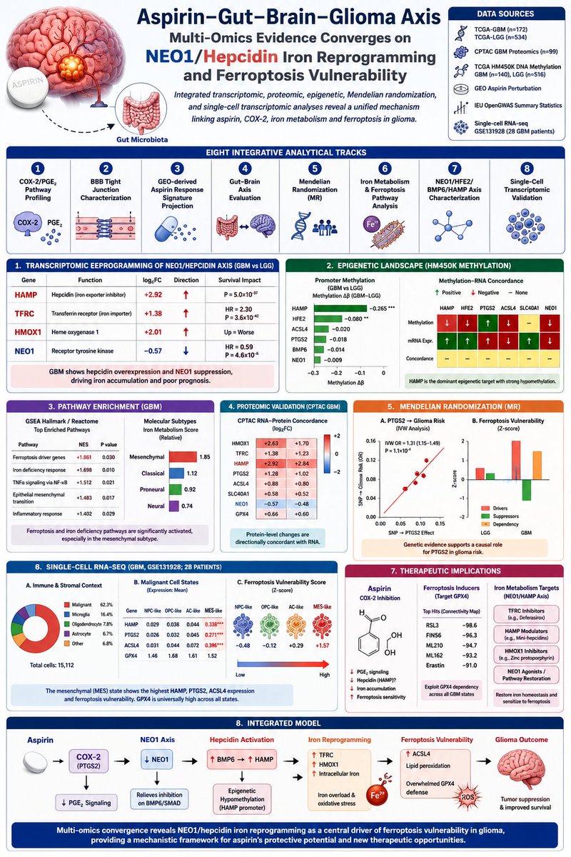
Aspirin–Gut–Brain–Glioma Axis: Iron Reprogramming Creates a Ferroptosis Opportunity
🧠 Can a common drug like aspirin influence glioma biology through iron metabolism and ferroptosis?
A comprehensive multi-omics study integrates transcriptomics, proteomics, DNA methylation, Mendelian randomization, and single-cell RNA sequencing to propose a unified mechanism linking COX-2 signaling, hepcidin-mediated iron reprogramming, and ferroptosis vulnerability in glioma.
Key Discovery: The NEO1–HAMP Iron Axis Is Rewired
The strongest signal across datasets was the dramatic activation of the hepcidin (HAMP) pathway:
⬆ HAMP (log2FC +2.92)
⬆ TFRC/transferrin receptor (log2FC +1.38)
⬆ HMOX1 (log2FC +2.01)
⬇ NEO1 (log2FC −0.57)
These changes create a state of intracellular iron accumulation and oxidative stress that may sensitize glioma cells to ferroptotic cell death.
Epigenetic Driver: HAMP Hypomethylation
Among all iron-regulatory genes, HAMP emerged as the dominant methylation target:
🔬 Δβ = −0.265
🔬 P = 1.4 × 10⁻⁴⁸
This suggests that glioblastoma actively unlocks hepcidin expression through epigenetic remodeling, reinforcing iron sequestration programs.
Ferroptosis Pathways Are Activated
Gene-set enrichment identified:
🔥 Ferroptosis driver pathway
NES = +1.861
🔥 Iron deficiency response
NES = +1.698
The transcriptomic landscape indicates tumors are simultaneously accumulating iron while activating defenses against ferroptotic collapse.
Single-Cell Validation
Analysis of 15,112 cells from 28 GBM patients revealed:
▪ Mesenchymal (MES) tumor cells show the highest HAMP expression
▪ PTGS2 (COX-2) enriched in MES state
▪ ACSL4 enriched in MES state
▪ Highest ferroptosis vulnerability score in MES cells
Meanwhile:
▪ GPX4 remained highly expressed across all malignant states
This suggests a broad dependency on GPX4-mediated ferroptosis suppression throughout GBM.
Aspirin as the Molecular Bridge
The study places PTGS2 (COX-2) at the center of the network:
Aspirin → COX-2 inhibition → iron metabolism modulation → ferroptosis susceptibility
Mendelian randomization supported a causal relationship between PTGS2 expression and glioma risk:
OR = 1.31
P = 1.1 × 10⁻⁴
providing genetic support for targeting this pathway.
Therapeutic Implications
The authors identify three potentially actionable vulnerabilities:
💊 Aspirin
COX-2 inhibition
Possible HAMP modulation
💊 GPX4 inhibitors (RSL3, FIN56)
Highest-ranked ferroptosis-inducing agents
Target a universal GBM dependency
💊 Iron metabolism targeting
TFRC
HAMP
HMOX1
NEO1 axis
The proposed model suggests that aspirin could function as a ferroptosis-sensitizing agent when combined with GPX4-directed strategies.
Why This Matters
This study moves beyond the classical anti-inflammatory view of aspirin and proposes a broader framework:
Aspirin → COX-2/PTGS2 → NEO1–HAMP iron reprogramming → ferroptosis vulnerability → glioma suppression
While still a preprint and not peer-reviewed, it provides one of the most comprehensive multi-omics maps connecting inflammation, iron metabolism, epigenetics, and ferroptosis in glioma.
Reference
Ma C, Zhang F, Wu F et al. Multi-Omics Integrative Analysis of the Aspirin-Gut-Brain-Glioma Axis: Transcriptomic, Proteomic, Epigenetic, Mendelian Randomization, and Single-Cell Transcriptomic Evidence Converges on NEO1/Hepcidin Iron Reprogramming and Ferroptosis Vulnerability. medRxiv (2026). DOI: 10.64898/2026.06.01.26354602.
Inspiring first annual Women’s Health Summit yesterday - women’s health talks and discussions among industry, investors, patients, scientists and clinicians - organized by Pillar in Boston. Energizing optimism in the room all day! #womenshealth
My continued biomedical education
#Atherosclerosis
The complex pro-atherosclerotic role of lipoprotein(a): a multiplicity of cellular targets
A nice synthesis of recent LPA research
👉MFSD5 as Endothelial Cell/Valvular Interstitial Cell receptor for LPA uptake
👉Direct pro-inflammatory effect of LPA on monocyte/Mφ, oxidized phospholipid-dependent & -independent
👉LPA pro-calcifying in Smooth Muscle Cell/VIC
👉Clinical evidence on LPA & coagulation/inflammation
@JuliaStJohn253@CO_Lipidology 2025
https://t.co/a98asX9viy
Last night, I celebrated what would have been Marilyn Monroe's 100th birthday in the most inspiring way. The new END Of cycle film is a MUST WATCH!!!!! A great film exploring the journey through pain. Free to watch: https://t.co/E5BeBux2Sc
Cancer as a Window into Mitochondrial Biology
DOI: 10.1016/j.cmet.2026.05.003
Journal: Cell Metabolism (2026)
Cancer may be the ultimate natural experiment in mitochondrial biology.
In this Cell Metabolism commentary, MacVicar and colleagues argue that tumors function as a "mitochondrial stress test," exposing the extraordinary adaptability of mitochondria under extreme evolutionary pressure. Rather than viewing mitochondria merely as ATP-producing organelles, cancer research reveals them as dynamic regulators of metabolism, genetics, inflammation, and cell fate.
Three major lessons emerge.
1. Metabolic plasticity drives cancer adaptation
Cancer cells continuously rewire mitochondrial metabolism in response to hypoxia, nutrient limitation, immune pressure, and therapy. Tumors exploit alternative carbon sources, reverse metabolic pathways, and reshape TCA cycle activity to maintain growth. Landmark discoveries involving IDH, SDH, and FH mutations established that mitochondrial metabolites themselves can function as oncogenic signals, linking metabolism directly to epigenetic regulation and tumorigenesis.
2. Mitochondrial DNA is an active participant in cancer evolution
The authors highlight a major shift in thinking: mtDNA mutations are not simply passengers. Large-scale cancer sequencing studies show non-random selection of mitochondrial mutations, particularly in respiratory-chain genes. Partial OXPHOS defects can create advantageous metabolic states that enhance tumor fitness, therapy resistance, and metastatic potential. This positions mtDNA as both a biomarker and a potential therapeutic target.
3. Mitochondria control death—and inflammation
The discovery that BCL-2 regulates mitochondrial apoptosis fundamentally transformed cancer biology. Today, drugs such as venetoclax exploit this vulnerability clinically. Yet the story is more complex: sublethal mitochondrial permeabilization ("minority MOMP") can promote DNA damage, invasion, and treatment resistance. Furthermore, mitochondrial DNA release activates cGAS-STING signaling, connecting mitochondrial dysfunction to innate immunity and inflammatory responses that influence cancer progression.
Perhaps the most exciting proposal is the creation of a Mitochondria-Cancer Atlas, an international initiative integrating mitochondrial physiology, multi-omics, mtDNA sequencing, proteomics, and clinical data across tumor types. The goal is to define cancer-specific mitochondrial states and identify therapeutic vulnerabilities invisible to conventional genomic analyses. Figure 1 outlines this framework, linking metabolic reprogramming, ROS signaling, mitochondrial genetics, inflammation, and cell death across diverse cancers.
The broader message extends far beyond oncology. Cancer exposes the full spectrum of mitochondrial behaviors, offering insights relevant to aging, neurodegeneration, metabolic disease, autoimmunity, and cardiovascular disorders. As mitochondrial biology and cancer metabolism continue to converge, the next decade may reveal a new class of precision therapies built around mitochondrial state rather than genetic mutation alone.
#Mitochondria #CancerMetabolism #mtDNA #OXPHOS #Immunometabolism #cGASSTING #Metastasis #CellMetabolism #SystemsBiology #AgingResearch
Can we predict mental health deterioration years before symptoms worsen?
Most psychiatric research focuses on diagnosis after symptoms appear. This new study takes a different approach: can we identify people at risk of worsening depression, anxiety, or cognitive decline years in advance using multimodal biological and behavioral data?
Using 157,733 participants from the UK Biobank, investigators integrated 2,911 plasma proteins, 2,126 brain imaging-derived phenotypes (IDPs), 77 lifestyle factors, and 103 mental health/cognitive questionnaires to build one of the largest predictive frameworks for future mental health deterioration.
The study combined PRS-PheWAS, Mendelian randomization, mediation analysis, and stacked machine learning to identify biological markers and evaluate predictive performance.
Key biological findings
Among hundreds of significant associations, two circulating proteins repeatedly emerged:
🧬 GDF15
🧬 HGF (Hepatocyte Growth Factor)
Both proteins were associated with depression, anxiety, and cognitive outcomes and were linked to structural brain features including the hippocampus, thalamus, cerebellum, and whole-brain volume measures. Mediation analyses suggested that specific brain imaging phenotypes statistically accounted for part of the relationship between circulating proteins and symptom severity.
Notably, 220 plasma proteins and 138 brain imaging phenotypes showed significant associations with at least one mental health outcome. Many signals converged on inflammatory, immune, and neurodegenerative pathways.
Mitochondria Don't Burn Lactate—They Vent It
Cell Metabolism (2026)
DOI: 10.1016/j.cmet.2026.02.020
For nearly three decades, the intracellular lactate shuttle (ILS) hypothesis has suggested that mitochondria directly oxidize lactate as a respiratory fuel. This new Cell Metabolism study challenges that paradigm and proposes a fundamentally different role for mitochondrial lactate.
The authors combined genetically encoded lactate sensors, redox reporters, mitochondrial physiology, CRISPR gene editing, proteomics, and in vivo two-photon imaging to track lactate dynamics inside mitochondria. Their conclusion is provocative:
Mammalian mitochondria are not primarily energized by lactate—they produce and export lactate.
Using a mitochondria-targeted lactate biosensor, the team identified a conserved mitochondrial lactate pool (~0.6–1.5 mM) across multiple cell types including HEK293 cells, hepatocytes, breast cancer cells, astrocytes, neurons, and cortical neurons in living mice. Blood lactate elevations rapidly increased mitochondrial lactate levels in vivo, demonstrating dynamic exchange between systemic metabolism and mitochondrial compartments.
Mechanistically, lactate crossed the inner mitochondrial membrane through a saturable transport system involving the mitochondrial pyruvate carrier (MPC). Both pharmacological inhibition and CRISPR deletion of MPC2 reduced mitochondrial lactate transport and altered lactate dynamics.
However, the critical discovery emerged when investigators tested whether lactate could fuel oxidative phosphorylation.
Pyruvate robustly activated the electron transport chain, increased mitochondrial alkalinization, and altered NADH/FAD redox signals. Lactate failed to do any of these. Despite entering mitochondria and encountering matrix LDH activity, lactate did not measurably energize respiration under physiological conditions.
Instead, mitochondria converted pyruvate → lactate within the matrix.
This reaction became especially prominent when matrix NADH accumulated. Under hypoxia, lactate production increased further, suggesting that mitochondrial lactate synthesis serves as a rapid redox-balancing mechanism.
The most striking observation was that blocking lactate export through MPC inhibition caused simultaneous accumulation of:
▪️ Matrix lactate
▪️ Mitochondrial H₂O₂
▪️ Oxidative stress signals
These findings support a model in which mitochondria use lactate production as a "metabolic vent" that dissipates excess reducing equivalents when electron transport becomes constrained. By converting pyruvate into lactate and exporting it, mitochondria lower matrix NADH burden and limit ROS generation.
The implications extend beyond bioenergetics.
The authors also detected extensive mitochondrial protein lactylation, including modification of enzymes involved in redox homeostasis and metabolic shuttles, linking mitochondrial lactate to signaling and post-translational regulation.
This work reframes lactate from being merely a glycolytic waste product—or even just a circulating fuel—to a mitochondrial stress-response metabolite.
In this model, lactate is not simply what cells make when oxygen is scarce.
It is what mitochondria deliberately generate when they need to protect themselves from excess reducing pressure and oxidative damage.
Take-home message:
Mitochondria are not passive consumers of lactate. Under metabolic stress, they actively produce and export lactate as a redox safety valve that limits ROS accumulation and preserves mitochondrial homeostasis.
#Mitochondria #Lactate #CancerMetabolism #ROS #RedoxBiology #Hypoxia #MPC #CellMetabolism #Aging #MetabolicFlexibility #OxidativeStress #MitochondrialBiology
🚀 Monomers can teach protein interactions.
A new study introduces Split and Merge Proxy (SMP), a surprisingly simple idea: take abundant monomeric protein structures, split them into artificial “pseudo-dimers,” and use them to pretrain protein interaction models before fine-tuning on real complexes.
Why does this matter?
Protein-protein interaction (PPI) prediction remains limited by the scarcity of experimentally solved dimer structures. While AlphaFold-Multimer and AlphaFold3 have transformed structural biology, high-quality multimer datasets remain orders of magnitude smaller than monomer datasets. SMP addresses this bottleneck by converting monomer structures into self-supervised interaction training data.
Key idea
1️⃣ Split a monomer into two fragments ("pseudo-receptor" and "pseudo-ligand")
2️⃣ Generate interaction labels directly from the original structure
3️⃣ Train models to reconstruct interactions between fragments
4️⃣ Fine-tune on real dimers
This creates 22,589 pseudo-dimers from existing monomer structures without requiring new experiments.
Results
🔹 Protein contact prediction
Homodimer300:
Top-L precision improved from ~0.69 to ~0.72
Heterodimer99:
Top-L precision improved from ~0.32 to ~0.37
Consistent gains across Top-1, Top-10, Top-25, Top-50, L/10, and L metrics.
🔹 Protein docking
Success rate improved:
EquiDock: 30%
SMP: 35%
Lower CRMSD and IRMSD
Higher DockQ scores across benchmarks.
🔹 Protein-protein interaction prediction
D-SCRIPT benchmark:
Recall ↑ 12.7%
F1 ↑ 5.9%
AUPR ↑ 3.0%
Similar gains observed on HIPPIE and cross-species prediction tasks.
Most intriguing finding
SMP occasionally outperformed AlphaFold-Multimer and AlphaFold3 on challenging CASP15 intertwined protein complexes.
For target T1176, SMP achieved:
✅ DockQ = 0.785
vs
❌ AF3 = 0.008
❌ AF-Multimer = 0.011
❌ Chai-1 = 0.009
The authors propose that random splitting naturally generates intertwined pseudo-complexes, helping models learn topologies that remain difficult for current structure predictors.
Why this matters
The broader message extends beyond protein interactions:
Data generation may be as important as model scaling.
Instead of collecting more expensive experimental complexes, SMP extracts interaction priors from the vastly larger universe of monomer structures. This is reminiscent of self-supervised learning in language and vision—creating useful supervision from existing data.
If extended to AlphaFoldDB-scale datasets, this strategy could provide a scalable route toward stronger protein complex prediction with minimal additional experimental cost.
Reference
Du H, Zheng X, Ren Y, et al. Improving protein and protein interactions using pseudo-dimers derived from monomeric proteins. Nature Communications (2026). DOI: 10.1038/s41467-026-73885-5.
#ProteinAI #AlphaFold #ProteinStructure #ComputationalBiology #MachineLearning #Bioinformatics #DrugDiscovery #AIforScience #ProteinInteractions #NatureCommunications
🧬 A Hormone Cell Atlas maps the human endocrine system at single-cell resolution
For over a century, endocrinology has viewed hormones through the lens of classical endocrine organs. But what if the human endocrine system is far more distributed than we imagined?
A new Science study integrates >14 million single cells and nuclei from 47 human tissues to build the first Hormone Cell Atlas, a comprehensive map of hormone-producing and hormone-sensing cells across the human body.
The scale is extraordinary.
Using a curated database of 162 hormones and hormone-receptor systems, the authors developed the hormone2cell framework to identify endocrine communication networks at cellular resolution. The resulting atlas reveals that endocrine signaling extends well beyond classical glands and is embedded throughout immune, vascular, epithelial, neural, and metabolic tissues.
Key discoveries
🔹 74% of cell types showed hormone-producing potential
Hormone synthesis is not restricted to pituitary, thyroid, adrenal, or gonadal cells. Endothelial cells, immune cells, adipocytes, and other tissues display distinct endocrine signatures.
🔹 93% of cell types expressed hormone receptors
Nearly every cellular compartment appears capable of receiving endocrine information, highlighting extensive hormonal integration across organs.
🔹 Unexpected immune-endocrine crosstalk
The team discovered that plasmacytoid dendritic cells express secretin (SCT), historically considered a gut hormone. Secretin expression increased during viral infection and interferon activation, suggesting a previously unrecognized immune-endocrine axis.
🔹 Adipocytes emerge as endocrine hubs
Single-cell integration across adipose depots revealed dynamic hormone production and hormone responsiveness during adipocyte differentiation, obesity, and depot-specific specialization.
🔹 Drug target mapping
The atlas predicts cellular targets of GLP-1, GIP, and glucagon receptor agonists. Interestingly, GLP1R and GIPR expression were observed in cardiac pacemaker cells and cardiomyocytes, offering mechanistic clues to cardiovascular effects of incretin-based therapies.
Why it matters
The Human Cell Atlas transformed our understanding of cellular diversity.
The Hormone Cell Atlas does the same for endocrinology.
Instead of isolated endocrine glands communicating with distant organs, the emerging picture is a dense network of cellular endocrine interactions spanning the immune system, metabolism, vasculature, and nervous system.
This resource provides a foundation for discovering new hormones, understanding endocrine disease mechanisms, predicting drug effects, and building a systems-level view of human physiology.
The endocrine system may be less like a collection of glands—and more like a body-wide communication network operating at single-cell resolution.
Reference
Fei L et al. A Hormone Cell Atlas maps the human endocrine system at cellular resolution. Science (2026).
DOI: 10.1126/science.aeb2672
#Science #SingleCell #HumanCellAtlas #Endocrinology #Hormones #GLP1 #Obesity #Metabolism #Immunology #SystemsBiology
@emollick Most people see this as an AI writing problem. It’s really a human-context problem: thin prompts produce average language. Better AI use is co-intelligence: the human brings context, tension, actors, and judgment; AI maps facts, mechanisms, and non-obvious angles before writing.