Di era resiko medis dianggap malpraktek, memulangkan pasien stabil dianggap menolak, menunda tindakan non emergency demi menolong yang emergency dianggap menelantarkan, disaat klaim jujur dianggap fraud. Ah sudahlah....
Day1 :
Abis nganggur 2 bln + jaga di tempat baru (artinya org2nya baru, budayanya baru, sistemnya baru, semuanya baru, a.k.a mulai dari 0 ya kak) + diresponsi DPJP dari jam 2-4 subuh = ☠️ bingung promax
Pantes ada aja org2 yg kepikiran resign pdhl baru masuk
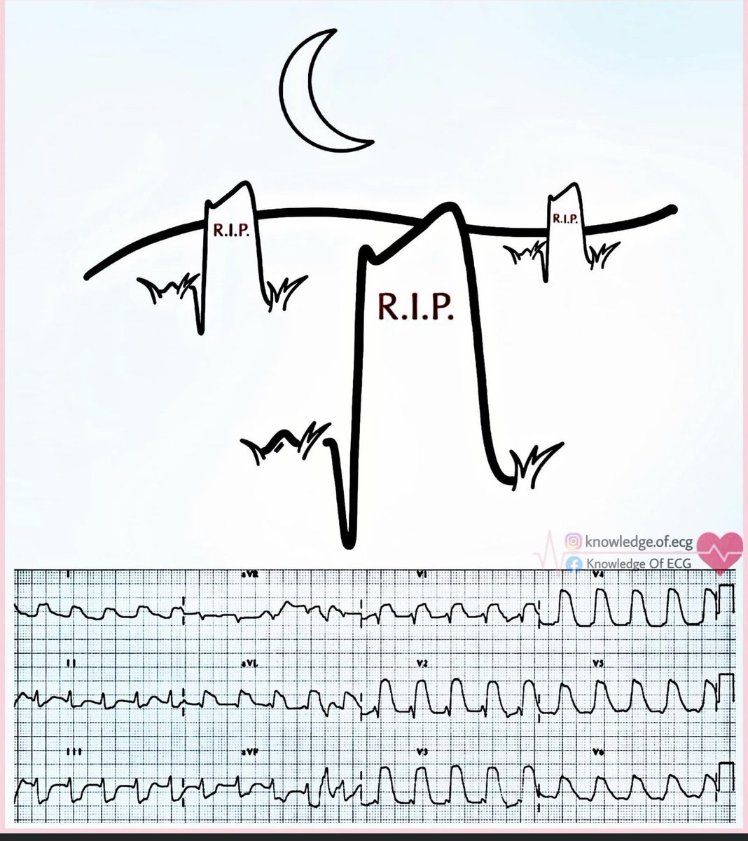
🚨 Breakdown of this ECG
✅Dx- Wide Complex Tachycardia Why This is VT (Not SVT with Aberrancy)
Let’s prove it 👇
1️⃣ Golden Rule
Any regular wide complex tachycardia = VT until proven otherwise.
• ~80% of regular WCT in adults = VT
• >90% if structural heart disease
Burden of proof is on SVT.
2️⃣ ✅ Rate ~200–220 bpm
Regular.
✅ QRS Duration
Clearly >160 ms.
Broad, monomorphic complexes.
Major red flag.
3️⃣ Precordial Analysis (Brugada Step 1)
Look at V1–V6:
No RS complexes.
All monophasic R waves → positive concordance.
📌 Brugada Criterion:
Absence of RS in all precordial leads = VT.
Diagnosis essentially made here.
4️⃣ RWPT (R-Wave Peak Time) Vereckei Criterion
Now look at V2 (or V1):
Measure from QRS onset → peak of R wave.
RWPT > 50 ms.
That means:
• Initial ventricular activation is slow
• Conduction is NOT via His-Purkinje system
• Favors myocardial origin
📌 RWPT >50 ms strongly supports VT.
In SVT with aberrancy:
Initial septal activation is rapid → short RWPT.
Here, it’s prolonged → VT.
5️⃣ Axis Clues
aVR shows a dominant R wave.
Suggests extreme axis deviation.
Extreme axis = strong VT marker.
6️⃣ Morphology Check
Does it resemble:
• Classic RBBB? ❌
• Classic LBBB? ❌
No typical bundle branch block pattern.
If it doesn’t fit standard BBB morphology → think VT.
SVT with aberrancy must resemble a known BBB.
This doesn’t.
7️⃣ AV Relationship
No clear consistent P–QRS relationship.
Likely AV dissociation.
AV dissociation = pathognomonic for VT.
⚡️ Electrophysiology: Why It Looks Like This ?
Most consistent with monomorphic VT.
Mechanism:
Scar-mediated re-entry.
Impulse:
• Originates in ventricular myocardium
• Spreads cell-to-cell
• Slow initial forces
• Wide QRS
• Abnormal axis
• Uniform morphology
Because it bypasses the His-Purkinje system:
→ Prolonged RWPT
→ Broad QRS
→ Extreme patterns
That’s the electrophysiologic basis.
❌ Why This Is NOT SVT with Aberrancy ?
To diagnose SVT with aberrancy, you need:
✔ Typical BBB morphology → NOT present
✔ RS complexes in precordial leads → absent
✔ Short RWPT (<50 ms) → NOT present
✔ Normal frontal axis → absent
✔ Clear atrial-driven rhythm → absent
SVT with aberrancy still uses fast Purkinje conduction.
So:
Initial forces must be rapid.
They are not.
🔥 Diagnostic Summary
✔ Regular WCT ~210 bpm
✔ QRS >160 ms
✔ No RS in V1–V6 (Brugada positive)
✔ RWPT >50 ms
✔ Dominant R in aVR
✔ Extreme axis
✔ Non-BBB morphology
✔ Probable AV dissociation
→ Monomorphic Ventricular Tachycardia.
🏆 Teaching Pearl
If unsure:
Treat regular WCT as VT.
Mislabeling VT as SVT and giving AV nodal blockers can cause hemodynamic collapse
#MedTwitter #MedX #ECG #Cardiology
Men's Volleyball 🥇
20 - 25
25 - 16
23 - 25
25 - 23
12 - 15
Indonesia 🇮🇩 2 - 3 🇹🇭 Thailand
Silver Medalist! 🥈
Misi untuk meraih Quattrick Gold harus terhenti di tangan tuan rumah.
Terimakasih sudah berjuang, we go again next year! 👊
#SEAGames2025
Why ST Elevation Isn’t Always a STEMI.
Every clinician has felt that moment…
You see an ST elevation on #ECG and your heart rate goes up before the patient’s does. STEMI? Cath lab activation? Thrombolysis?
But here’s the reality:
Not every ST elevation means ACS
A 🧵
Scratch test for organomegaly. First do a "control". Won't hear scratching until very close to scope. Next put scope over organ (eg, spleen). Now you'll hear scratching when you reach edge of the organ (much farther from scope vs control). The organ "carries" the sound to scope.
Differential Diagnosis of ST-Segment Elevation
ST-segment elevation on ECG isn’t always a heart attack. Here’s a breakdown of cardiac and non-cardiac causes every clinician should keep in mind. 🧵
Baru kali ini nemu pasien neuropati DM betul2 sesuai teori: "Gloves and Stocking"
👨🏻 : kebas dok dari ujung jari sampai mata kaki (sambil jarinya gerak nunjuk batasnya, kayak batas kaos kaki)
👨🏻⚕️ : oh iya pak kayak pake kaos kaki ya😅
What effects do LBBB, RBBB and LVH with repolarization abnormalities have on exercise stress testing interpretation?
⬜ LBBB → ST changes are unreliable due to abnormal repolarization. False-positive ST depressions may appear in V5-V6, I, aVL. Instead, use nuclear stress imaging or stress echo for accurate assessment.
⬜ RBBB → Less impact than LBBB, but ST-T changes in V1-V3 are not ischemic. Focus on inferolateral leads (V5-V6, II, III, aVF, I, aVL) for valid ischemia detection.
⬜ LVH with repolarization abnormalities → ST depressions & T-wave inversions (strain pattern) are common, especially in lateral leads (I, aVL, V5-V6). These changes often lead to false positives, making imaging crucial for confirmation.
😍