Primary PCI: Opening the Artery Is Not the Whole Story
In STEMI, the angiogram may show a beautiful epicardial result after PCI.
But the myocardium can still be injured.
(Why)
An occluded coronary artery contains more than thrombus. It may contain atherosclerotic debris, thrombotic material and disrupted plaque. During PCI, some of this material can embolize downstream.
The result can be:
1️⃣ Distal embolization
2️⃣ Microvascular obstruction/no-reflow
3️⃣ Side-branch occlusion
4️⃣ Reperfusion-related injury
So the goal of primary PCI is not simply:
(Open the artery)
It is:
Restore epicardial flow while preserving myocardial perfusion.
How PCI evolved
Early STEMI intervention relied heavily on balloon angioplasty.
The problems were familiar:
Residual stenosis
⬇️
recoil
⬇️
reocclusion
⬇️
restenosis
Coronary stents changed this by providing a scaffold, reducing residual stenosis and recoil and improving acute vessel patency.
Modern primary PCI therefore generally uses stent implantation when appropriate, rather than balloon angioplasty alone.
But the 2016 figure needs an important modern update.
Some strategies shown in the original diagram are not routine contemporary STEMI practice:
Routine thrombus aspiration is not recommended. Large randomized trials failed to show clinical benefit, and routine aspiration may increase stroke risk. Selective/bailout aspiration can still be considered when substantial thrombus persists, particularly after other PCI maneuvers.
Likewise, strategies such as routine deferred stenting or distal protection have not become standard approaches for every STEMI patient.
Modern PCI has shifted toward:
Rapid reperfusion + appropriate stenting + meticulous lesion treatment + prevention/management of no-reflow + selective use of adjunctive technologies.
And for complex ACS lesions, contemporary guidelines recommend IVUS or OCT-guided PCI to improve procedural results and reduce ischemic events.
Don't stop at:
(Did I achieve TIMI 3 flow)
Also ask:
(Did I restore effective myocardial perfusion)
Because an open epicardial artery does not automatically mean that the myocardium is adequately reperfused.
Source: Ndrepepa G, Kastrati A. Mechanical strategies to enhance myocardial salvage during primary percutaneous coronary intervention in patients with STEMI. EuroIntervention. 2016;12:319–328.
Updated with the 2023 ESC ACS Guidelines and 2025 ACC/AHA ACS Guideline.
STEMI: Every Minute of Ischemia Matters
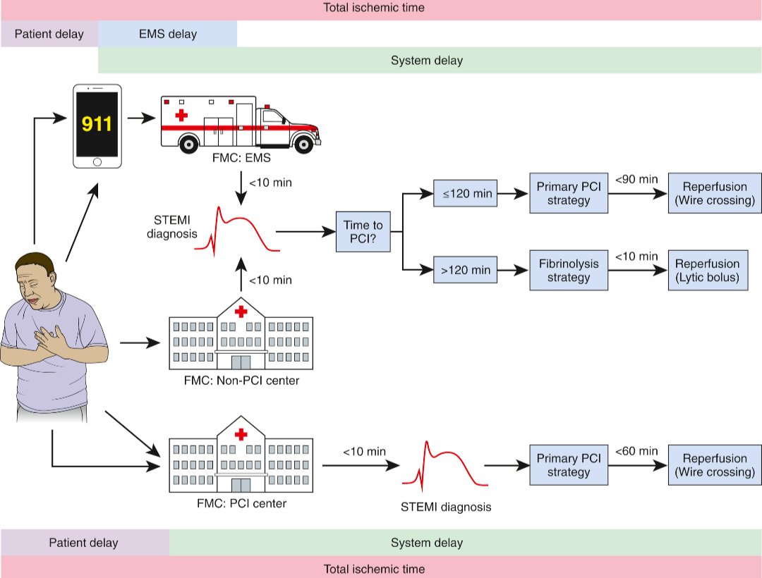
In STEMI, the clock starts before the patient reaches the cath lab.
Total ischemic time includes:
Patient delay + EMS/system delay = total ischemic time
The goal is not simply door-to-balloon.
It is to minimize the entire interval from symptom onset to coronary reperfusion.
The practical decision
Once STEMI is diagnosed, ask:
Can primary PCI be achieved within 120 minutes from diagnosis/FMC?
✅ If yes, the patient should be taken for primary PCI.
❌ If timely PCI cannot be achieved within 120 minutes, fibrinolysis should be considered when appropriate and there are no contraindications, followed by transfer to a PCI-capable centre. The 2023 ESC ACS guideline continues to use this 120-minute threshold.
For systems capable of rapid primary PCI, contemporary U.S. guidance emphasizes even faster targets: the 2025 ACC/AHA ACS guideline recommends a FMC-to-first-device goal of ≤90 minutes for patients transported directly to a PCI-capable hospital, while ≤120 minutes remains an important threshold when longer transfer is involved.
Why this matters
⚪ Every unnecessary delay means prolonged myocardial ischemia.
⚪ The 2025 ACC/AHA guideline notes that for patients undergoing primary PCI, each additional 30 minutes of treatment delay has been associated with an increase in relative 1-year mortality risk.
Source: Ibanez B, et al. 2017 ESC Guidelines for the management of acute myocardial infarction in patients presenting with ST-segment elevation. Eur Heart J. 2018;39:119–177.
حالة بسيطة لعرض علاج معروف:
وصل شاب عمره 25 سنة إلى الطوارئ بعد حادث سيارة
في البداية كان يتكلم عادي وواعي
لكن خلال فترة قصيرة انخفض مستوى وعيه وصار GCS = 8
مع وجود استفراغ، ارتفاع ضغط الدم وبطء في النبض.
أظهر التصوير وجود وذمة دماغية وارتفاع في الضغط داخل الجمجمة
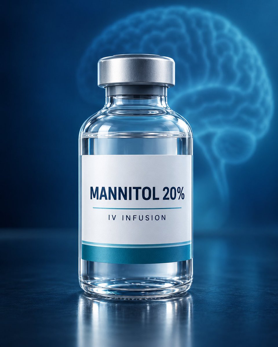
انكتب له Mannitol IV حسب حالة المريض وبروتوكول المستشفى.
كيف يشتغل المانيتول؟
باختصار يسحب الماء من أنسجة الدماغ إلى الدورة الدموية، ثم تتخلص منه الكلى عن طريق البول؛ مما يساعد على تقليل الوذمة وخفض الضغط داخل الجمجمة.
المهم لك كممرض قبل إعطائه يجب الانتباه إلى ضغط الدم، حالة السوائل، كمية البول ووظائف الكلى؛ لأن المانيتول قد يزيد انخفاض الضغط والجفاف ويؤثر في الكلى.
في هذه الورقة العلمية موجود ارشادات لاستخدام اهم العلاجات للوذمة الدماغية وارتفاع الضغط داخل الجمجمة.
الرسالة الأساسية هي أن العلاج يجب أن يُختار وفق سبب وذمة الدماغ وحالة المريض، وليس بطريقة واحدة لجميع المرضى.
https://t.co/lmeQzDqkKZ
Correct answer: B. Hematoma expansion
The satellite sign is a non-contrast CT marker in spontaneous intracerebral hemorrhage (ICH). It describes one or more small, separate hemorrhagic foci adjacent to the main hematoma.
Its presence has been associated with an increased risk of hematoma expansion (HE), which is clinically important because expansion is associated with neurological deterioration and worse outcomes.
A meta-analysis of 5 studies involving 1,493 patients found that the satellite sign had approximately 50% sensitivity and 71% specificity for predicting hematoma expansion.
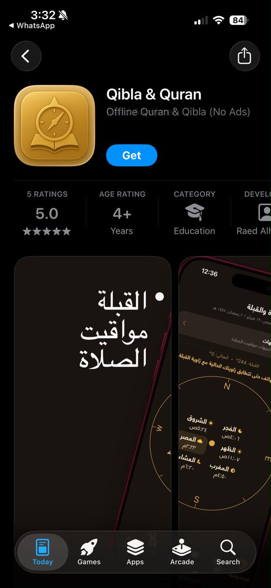
بابا اشتغل على هذا التطبيق شهور وتعب عليه😢
وكل اللي أبيه إني أفرحه وأنشره للأجر دعمكم بيفرق معه كثير.
فيه قرآن، مواقيت الصلاة والقبلة وبدون اعلانات وموجود على
Apple store
Goggle play
جربوه وإذا عجبكم لا تبخلون عليه بنشره بين أهلكم وأصحابكم
الله يكتب أجر كل شخص ساهم في نشره 🤍