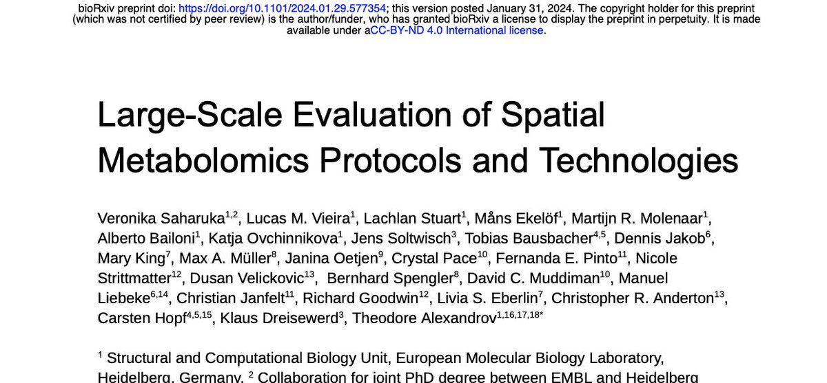
I’m happy to present our new preprint by @VSaharuka et al which helps answer the key question in #spatial#metabolomics and #imaging#mass#spectrometry: which molecules are detectable and which protocols shall one use? https://t.co/WtA5htRwTM 🧵 1/n
Really great work I had a chance to participate in with @AllisonEsselman. We did spatially targeted MALDI imaging of hundreds of discrete glomeruli. We found them all with automated segmentation of label-free autofluorescence microscopy.
So proud of the amazing work and wonderful presentation by Ally Esselman (grad student @VanderbiltChem) at #ASMS2023. Thanks to our collaborators and friends in the @vandeplaslab
Two incredible oral presentations coming from the Spraggins lab at #ASMS2023@AllisonEsselman will unravel the molecular distribution of glomeruli using microscopy-directed high spatial resolution IMS, while jackie dives deep into the molecular distribution of S. aureus abscess
Happy (belated) New Year! Let's get the year started off right with some cool collaborative informatics work associated with the @_hubmap project!
https://t.co/TdzmUABZvy
Excited to share my most recent manuscript on lens aging! We used DIA MS Proteomics to identify key age-related protein changes, identifying a proteome remodeling event concurrent to enhanced risk of cataract formation at approximately 50 years. https://t.co/nNV8gZuHGh
@seejbrown When I saw your post yesterday I almost wrote something like "get a microscopy image! Interpreting chemical images from IMS with morphology from microscopy is a big win (almost a necessity IMO)" Looks like you got on the right path quickly! Good luck!
Thrilled about imaging? We are hiring a #PhDposition and #postdocposition in signal processing & machine learning at @tudelft.Join us in building 3-D molecular atlases to decipher cellular diversity in human organs.Please share.https://t.co/aRzBqWogLY https://t.co/0NGquxSeEl #PhD
Come see our #ASMS2022 posters to learn about our microscopy data integration tools and our cutting edge sample preparation for high-res MALDI IMS. If you want deep dive on our multimodal MALDI IMS: our approach and tools will be taught at IMSS in Baltimore. @IMSSNews
Final day #ASMS2022! Let's end it with a bang! We've got two excellent posters by Heath (@NHeathPatterson) and Martin, as well as a talk by collaborator and friend of the lab Lukasz (@lgmigas). Make sure you check it all out! Hope to see you there!
Viv is published @naturemethods! Our #bioimaging library enables the creation of web-based vis and analysis tools to complement desktop software also built upon open standard #OMETIFF & #OMENGFF from @openmicroscopy. https://t.co/5HbuKhasnM
https://t.co/GXIw1TV6JG
The output images are pyramidal OME-TIFF and work well in @QuPath@openmicroscopy and Vitessce. Vitessce demo: https://t.co/rTwjv3Xbga Besides images, wsireg can "attach" point-sets and additional images to those registered and seamlessly transform them.