Dad, Skull Base and Cerebrovascular Neurosurgeon, Entrepreneur, Researcher, and Philanthropist. Founder and President of @MyRightToHear and @MSLogger_Inc.
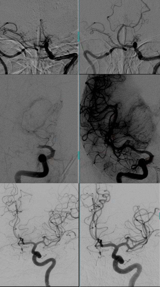
Thrombectomy for acute ischemic stroke is one of the most rewarding interventions in medicine. It involves the removal of a blood clot occluding a brain vessel, restoring blood flow and preventing a potentially devastating stroke. Many patients are able to regain much, or even all, of their neurological function afterward.
The procedure is extremely time-sensitive. It is estimated that nearly 2 million brain cells (neurons) die every minute during an acute large vessel stroke, which is why rapid diagnosis and treatment are critical.
These angiographic images demonstrate restoration of blood flow after thrombectomy in different cerebral vessels, including the basilar artery, middle cerebral artery (MCA), and anterior cerebral artery (ACA).
#Thrombectomy #Stroke #StrokeTreatment #NeuroIntervention #Brain #Surgery #Neurosurgery #Neurology #Radiology #Health #Medicine
One of the most fascinating anatomy exposures in neurosurgery is seen during the Far Lateral approach, which provides access to the anterolateral aspect of the spinomedullary junction. In this view, you can appreciate the lower cranial nerves (IX–XII), each responsible for critical functions such as swallowing, speech, taste, and tongue movement. The vertebral artery and the course of the posterior inferior cerebellar artery (PICA) are also beautifully demonstrated. Each one of those structures has to be handled delicately to avoid devastating injury.
This case involved clipping of a wide-neck ruptured PICA takeoff aneurysm that was not a suitable candidate for endovascular treatment.
#Neurosurgery #SkullBase #FarLateral #CranialNerves #Neuroanatomy #Anatomy #SurgicalAnatomy #Brain #Surgery #Neuroscience #Neurosurgeon
In patients with Moyamoya disease, twig-like MCA, and other causes of severe MCA stenosis, evaluation of vasomotor reactivity (VMR) is essential. VMR assesses the ability of cerebral blood vessels to increase blood flow in response to increased demand or vasodilatory stress. One way I use to assess this is with CT perfusion imaging before and after administration of Acetazolamide (Diamox), as demonstrated in this case.
On the right, after Diamox administration, brain perfusion becomes critically diminished (seen in red). This occurs because the diseased vessels are often already maximally dilated at baseline. After Diamox, the normal vessels dilate further and “steal” blood flow away from the compromised territory, a phenomenon known as cerebrovascular steal.
This indicates severely impaired cerebrovascular reserve, placing the patient at increased risk for cerebral ischemia and stroke, and suggests that revascularization treatment with bypass surgery is indicated to reduce future stroke risk.
#Moyamoya #Neurosurgery #Brain #Surgery #Bypass #Stroke #CVA #Neurology #Neuroscience #Health #Neurosurgeon #Medicine
A challenging case of a ruptured anterior vermian arteriovenous malformation (AVM) in a 10-week pregnant teenager. The AVM ruptured twice, resulting in significant ataxia and hydrocephalus. The patient also faced major social barriers, with no paperwork, insurance, or medical coverage available.
Treatment involved staged management with preoperative embolization followed by microsurgical resection. To preserve as much of the vermis as possible, the operation was performed through multiple skull base corridors. I began with a telovelar approach to address the feeders from the PICA territory, followed by a supracerebellar infratentorial approach to disconnect the SCA feeders. A limited transvermian approach through the superior vermis was then used to complete the resection of the AVM.
Postoperatively, the patient developed cerebellar mutism, which lasted approximately 2 weeks before gradual recovery of speech. She is currently undergoing rehabilitation for both her ataxia and speech deficits, with a viable ongoing pregnancy.
Complex cerebrovascular surgery requires a strong understanding of skull base approaches and surgical corridors. Radiosurgery is often ideal for deep AVMs; however, the history of multiple consecutive ruptures, pregnancy, and lack of medical coverage excluded that option.
#AVM #ArteriovenousMalformation #Neurosurgery #Cerebrovascular #BrainSurgery #SkullBaseSurgery #Microsurgery #Neurovascular #PICA #SCA #CerebellarAVM #Neurosurgeon #Brain
Trigeminal neuralgia, historically known as the "suicide disease", is considered one of the most painful forms of facial pain. It most commonly occurs when a nearby artery — usually the superior cerebellar artery (SCA) — compresses the trigeminal nerve, the nerve responsible for facial sensation. Over time, the constant pulsation against the nerve leads to nerve injury and severe recurrent facial pain.
Microvascular decompression (MVD) is the most definitive treatment for trigeminal neuralgia. It involves separating the artery from the nerve and placing a small piece of Teflon between them to act as a cushion. This operation remains one of the most rewarding procedures in skull base surgery. Around 90–95% of patients experience immediate pain relief after surgery, and most are discharged home within 1–2 days.
#TrigeminalNeuralgia #SkullBaseSurgery #FacialPain #MicrovascularDecompression #Neurology #Neurosurgery #Brain @#Surgery #Surgeon #Neurosurgeon #SkullBase
A brain aneurysm is a weakened bulge in a brain artery that can rupture and cause life-threatening bleeding. As a dual-trained neurosurgeon, I carefully determine whether a patient is best treated with endovascular techniques or microsurgical clipping.
Aneurysm clipping remains a fundamental skill in neurosurgery. For selected aneurysms, microsurgical clipping provides a durable, permanent treatment with a very low risk of recurrence.
This patient presented with a ruptured anterior communicating artery (ACom) aneurysm. Adequate exposure is critical in these cases, and here the entire ACom complex was well exposed before clip placement, allowing for safe aneurysm obliteration. The small perforators were also dissected away and preserved.
#Neurosurgery #BrainAneurysm #Microsurgery #Neurosurgeon #VascularNeurosurgery #Neuroscience #Aneurysm #Brain #Health
Extradural clinoidectomy is a key skull base technique that involves removal of the anterior clinoid process before opening the dura.
This approach is important in the treatment of select anterior skull base pathologies, like meningiomas. By working extradural first, the tumor can be devascularized early, making resection safer and more controlled. It also allows for early decompression of the optic nerve and provides improved visualization of critical neurovascular structures once the dura is opened.
#SkullBaseSurgery #Neurosurgery #Meningiomas #OpticNerve #NeurosurgicalApproach #SurgicalAnatomy #NeuroOncology #SkullBase
Two different patients. Two very different operative environments.
These intraoperative images of the brain during aneurysm clipping show the difference between operating on an unruptured versus ruptured aneurysm.
Ruptured brain aneurysms are fatal in about 50% of cases. Smoking can increase aneurysm rupture risk by up to 3–6x, and hypertension is one of the strongest modifiable risk factors.
If you have two or more first-degree relatives with brain aneurysms, the risk of harboring an aneurysm may reach 8–12%, and screening is necessary. Early detection can save lives.
@BAFOUND
#BrainAneurysm #Neurosurgery #AneurysmAwareness #SurgicalTechniques #BrainHealth #Brain #Aneurysm #Bleeding #BrainBleed #Neurology
Skull base surgery demands a deep understanding of lateral skull base anatomy.
In this case, a large tentorial meningioma was approached via a retrolabyrinthine approach, with meticulous temporal bone drilling personally performed to access both the infra- and supratentorial compartments.
This approach provides safe exposure, facilitates tentorial division, and enables complete tumor resection while preserving hearing. This is one of the many skills I owe to Dr. @jacquesmorcosmd!
The patient initially presented with seizures and remained neurologically intact and seizure-free following surgery.
#Meningioma #Neurosurgery #SkullBaseSurgery #Neuroanatomy #Brain #SkullBase #Surgery #Surgeon
Left-sided Moyamoya disease.
One of the main brain arteries (MCA) is missing on the left side → not enough blood flow → multiple strokes.
We created a new route for blood supply to the brain by connecting a scalp artery (STA) to a brain artery in a bypass surgery.
Low blood flow doesn’t just cause strokes, it can affect memory, focus, and thinking.
Restoring blood flow can change everything.
#MoyamoyaDisease #BrainHealth #BypassSurgery #StrokeAwareness #BrainSurgery #Neurosurgery #Stroke #Brain
Surgical view of a large brain meningioma. Meningiomas are the most common benign brain tumors. Because they are typically slow-growing, they can reach a significant size before causing symptoms. In this case, the patient presented with headaches, and imaging revealed a large meningioma originating from the falx.
To achieve complete resection and minimize the risk of recurrence, it is critical to remove not only the tumor but also its site of origin, in this case, part of the falx, and occasionally the bone if involved.
The patient had an excellent outcome and was discharged home within 48 hours of surgery.
#BrainTumor #Meningioma #Neurosurgery #BrainSurgery #BrainHealth #BrainSurgeryRecovery #MeningiomaSurgery #Neuroscience #Surgery
Brain bypass surgery is one of the most demanding and delicate skills in cerebrovascular neurosurgery. It is the art of connecting and reconnecting tiny blood vessels to restore or augment blood flow to the brain when natural circulation is compromised.
Note the microsutures used in this operation, 10-0 sutures that are only one fourth the diameter of a human hair. The image on the right demonstrates the robust new blood supply created by the bypass
This particular patient had Moyamoya disease, a progressive condition in which the major blood vessels supplying the brain gradually close off, leading to recurrent strokes. She had already suffered multiple strokes before surgery. She was discharged home 2 days after surgery with no more strokes.
#BrainBypassSurgery #MoyamoyaDisease #Neurosurgery #CerebrovascularSurgery #StrokeRecovery #VascularNeurosurgery #BrainSurgery #Neuroscience #Microsurgery #Neurosurgeon #Stroke
The view of a large brain arteriovenous malformation (AVM) prior to a successful removal. Note the complex network of dilated, abnormal vessels. This is just the surface, this particular AVM was also 5 cm deep.
AVMs are congenital tangles of abnormal blood vessels in the brain. Because of the high blood flow through them, they carry a risk of rupture that can lead to life-threatening brain hemorrhage. Depending on their location, AVMs may also cause seizures, headaches, or other neurological deficits.
Treatment options may include observation, microsurgical resection, endovascular embolization, radiosurgery, or a combination of these approaches. The best treatment plan is individualized and depends on several factors, including the AVM���s size, location, vascular architecture, and the patient’s overall condition.
#AVM #Neurosurgery #BrainHealth #AVMTreatment #Neurology #BrainAVM #CerebralAVM #Surgery #VascularNeurology #AVMSurgery #Brain
One of the most impactful periods of my career was training under Dr. @jacquesmorcosmd as his fellow. He is not just the GOAT in skull base and cerebrovascular #neurosurgery but also one of the most extraordinary human beings I've ever met. His guidance, teaching, and mentorship were profound honors that helped shape who I am—and who I strive to become. I hope to carry forward his legacy with the same dedication and integrity he exemplifies.
At @MyRightToHear, we have established the first #charitable cochlear implant program in #Lebanon, which will serve #children with #hearing loss of all backgrounds: Lebanese, Syrian Refugees, and Palestinian Refugees. The first phase of the program included 10 children, and was great success. All children are now going through speech and hearing rehab to be able to talk after they could hear for the first time.
This was a result of our strong collaboration with the Syrian American Medical Society (SAMS), and the generous support of @TheTradeDesk and @medel.