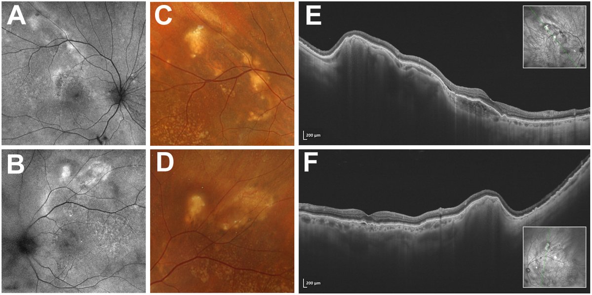
Ultra-widefield color fundus photography (Optos RGB) and swept-source OCT B-scans (Intalight DREAM OCT) were obtained in an 81-year-old White male patient with bilateral idiopathic sclerochoroidal calcification, showing yellow-orange and hyperautofluorescent mass lesions along the superotemporal vascular arcades (A–D). OCT confirms that the primary site of calcium deposition is intrascleral (E, F) with a mountain-like undulating surface, with compression and draping of the overlying choroid. Because the calcium deposition appears confined to the sclera, “scleral calcification” may represent a more precise description for this entity. Serum calcium, phosphate, and parathyroid hormone were within normal limits. This unique OCT configuration can assist in differentiating scleral calcification from other masquerading mass lesions.
https://t.co/7spN7aP54j
#ophthalmology #retina
OCT AND OCT-A FINDINGS IN GIANT CELL ARTERITIS: PAMM as a Specific Ischemic Marker
Chapron, Louise MD; et. al.
Retina 46(6):p 1065-1071, June 2026
https://t.co/MbtkDtzLGu
#retina
It seems like writing will no longer be an important part of the research process. It can easily be outsourced to AI.
What's going to matter is the way researchers curate their materials.
Claude Code was able to do a good job partially because I already had a well-curated list of materials, and I knew the direction of the argument.
That said, researchers will still need to learn writing because it helps you think through problems.
🔍New Study Alert!
Longitudinal Progression of Myopic Maculopathy in a Long-Term Follow-Up of a European Cohort: Imaging Features and Visual Outcomes
📅 Aug 2025 | DOI: 10.1016/j.oret.2025.02.015
🔗Link: https://t.co/QRm0KxeACt
#myopicmaculopathy#visualfunction#axiallength