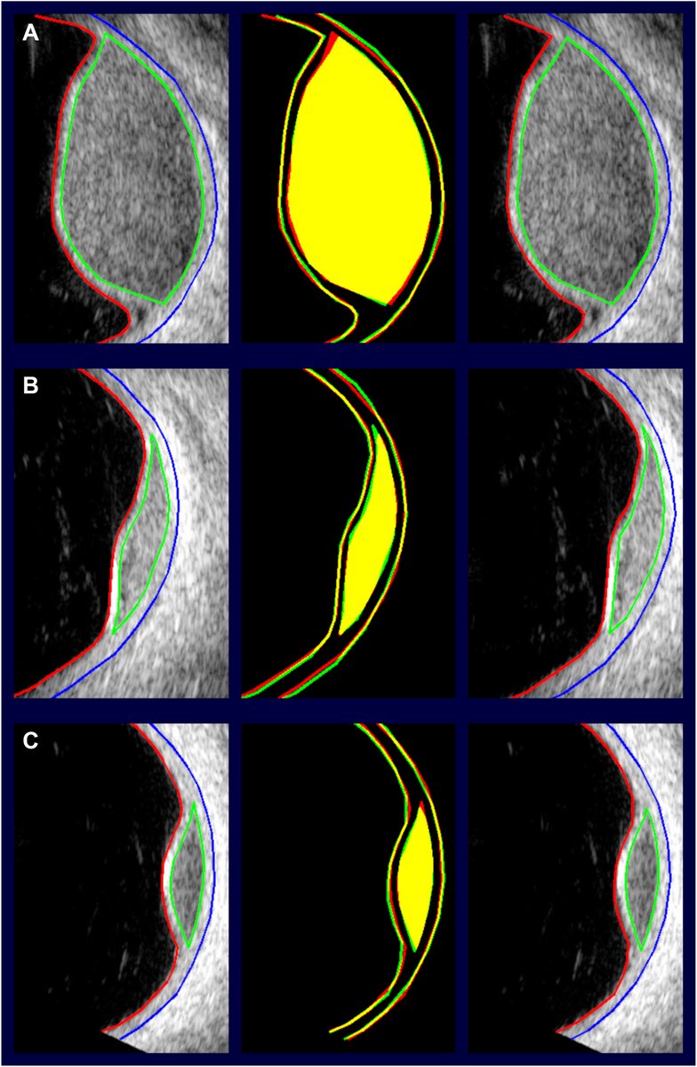
Comparative Diagnostic Efficacy of Swept-Source OCT and Scheimpflug Imaging in Clinically Unaffected Eyes of Very Asymmetric Ectasia
https://t.co/E8DARXqwBQ
#ophthalmology
A 64-year-old woman with a history of chloroquine-related cardiac toxicity (biopsy-proven and consistent with atrioventricular nodal lysosomal toxicity) was referred for retinal evaluation. Visual acuity was 20/70 in both eyes. The cumulative chloroquine dose was approximately 182 grams. (A) Color fundus photography shows diffuse pigmentary retinopathy with predilection for the inferior quadrants. (B) Fundus autofluorescence illustrates severe atrophy, especially involving the inferior macula with notable sparing superior. (C) OCT shows diffuse outer retinal atrophy with relative central ellipsoid zone preservation in both eyes. Genetic testing for inherited retinal degeneration was negative. Findings are consistent with severe chloroquine retinal toxicity, thought to result from lysosomal dysfunction with impaired autophagic degradation within the retinal pigment epithelium and photoreceptors.
https://t.co/HDVT2EOJc7
#ophthalmology #retina
Scleral depression can feel challenging at first, but repetition and a systematic approach help residents build confidence and improve peripheral retinal examinations.
Read: https://t.co/xJTD0Mk7Xb
#YOInfo#AAO#YO
40% of U.S. doctors are already using AI to make real-time clinical decisions. The catch? Most of these tools learned medicine from the open internet.
Stephen D. McLeod, MD, CEO of the American Academy of Ophthalmology, makes the case for something better: an AI built on eye care's own gold-standard guidance.
A clear-eyed take on where these tools help, where they fail, and what comes next.
Read: https://t.co/7QEafPcw1j
#AAO #AIMedicine #EyeCare #EyeNetMag #Ophthalmology
El truco para aplicar sombra de #ojos después de los #50 : evita que se acumule en los pliegues y lucen más frescos y luminosos: https://t.co/k1glYgSyZ7
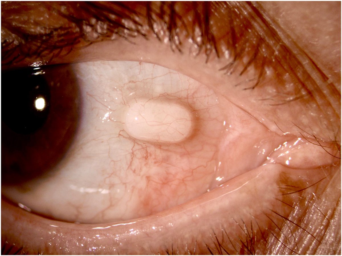
A patient presented with an unusual case of a nasal subconjunctival Mydriasert insert in the right eye, detected 3 weeks after uneventful cataract surgery. The insert had been placed pre-operatively to achieve mydriasis, and sub-Tenon’s anesthesia was performed in the inferonasal quadrant. At the routine postoperative visit, the patient was asymptomatic and satisfied with the visual outcome but noted a small nasal conjunctival swelling. Slit-lamp examination revealed the Mydriasert insert within the subconjunctival space, which was removed easily under aseptic conditions at the slit lamp. The most likely mechanism was migration of the insert through the sub-Tenon entry site created during anesthesia. This case highlights the importance of confirming removal of the Mydriasert insert before surgery and carefully inspecting the conjunctival fornices thereafter to prevent retention and possible postoperative complications.
From “Subconjunctival Retention of a Mydriasert Insert after Cataract Surgery” by Filomena Palmieri, MD and Vincenzo Maurino, MD. Published by Ophthalmology online on January 7, 2026.
Read: https://t.co/VP7gjHjQDK
#AAO
Eventual atrophy-associated vision loss from a choroidal nevus: 13 years of follow-up. A 73-year-old woman with an 8-year history of a stable choroidal nevus at the macula presented with distortion (visual acuity 20/25) (A). Fluorescein angiography showed pinpoints of leakage from nevus-related retinal pigment epithelium (RPE) dysfunction (B). OCT revealed subretinal fluid, cysts, shaggy photoreceptors, and subretinal hyper-reflective photoreceptor debris (C). There was no change after 3 intravitreal bevacizumab injections, also suggesting absence of choroidal neovascularization. The patient was observed carefully. Five years later, there was development of subfoveal atrophy (visual acuity 20/60) (D-E). This case illustrates an underrecognized pathway of nevus-driven RPE degeneration leading to atrophy and vision loss.
https://t.co/0agjOU89Nh
#ophthalmology #retina
Managing #glaucoma isn’t just about pressure — it’s about patterns, progression and precision. Five key pearls help sharpen clinical judgment and avoid common missteps. #YOInfo https://t.co/id7I1qtiU5
A patient from Congo traveled to Paris to undergo cataract surgery. One month after an uneventful procedure, the patient was referred to our department due to the appearance of multiple firm scleral nodules (A). Clinical examination revealed asymptomatic distal ulcerations on the fingers (B) and multiple subcutaneous nodules. Slit-lamp examination revealed complete corneal anesthesia with markedly hypertrophied nerves (arrowheads, C) that appeared moniliform on in vivo confocal microscopy (arrowheads, D). Given the combination of ocular findings with asymptomatic digital involvement, subcutaneous nodules, and underlying immunosuppression (HIV+), leprosy was suspected. Skin biopsy and bacteriological examination confirmed multibacillary leprosy, consistent with a borderline form of leprosy.
https://t.co/aJGjngolxC
#ophthalmology
La #migraña no es solo un dolor de cabeza.
A menudo quienes la sufren se enfrentan a comentarios o frases que minimizan lo que sienten, situaciones que no reflejan la verdadera carga de esta enfermedad.
Confidence in #glaucoma care comes from recognizing subtleties — when to escalate, when to monitor and how to avoid diagnostic traps that can delay treatment. #YOInfo#Ophthalmology https://t.co/fRWKjQSlYI
What's new in #ophthalmology this week?
From #retinal detachment risk with floaters vs flashes to the effects of long #COVID-19 ocular infections, here's what you need to know: https://t.co/QjAMt47Wqv.
#AAO#ophthalmiceducation