Our article is out on cytology whole slide imaging! @path_DOan_lie@MikeThrallMD
Assessing the quality of cytopathology whole slide imaging for educati... https://t.co/5tSmJSIQYs #PathTwitter#PathArt
Thyroidectomy revealing classic papillary thyroid carcinoma. Papillary structures of the tumor are seen below normal thyroid tissue. #PathArt#PathTwitter#MedTwitter#CytoPath
Thyroidectomy revealing an oncocytic follicular carcinoma. Hurthloid tumor cells are seen in a follicular growth pattern. Extensive invasion was identified (not shown). #PathArt#PathTwitter#Pathology
Transurethral resection of a bladder tumor revealing a high-grade papillary urothelial carcinoma. Fibrovascular core juxtaposed with tumor epithelial cells. #PathArt#PathTwitter#Pathology
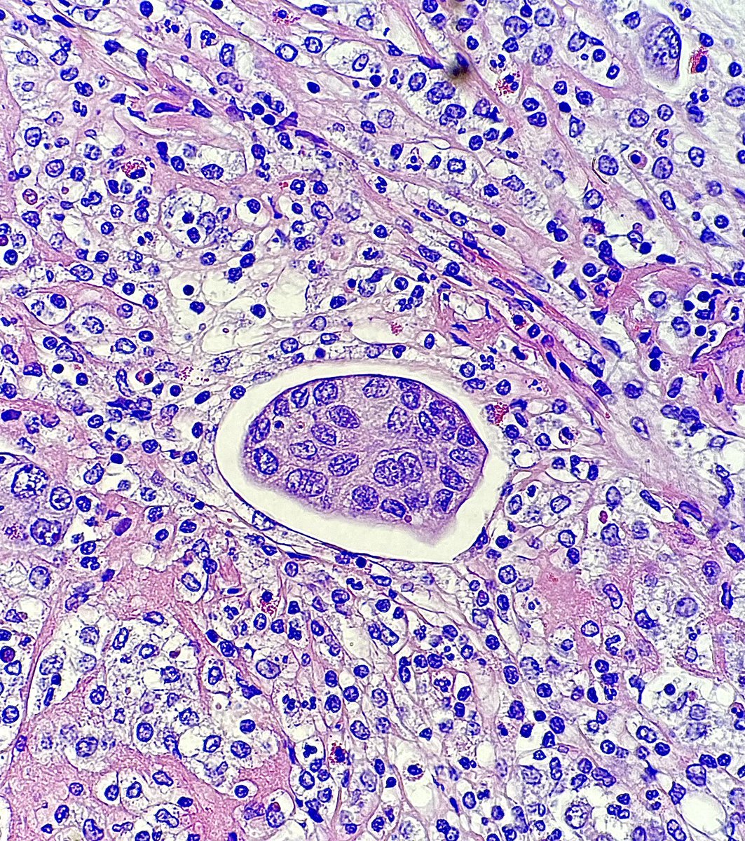
Lung lobectomy revealing a mycetoma filled with the dimorphic fungal pathogen Coccidioides immitis. Ruptured spherules are seen releasing endospores. Hematoxylin & eosin stain. #PathTwitter#PathArt#Pathology#Microbiology#Fungi
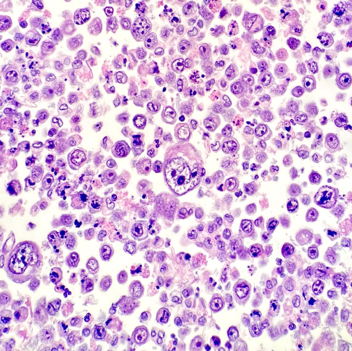
Endobronchial ultrasound guided transbronchial needle aspiration of a mediastinal lymph node revealing Histoplasma capsulatum. Histiocytes filled with small intracellular yeasts were seen on Diff-Quick stain. #PathTwitter#CytoPath#Microbiology#Pathology#PathArt
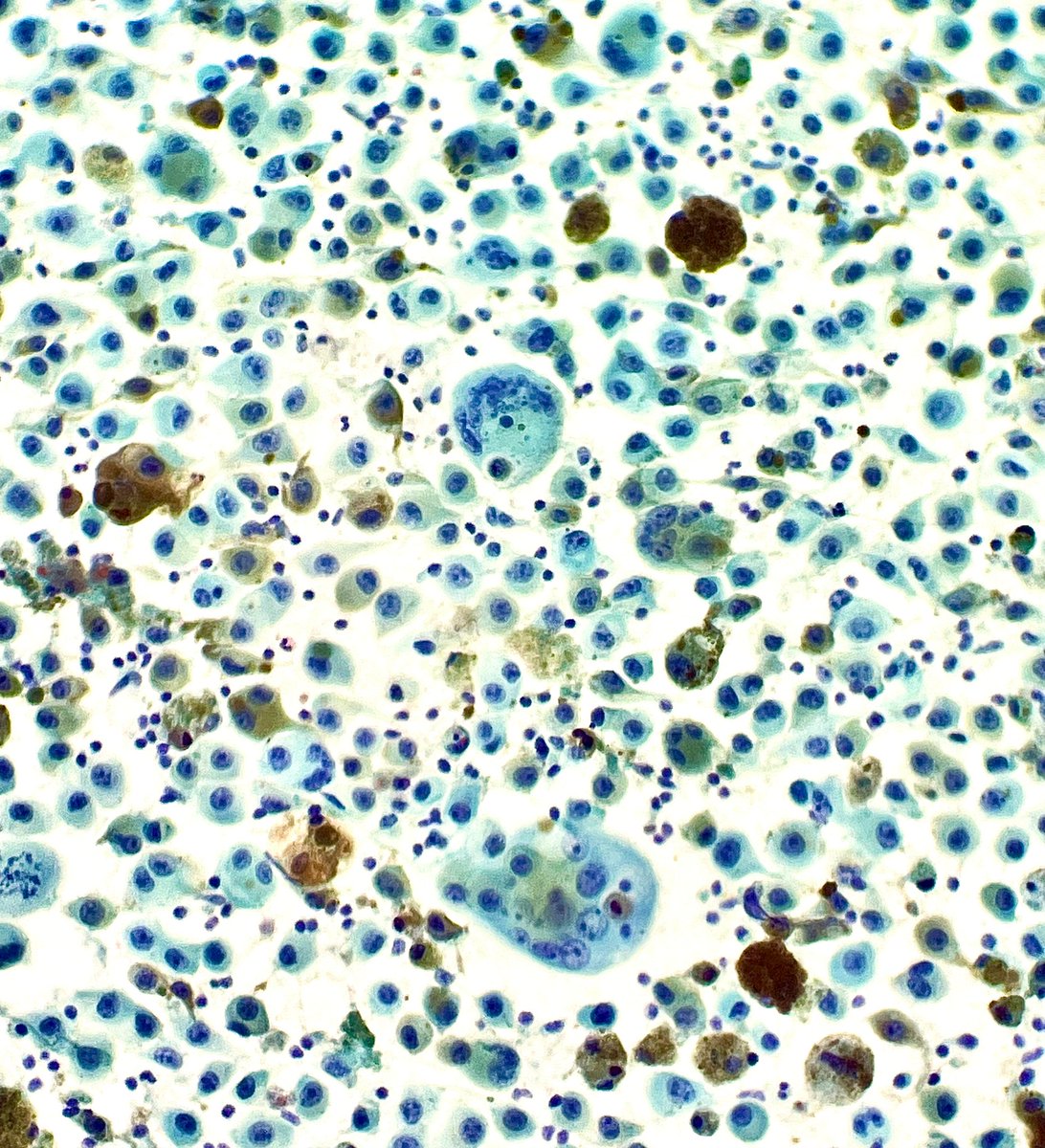
Fine needle aspiration of a lymph node containing metastatic melanoma. Pigmented, pleomorphic tumor cells are seen on Papanicolaou stain. #PathTwitter#Pathology#CytoPath#Melanoma
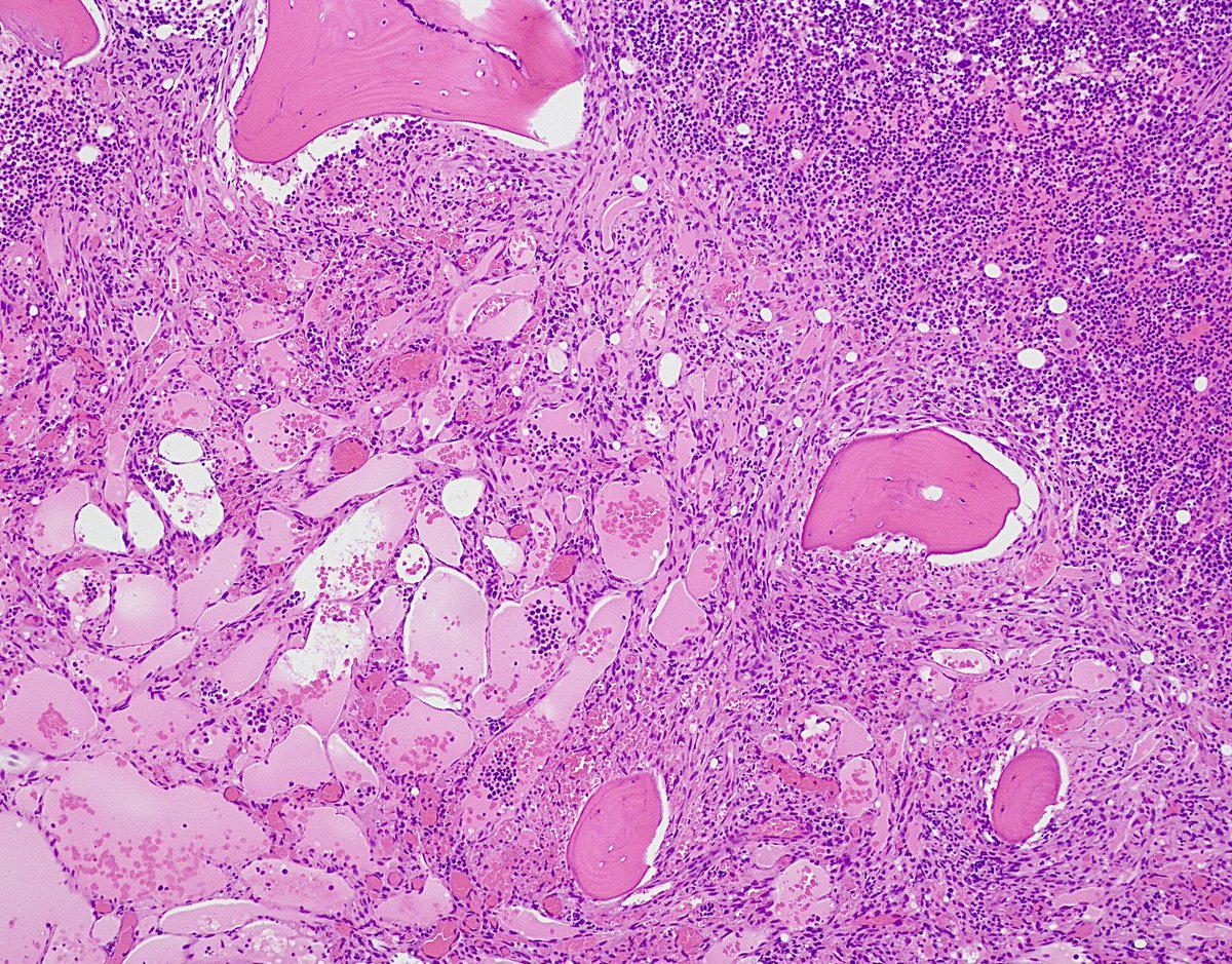
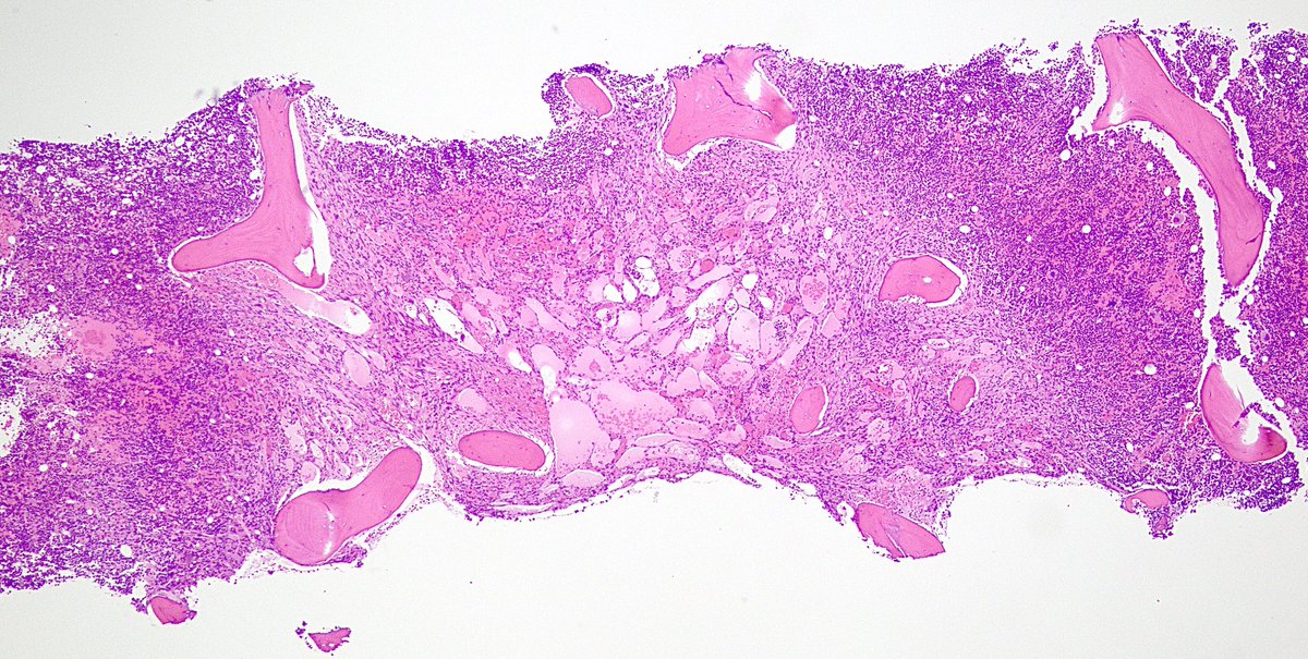
Bone marrow biopsy of a patient with untreated HIV revealing involvement by Kaposi sarcoma. Spindled tumor cells are seen forming irregular, dilated vascular spaces. HHV-8 immunostain was positive in tumor cells. #PathTwitter#PathArt#Pathology#BSTPath