❗️Important info hidden in the long thread❗️
Download the new QuPath v0.6.0 *release candidate* for the easiest way to try this out 💻🔬🤖
https://t.co/S6aFegoUVX
We are excited to announce our expanded partnership with @SynapticSystems. They now offer primary ABs with #abberior STAR dyes—labels optimized for STED microscopy, providing precise neuronal protein detection with strong signals and low background fluorescence.
Read more > https://t.co/a3Y4uzS0LB
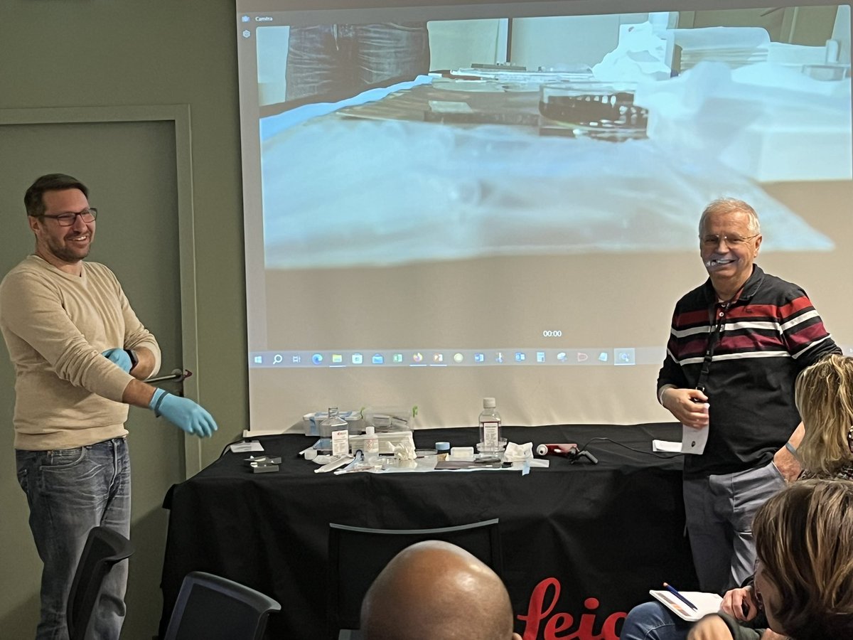
Yesterday we enjoyed the 1st FBI Alsace Node meeting organized at the @IGBMC about "How the platforms’ technologies serve your research?"
Great opportunity to meet & discuss with our new node members and their users 👥🔬
FLIM unmixing is a powerful method for Live cell multicolor imaging.
In a recent article from the team led by Ludovic Galas, SPY650-FastAct, SPY620-DNA and SiR700-actin were used to illustrate the principle using a @LeicaMicro FALCON system and the intuitive phasor plot separation of fluorophores.
With a single excitation line, 2 or 3 probes are acquired with a perfect temporal synchronicity. This is the ideal tool to study e.g. organelles interactions.
The movie illustrates a 3 color scheme with SPY620-DNA (nucleus, red hot LUT), SiR700-actin (F-actin, magenta LUT) and LBL-Dye M715 (mitochondria, Yellow LUT)
Article link: https://t.co/8bax7Z899d
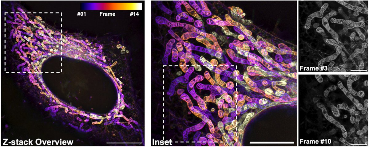
After countless requests, we're thrilled to introduce PK Mito Orange FX - a fixable variant of PKMO. PKMO FX allows immunolabeling and CLEM approaches.
Happy to have contributed to this project led by @ZhixingChen2 and Christian Jüngst.
#mitochondria
https://t.co/nC6gX3LBGt
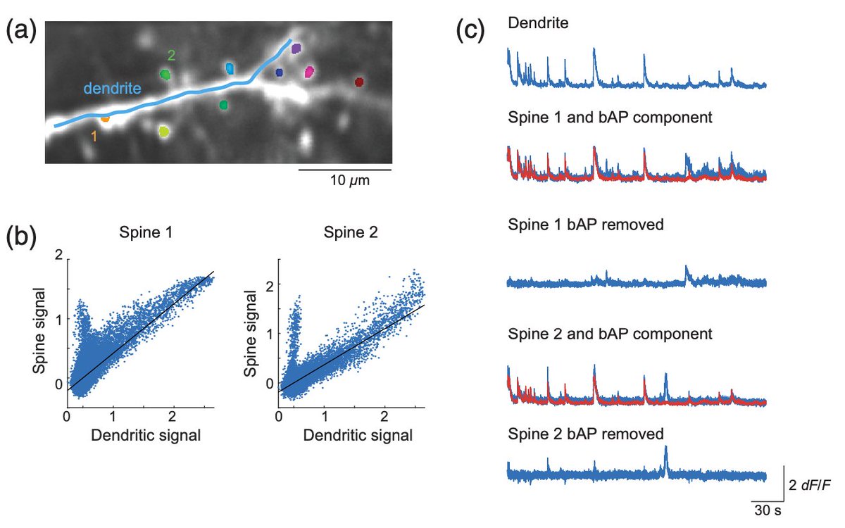
New analysis software for imaging data in dendrites, spines, and synapses. "Comprehensive software suite for functional analysis and synaptic input mapping of dendritic spines imaged in vivo" Open source. https://t.co/bsVrXCE8l8 Look at that bAP subtraction! (1/5)
4th Summer School Advanced Tools for Data Analysis in Neuroscience, Sept 12th to Septh 21th, 2024 Analyse data and meet colleagues from european countries. No fees , and accomodation/lunches offered to all students
https://t.co/WvJvFBvVQX
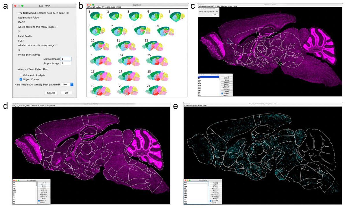
FASTMAP is an #opensource plugin for ImageJ. It allows for registering biological images within brain atlases. It also segments biological labels of interest within brain regions. @DTerstege Read about FASTMAP in this week's post on OpenBehavior:
https://t.co/YicbxW2xYU
📢Anne-Sophie Mace (@institut_curie) will present the FBIAS, France BioImage Analysts, which aims at building a nation-wide remotely operating core facility for bioimage #analysis service !
https://t.co/xbwPL8pIQD
📆March 26 at 14:00 CET online
Registration down below ⤵️
Our featured image this week is from @t_dhellemmes and @JTeillon. It shows the Relaxin-3 neurons in an adult mouse brain, cleared using Adipoclear and imaged on a light sheet microscope: https://t.co/rFpFMeKlmm
Excited to share Spotiflow, our new spot detection method for image-based spatial transcriptomics that facilitates the analysis of large iST images. Lead by @Albertdm99 jointly w @GioeleLaManno@epflSV@EPFL_Imaging@EPFL_BioE https://t.co/UKooR3wV0V
https://t.co/n3ZNfNaMvG
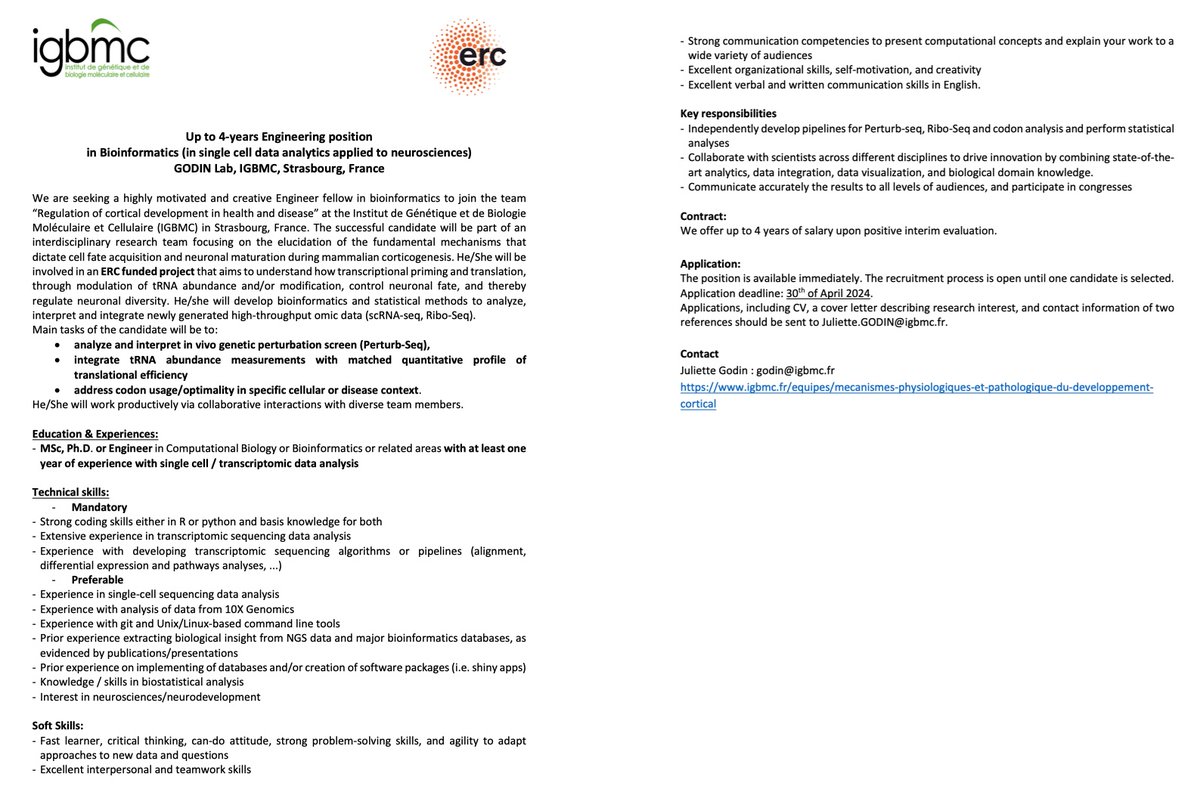
We are hiring ! A up to 4-years Engineering position
in Bioinformatics (in single cell data analytics applied to neurosciences) is available in my lab, @IGBMC, Strasbourg, France, please RT.
Update: most of the Contact-FP probes are now available on @Addgene. We hope folks will try them out and let us know what you think! https://t.co/BK3kz3XoqU
Are you a microscopist/biologist seeking new ways to visualise subcellular compartments? 🔬Our new paper in @nanoscale_rsc shows how fluorescent esters can give you valuable morphological insights into cellular architecture 🦠https://t.co/wjfxr5ZFXU (1/6)
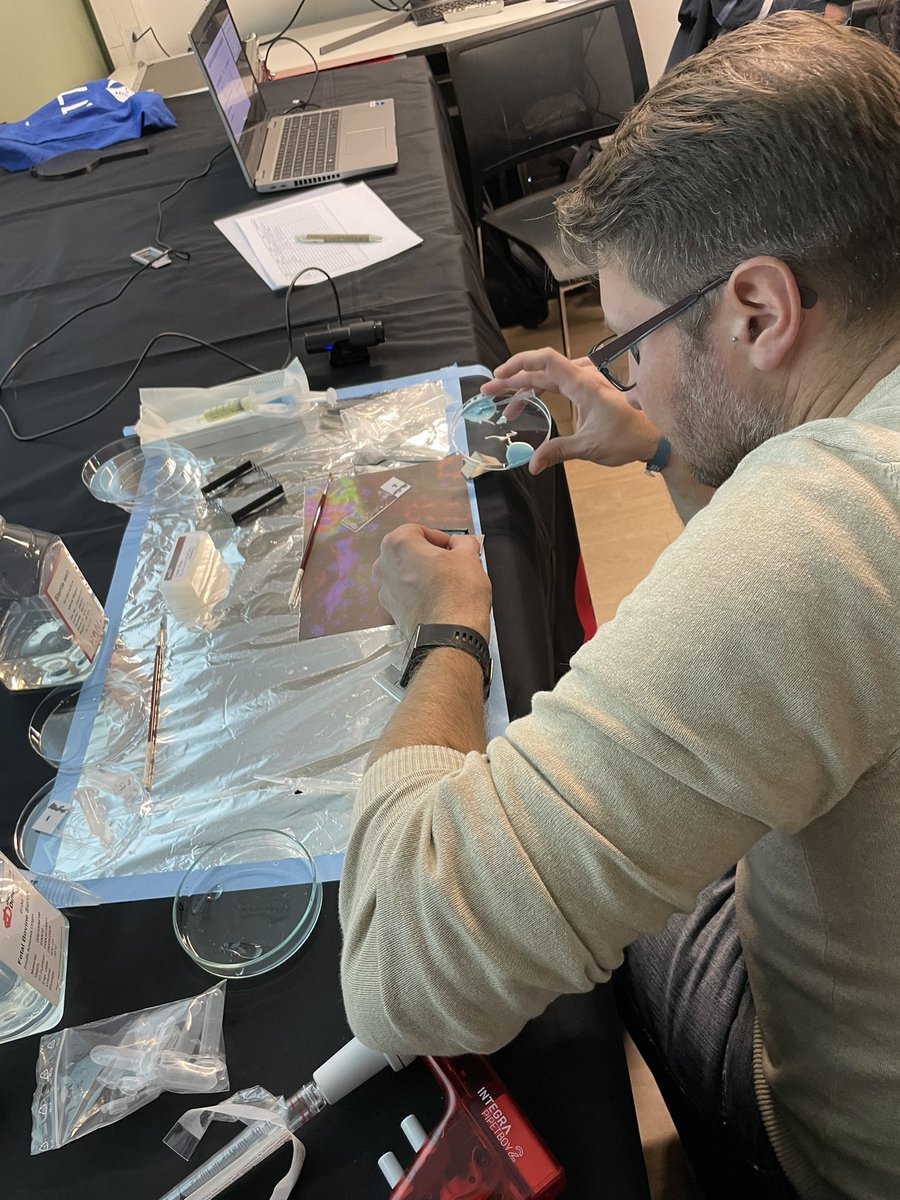
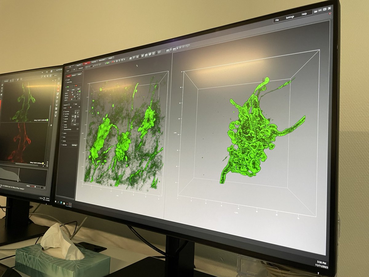
Looking @ synapse structures with Pierre Hener & Yves Lutz with #Expansionmicroscopy on mouse brain tissue slices. For this afternoon #MiFoBio2023 on STELLARIS 8 STED. Wonderful samples and great insights ! 🤩