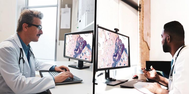
Innovating #digitalpathology with German precision! Digitizing workflows & enhancing cancer diagnostics with easy-to-use microscopes, scanners & software.
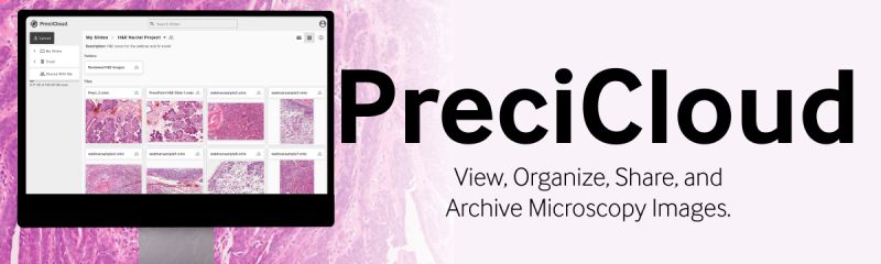
Smarter Workflows Start with PreciCloud!
The new features of PreciCloud take your #DigitalPathology workflow to the next level. PreciCloud, our secure, web-based platform for managing and sharing whole slide images, now offers:
🔍 Split View – Accurately compare different slides side-by-side at the same time.
🖊️ Import Annotations – Add your existing work seamlessly into PreciCloud.
📁 Group Annotations – Stay organised with logical grouped annotations for enhanced labeling and counting.
🛠️ New Annotation Brush Tool – Use the new brush tool to quickly annotate large or small areas. Accurately reduce or enlarge the area with exacting precision.
If you work with #WholeSlideImages and need to collaborate with peers, obtain second opinions, or teach students remotely, PreciCloud provides you with the tools to do it all—securely, from any device, anywhere in the world.
Ready to upgrade your digital pathology experience?
👉 https://t.co/K2H1ig3EOk
#Telepathology #AnnotationTools
Want to see how researchers are pushing the boundaries of digital pathology?
Explore scientific publications from experts around the world—real studies, real insights, and real applications.
If you're interested in how digital pathology is being used, tested, and improved in clinical and research settings, visit
https://t.co/q5Qf3QMA8Q
#DigitalPathology
#VirtualMicroscopy
#MedicalResearch
When time is critical, you need technology that keeps up. ⏰
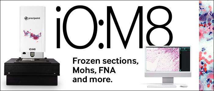
With the iO:M8 Digital Live Microscope, you gain:
✅ Faster intraoperative consultations – enabling confident decisions during surgery without delay.
✅ Instant second opinions – connecting with experts in real time, no matter where they are.
✅ More efficient frozen section handling – reducing turnaround time and supporting better patient outcomes.
You stay in control, even under pressure—because streamlined collaboration and rapid access to insights aren't optional.
👉 Learn how iO:M8 works for you: https://t.co/41l12M61k3
#DigitalPathology #IntraoperativeConsultation #FrozenSection #SecondOpinion
How Is Interventional Pathology Changing Patient Care?
Pathology is no longer just about analyzing slides in a lab. It’s now actively shaping patient outcomes in real time. Interventional pathology is changing the way diagnostics and treatment decisions are made at the point of care. As a result, treatment plans and medicine have become faster, more precise, and patient-centric.
What’s Driving This Shift?
🔬 Rapid On-Site Evaluation (ROSE) – Immediate pathology insights during biopsies ensure accurate sampling, reducing repeat procedures.
🔬 Minimally Invasive Procedures – Precision-guided biopsies minimize patient discomfort while maximizing diagnostic accuracy.
🔬 AI & Digital Pathology – AI-driven image analysis and telepathology enable real-time consultations, even across borders.
🔬 Personalized Medicine – Molecular profiling at the time of biopsy allows for faster, targeted treatments, especially in oncology.
🔬 Faster Decision-Making – Direct collaboration between pathologists, radiologists, and interventionalists streamlines workflows for quicker, more effective treatment plans.
The Impact on Patient Care
⚡ Faster diagnosis.
🥼 More precise treatments.
👨🏼⚕️ 👩🏽⚕️ Access to expertise expands beyond hospital walls.
As interventional pathology continues to evolve, the role of pathologists is becoming even more proactive in shaping patient outcomes.
#InterventionalPathology #DigitalPathology
Advancing Digital Pathology with PreciPoint 🔬
Gesundheit Nord - Klinikverbund Bremen has taken a big step into the world of #digitalpathology with our iO:M8 - digital live microscope for rapid on-site evaluation. As one of the most advanced solutions for remote-controlled pathology, iO:M8 transforms the way pathologists work—delivering precise, real-time #frozensection diagnoses from any location.
Dr. Samy Hakroush, Head of the Institute of Pathology at Bremen-Mitte Hospital, shares:
“By working together with PreciPoint, we have integrated a system that is one of the most advanced solutions for remote-controlled pathology.”
With iO:M8 digital live microscope, Bremen-Mitte is now among the most modern pathology facilities in northern Germany, ensuring:
✅ Faster and more accurate diagnostics
✅ Flexible, remote pathology workflows
✅ Uncompromised quality in patient care
We are proud to support Klinikum Bremen-Mitte in driving digital transformation in pathology.
🔬 Ready to elevate your pathology workflow? Get in touch with us today!
🔬 Exciting News!
PreciPoint Joins the NVIDIA Inception Program! 🚀
PreciPoint is now part of the NVIDIA Inception program - an initiative designed to empower startups with cutting-edge technology services, solutions, expertise, and industry collaboration.
This collaboration marks a significant milestone in our mission to drive digital pathology forward. With this partnership, we accelerate the development of solutions that enhance telepathology workflows, AI-powered diagnostics, and real-time remote evaluations - expanding what’s possible in digital pathology.
Stay tuned as we push the boundaries of digital pathology!
#AI #NVIDIAInception
Some impressions from this year's USCAP - United States and Canadian Academy of Pathology in Boston. We’re thrilled and look forward to the exchange with many international pathologists, researchers and lab professionals! Great to be here!
#USCAP2025#DigitalPathology
🔬 Addressing Pathologists' Biggest Challenges at USCAP 2025
Pathologists face increasing pressure—faster diagnoses, complex cases, and limited resources.
The demand for real-time consultation, seamless remote access to slides, and AI-powered image analysis has never been greater. Yet, inefficient workflows and accessibility gaps continue to slow down pathology services.
At USCAP 2025, we’re bringing solutions that empower pathologists to work smarter.
📍 Stay tuned for details!
Let’s close the care gap and redefine pathology together. Join us at USCAP 2025 to see how technology can work for you.
#USCAP2025 #DigitalPathology #RemoteMicroscopy
Alternative Session on March 18!
If you couldn't join us on March 12, here's another opportunity to gain practical, expert-led insights into managing technical challenges in Telecytology with Handy Oen and James Brown.
What’s at Stake?
In Telecytology workflows, one software glitch, poor image resolution, or system lag could mean a missed or delayed diagnosis—affecting real patient outcomes. Are your workflows resilient enough to handle disruptions?
What Will This Webinar Be About:
🔹 The most common technical risks that disrupt Telecytology & ROSE and how to prepare for them.
🔹 Lessons from real-world failure cases where technical breakdowns led to operational inefficiencies.
🔹 The A.C.E. Strategy – an easy-to-apply method for Assessing, Coordinating, and Eliminating risks.
🔹 How Continuous Quality Improvement (CQI) & CAPA can turn weaknesses into workflow strengths?
🗓 Date: Tuesday, March 18, 2025
⏰ Time: 17:00 - 17:45 CET
📍 Location: https://t.co/M11sxCCoc3
🎤 Meet the Speakers:
- Handy Oen: Expert in digital pathology, contingency planning, and telecytology workflows
- James Brown: Specialist in technical infrastructure and workflow optimization
💡 Expert-Led Q&A: Bring your questions, and we’ll bring solutions.
Don’t miss this chance to strengthen your telecytology workflow—secure your spot today.
#DigitalPathology #PatientSafety
When Technical Failures Threaten Patient Care in #Telecytology – What’s Your Plan?
Telecytology is a game-changer for Rapid On-site Evaluations (ROSE), helping improve diagnostic speed and accuracy. However, technical failures—whether software crashes, poor image resolution, or network disruptions—can jeopardize patient outcomes and lead to delayed diagnoses.
💡 How can you safeguard your telecytology workflow from these risks?
Join our expert-led webinar to:
✅ Identify the most common technical pitfalls in Telecytology & ROSE.
✅ Demonstrate real-world cases where system failures led to diagnostic delays.
✅ Introduce the A.C.E. Strategy – a structured, actionable framework to Assess, Coordinate, and Eliminate technical risks.
✅ Share how Continuous Quality Improvement (CQI) & Corrective and Preventive Actions (CAPA) can create a robust telecytology system that prevents future issues.
🗓 Date: Wednesday, March 12, 2025
⏰ Time: 16:00 - 16:45 CET
📍 Location: https://t.co/MnsIvtKB9i
Expert Speakers:
- Handy Oen, Cytology Supervisor(Regional Labs), Memorial Sloan Kettering Cancer Center, United States
- James Brown, CEO, PreciPoint International Inc., United States
Interactive Q&A: Ask our experts your toughest workflow questions!
Don’t leave your telecytology workflow vulnerable—register now and take control!
#DigitalPathology #PatientSafety
Picture this: You’re on the go, but your team needs your expert opinion on a critical slide. Instead of waiting hours—or even days—you log in, review, and share your insights in minutes. ⏳
Sounds futuristic? It’s not. It’s PreciCloud.
Imagine a world where accessing, managing, and analyzing your pathology slides is as seamless as clicking a button. That world is here.
✨ This is PreciCloud – Your Gateway to Digital Pathology Excellence.
PreciCloud empowers pathologists and labs with:
☁️ Secure Cloud-Based Slide Management: Access your slides anytime, anywhere, on any device.
💻 Collaboration Made Easy: Share slides with colleagues effortlessly for consultations and second opinions.
🥼 Enhanced Workflow Efficiency: Streamline your operations and save valuable time.
📏 Scalable for Your Needs: From small teams to large-scale labs, PreciCloud grows with you.
But don't just take our word for it. Experience the difference for yourself 👉 https://t.co/K2H1ig3EOk
#DigitalPathology
#PreciPoint
#PreciCloud
🧐 Struggling to Keep Your Objectives Clean?
Immersion oil is essential for high-resolution microscopy, but cleaning it off your objectives can feel daunting.
Improper cleaning methods can damage your lenses and compromise image quality, but with the right steps, you can maintain the longevity and performance of your equipment!
✨ Here are a few quick tips:
🔬 Always use a lens-safe cleaning solution.
🔬 Avoid abrasive materials—your objectives deserve a gentle touch!
🔬 Clean immediately after use to prevent oil buildup.
For a detailed guide, including the do’s and don’ts, check out our blog post: How to Clean Immersion Oil Off Your Objectives?
👉 https://t.co/jfP4tOnS4x
🔗 Ensure your lenses stay crystal-clear for every observation.
Follow us and stay connected for the latest insights, expert tips, and practical tricks in the world of digital pathology!
#DigitalPathology
#PreciPoint
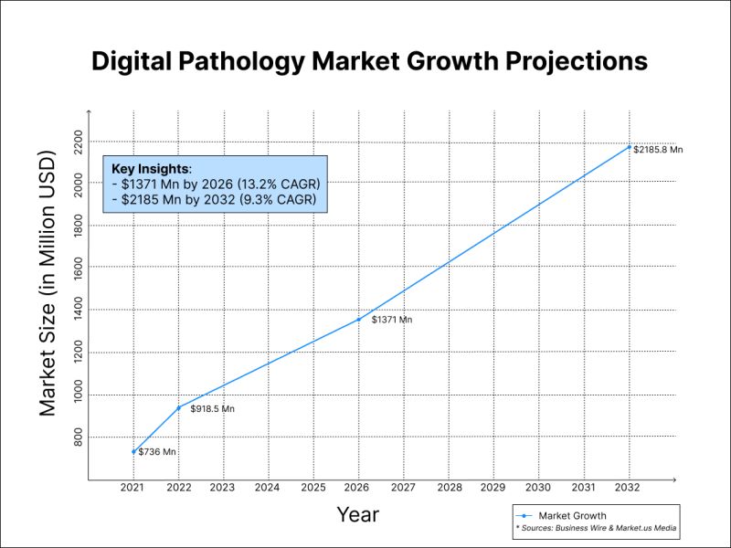
The digital pathology market is on a remarkable growth trajectory!
Here are some key insights shaping the future of this field:
🔹 $1371 million by 2026
The market is expected to grow from $736 million in 2021 to $1371 million by 2026, with a compound annual growth rate (CAGR) of 13.2%.
🔹 $2185.8 million by 2032
Other research highlights growth from $918.5 million in 2022 to $2185.8 million by 2032, with a CAGR of 9.3%.
At #PreciPoint, we are committed to empowering labs with innovative digital solutions to meet the demands of this growing industry. From seamless slide digitization to efficient remote workflows, we help labs stay ahead of the curve.
💡 Are you ready to embrace the future of pathology? Discover how we can support your digital transformation:
https://t.co/IiknHFK3SW
#Digitalpathology
#Digitization
🎉✨ Happy New Year 2025! ✨🎉
This past year, we continued to innovate, connect, and push boundaries in digital pathology, empowering pathologists worldwide with cutting-edge solutions and unwavering support. Your trust and collaboration have been instrumental in driving our mission forward. 🙌
Here’s to 2025 filled with discoveries, breakthroughs, and new milestones —together, we’ll achieve even greater heights. 🌟
🚀 Let’s make 2025 extraordinary!
#HappyNewYear2025
#DigitalPathology
🔬 Frozen Section Diagnostics in the Digital Era
In the world of pathology, frozensection procedures are often the key moments in surgical decision-making. Yet, traditional workflows come with challenges: geographical constraints, time pressures, and the ever-increasing complexity of cases.
Enter the digital era, where innovative solutions like livemicroscopy and digital tools are paving the way for efficient, remotediagnostics.
Here’s what’s changing:
🔍 Efficiency Boost: No more rushing across town. Pathologists can now assess frozen sections remotely, saving precious time and resources.
🌎 Bridging the Geographical Gap: Advanced digital solutions allow seamless collaboration between rural sites and centralized labs, ensuring every patient gets timely, accurate results.
📈 Scalability: Starting with frozen sections as a pilot project can serve as a gateway to fully digitizing pathology workflows, ensuring a smooth transition with quick wins along the way.
👩⚕️ Multidisciplinary Collaboration: These tools don’t just empower pathologists—they enhance communication with surgeons and oncologists, driving better outcomes for patients.
As the field evolves, it’s clear that embracing digital tools isn’t just about keeping up with technology—it’s about reimagining the future of patient care.
What are your thoughts? Are there challenges you foresee in your practice or benefits you've already experienced? 💬
#frozensection #DigitalPathology #PathologySolutions
Is Your Lab Ready for the Shift to Virtual Microscopy? 🔬
The transition to virtual microscopy promises a world of efficiency, collaboration, and innovation—but like any big change, it comes with its challenges.
🚧 Key hurdles labs face:
* High Initial Costs – Upgrading to virtual systems can feel like a steep investment.
* Data Management – Digital slides generate massive amounts of data, requiring robust storage and security solutions.
* Resistance to Change – Adapting to new workflows and technologies can take time for teams accustomed to traditional microscopy.
* Interoperability Issues – Ensuring systems and software work seamlessly together can be a headache without the right strategy.
But here’s the good news: overcoming these challenges is possible with the right guidance, technology, and training. Virtual microscopy isn’t just a trend—it’s the future.
📈 The benefits? Faster diagnoses, improved collaboration, and enhanced efficiency that will redefine lab workflows.
👉 Dive into the full article to explore solutions: https://t.co/t2uiKOim7K
💡 Curious about how your lab can navigate these challenges successfully? Follow us to stay connected and discover more insights!
#VirtualMicroscopy #DigitalPathology
🌍🔬 Happy International Pathology Day! 🔬🌍
Today, we celebrate the work and dedication of pathologists worldwide who play a critical role in advancing healthcare. From early diagnosis to precision treatments, your expertise makes a profound difference in patients' lives every day.
Pathology is at the heart of medical progress - empowering discoveries, improving patient outcomes, and paving the way for the future of personalized medicine. As we honor the achievements and innovations in the field, let's also look forward to how digital pathology is transforming workflows, enhancing efficiency, and enabling deeper insights.👩⚕️🔬👨⚕️
Thank you to every pathologist for your invaluable contributions and unwavering commitment to excellence.
#InternationalPathologyDay #DigitalPathology