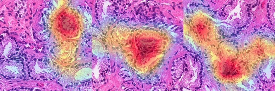
Train a contrastive model on histology from every organ and it learns something useless: telling a lung from a liver. Our new paper, SPADE, forces attention onto real molecular-morphological signal instead.
Paper: https://t.co/DnGgVEenW2
Code: https://t.co/69yJcFLdFo
We tend to evaluate diagnostic AI as a standalone classifier: feed it an image and get a score. But in ultrasound imaging, that ignores the person holding the probe.
Our new commentary in Annals of Thyroid dives into this issue.
https://t.co/PFnfBf1mfs
🚨 New paper in @IEEE_TMI & @IEEEorg
Can we predict no-reflow immediately after thrombectomy instead of waiting up to 24 hours for MRI?
DSA-NRP uses intra-procedural angiography to identify high-risk stroke patients in real time.
@UCLAHealth#Stroke#Neurointervention#AI
Thick-slice CT scans are common but may limit diagnostic accuracy due to lower spatial resolution.
This study developed an AI model to convert thick-slice CT into synthetic thin-slice CT, showing comparable quality to real thin-slice CT & improving diagnostic accuracy for #pneumonia + lung nodules, potentially offering a practical alternative in resource-limited settings.
https://t.co/7KSzsNZ2XA
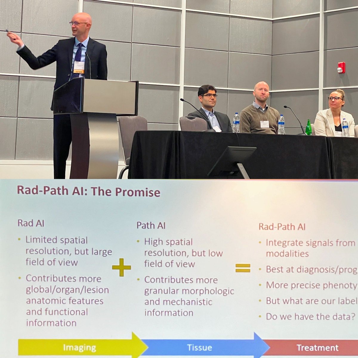
Tonight Distinguished Professor Denise Aberle rebooted our @RadiologyUcla research seminar series with a lecture on her research journey that led to her induction into @theNAMedicine. It was electric.
The wonderful staff at our Ackerman Blood & Platelet Center make donating easy & enjoyable. Please consider donating!
Give Blood. Save Lives. Donate Today! 🩸
📱: 310.825.0888 x2
✉️: [email protected]
🌐: https://t.co/ctkdcs1ifC
Grateful and honored that I could be a part of this amazing panel of speakers (still starstruck), even if I had to be there virtually. @FredNatLab @hood_college. Talk posted here: https://t.co/hE0uFU4OwB
Synthesizing contrast agents using #AI is a problem that I'm not sure we'll ever be able to demonstrate works convincingly, but that doesn't stop us from trying! Here's our recent paper in IEEE BME showing state-of-the-art results in brain cancer patients…https://t.co/sKDas3ANSq
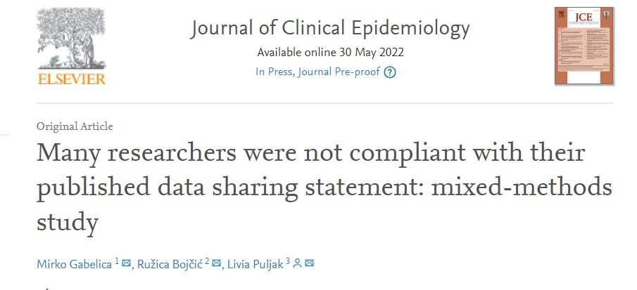
Our new study shows that data availability statements are not very useful; 1670 (93%) authors who indicated that data are available on request either did not respond or declined to share their data with us. Journal of Clinical Epidemiology: https://t.co/4IT2Dgphl4
Our new paper on active learning in digital pathology describes a framework for training #AI with less labeled data by automatically learning to distinguish between easy samples, hard samples, and noisy samples. Nice work @Wenyuan1991!…https://t.co/lBQNzOBuBy
Tumor board preparation requires a lot of time from a lot of people. We built a tool that makes the process easier and recently demonstrated a 45% time savings for pathologists covering our musculoskeletal TB.
Preprint: https://t.co/5QtM2OaXU1 https://t.co/dG6TxQzA4B
Our new paper in Medical Image Analysis describes a conditional generative adversarial network (GAN) that can be used to improve histology image segmentation for digital pathology applications. Nice work @Wenyuan1991 and co-authors!…https://t.co/lrdJpSLOD6
New in @RadiologyUcla Proceedings: Electronic Integrated Diagnostic Report for Presenting Results of Breast Imaging and Breast Biopsy https://t.co/F9NVEPhEHO #mammo#RadQI#HITRad#FOAMrad
In a cohort of >40k patients, our #ai EHR-based algorithm achieved a PRAUC of 0.76 for predicting depression three months out. It's a challenging problem to tackle retrospectively, but there's clearly signal that can be used to focus screening. https://t.co/8wGByyrLHE
Federated learning with patient data is poised to accelerate healthcare innovation. It's a great opportunity to involve patients and allow them to receive some kind of benefit from sharing their data.
https://t.co/BQhI6AhGBB
After analyzing 16k prostate biopsy cores, we found that 95% of cancer is detected within 10mm of the MR target! Results suggest that cancer detection rates can be maintained while taking fewer cores. Nice job @alex_raman@ksarma! https://t.co/eDqrQXh8uL
https://t.co/m6v7ZVVNXU
Many acute stroke patients arrive in the ER with with no information on when the stroke began, limiting treatment options. We developed a new #AI method for classifying stroke onset time using medical images. Nice work @polsonjen and @DarthDisney123!
https://t.co/5QsM83qvm7
Our multi-resolution model for histology analysis is now online. "The model obtained an AUROC of 99.4% and an AP of 99.8% for cancer detection on an external dataset." https://t.co/RMDrXqUMkh. Link to PDF on https://t.co/m6v7ZVVNXU. @Digi_Pathology @BillSpeier @Jiayun86541373