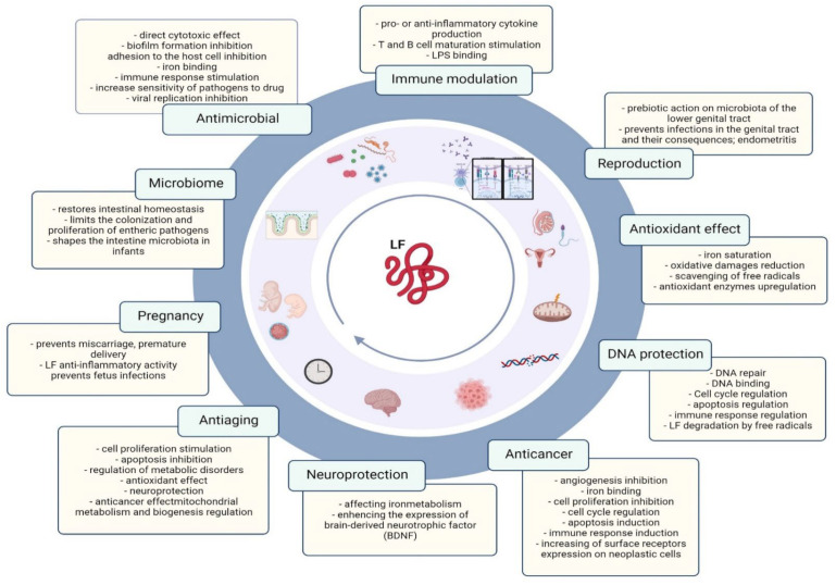
The Lactoferrin paradox:
How can one molecule selectively kill more pathogenic strains of bacteria, while at the same time stimulating the growth of beneficial Bifidobacteria?
It has to do with its structural motif containing cysteine residues, leading to a disulfide bridge
As it turns out, bifidobacteria evolved to have a receptor binding the glycoprotein, which seems to act as a growth stimulant
LF can provide carbons to be used in anabolic growth processes. This is only happening if the disulfide bridge is not reduced
One the other hand, by interacting with LPS on the outer membrane LF can destabilize the membrane of gram-negative bacteria, which leads to death of said bacteria
Been an interesting find to an interesting question
The diagram below illustrates the coordinated disinformation campaign against the Qatar Foundation.
The Foundation for Defense of @FDD Democracies (FDD), the Brandeis Center, Jewish Insider, and other entities within their network sought to create controversy over the U.S. @usedgov Department of Education's delayed release of the latest Section 117 data. They have been manipulating this data to accuse the Qatar Foundation (QF) of improper spending in U.S. higher education. On August 31st, FDD's Craig Singleton published a negative op-ed on this topic in the @WSJ Wall Street Journal. On the same day, the Brandeis Center claimed to have sent an open letter to Secretary McMahon, accompanied by a FOIA request for the records. Additionally, lawyers from the Brandeis Center published an anti-QF op-ed in the Chicago Tribune that Friday morning. @TheJusticeDept
Their goal appears to be eliciting reactions from House Education Republicans and, more broadly, spreading disinformation that serves the interests of their controllers.
drinking the first milk produced by a cow after giving birth sounds like the strangest supplement on earth until you read the immunology research showing it contains 100x more immunoglobulins than regular milk and seals the gut lining faster than any probiotic ever tested. then it sounds like liquid armor for your immune system
colostrum is the fluid mammals produce in the first 48 hours after birth. its biological purpose is to transfer the mother's entire immune blueprint to the newborn. it contains immunoglobulins (IgG, IgA, IgM), lactoferrin, growth factors (IGF1, TGF beta), and proline rich polypeptides that modulate immune function. your body recognizes bovine colostrum because the immunoglobulins are structurally identical to human ones
• seals intestinal permeability ("leaky gut") by 3x more effectively than any probiotic strain tested in a head to head comparison study
• increases secretory IgA by 79% in athletes during heavy training. IgA is the antibody that lines your respiratory tract and is your first defense against airborne pathogens
• reduces NSAID induced gut damage by protecting and repairing the mucosal lining. athletes and chronic pain patients who take ibuprofen regularly need this
• contains lactoferrin which binds to iron and starves pathogenic bacteria while feeding beneficial bacteria. a natural prebiotic and antimicrobial simultaneously
• increases lean muscle mass and reduces body fat in trained athletes over an 8 week study. the IGF1 content supports anabolic signaling
• reduces symptoms of upper respiratory infection by 3x in a trial of adults during cold and flu season
the supplement industry sells probiotics as the gut health solution. probiotics introduce bacteria. colostrum repairs the wall those bacteria live behind. if your gut lining is compromised, adding probiotics is like putting furniture in a house with no walls. fix the structure first.
take 5 to 10g of bovine colostrum powder daily on an empty stomach. morning is ideal, 30 minutes before food. look for "first milking" on the label (highest immunoglobulin concentration). sovereign laboratories and ancestral supplements both make reliable versions. $30 to $45 per month.
the first food every mammal on earth receives is colostrum. nature's first delivery to every newborn is an immune system in liquid form. and you can buy it in a jar.
Un desarrollador ucraniano creó un agujero negro en su terminal para obligarse a tomar descansos.
Cuanto más trabajas sin parar, más crece y deforma tu código con su lente gravitacional. Descansas y se encoge.
Not all exercise is equal for disease prevention. Research tracking nearly 3,000 proteins in blood reveals that sprint-interval exercise releases factors specifically protective against type 2 diabetes and obesity in ways moderate exercise doesn't.
Researchers tracked proteins in blood before, immediately after, and 3 hours following two types of exercise in young, healthy males. Sprint-interval exercise consisted of six 30-second all-out cycling bursts with 4-minute rests between sets. Moderate-intensity exercise was 90 minutes of continuous cycling.
The difference was striking. Sprint intervals altered 714 proteins immediately after exercise, more than 98% of which increased. Moderate-intensity exercise changed only 7 proteins at the same timepoint. The number increased to 19 after 3 hours, but remained dramatically lower than the sprint response.
Think of your bloodstream as a communication highway. After intense exercise, it floods with signals that travel to different organs. The study identified proteins originating from muscle, fat tissue, liver, brain, immune cells, pancreas, and other organs.
Skeletal muscle proved particularly sensitive to intensity. The researchers isolated human muscle cells and electrically stimulated them to mimic different exercise types. Simulated sprint exercise released 212 proteins into surrounding fluid. Simulated moderate exercise released only 9.
But muscle wasn't working alone. Sprint exercise increased proteins from the pituitary gland that regulate stress responses, factors from the brain involved in blood vessel relaxation, and immune proteins that modulate inflammation. The pattern suggested coordinated signaling across multiple organ systems.
The study also examined exercise metabolites, small molecules involved in energy production and cellular signaling. Sprint exercise immediately increased lactate, pyruvate, malate, and the obesity-suppressing compound N-lactoyl-phenylalanine (Lac-Phe). Moderate exercise showed a delayed response, with fatty acids rising primarily at the 3-hour mark.
To understand where these signals end up, researchers exposed human fat cells to blood plasma collected after each exercise type. Plasma from sprint exercise triggered extensive changes to fat cell gene activity, affecting 1,128 genes. Plasma from moderate exercise changed only 25 genes.
The remodeled fat cells showed activation of pathways controlling hormone responses, nutrient sensing, and fat breakdown. Several immune signaling receptors increased, suggesting exercise plasma primes fat tissue to respond to inflammatory signals differently.
The findings held up in real tissue. When researchers biopsied abdominal fat before and 3 hours after a maximal treadmill test, 418 genes overlapped with those changed by sprint plasma in isolated cells. This confirms circulating factors from intense exercise genuinely alter fat tissue biology.
Not all changes disappeared with training. After 8 weeks of regular exercise, the same intensity-dependent patterns persisted when participants repeated the acute tests. Growth hormone, von Willebrand factor, and POMC still increased more after sprints than moderate sessions.
The clinical relevance became clear when researchers cross-referenced exercise-responsive proteins with a database tracking 53,026 people for disease outcomes. They identified 143 proteins increased by exercise that associated with lower disease risk.
Of the 33 proteins specifically protective against type 2 diabetes, metabolic disorders, and obesity, 32 were elevated by sprint-interval exercise. Only 3 were elevated by moderate-intensity exercise. Proteins like ADGRG2, FGFBP1, and MXRA8 consistently showed strong protection across multiple metabolic conditions.
This isn't an argument against moderate exercise. The sustained energy demands of longer duration activity clearly stimulate different adaptive pathways, particularly in the liver. Proteins like IGFBP1 and follistatin increased exclusively after moderate exercise, likely reflecting sustained shifts in insulin and glucagon signaling.
The study reveals exercise intensity as a distinct variable that determines which protective signals reach your tissues, which organs respond, and potentially which disease risks decrease. The time-efficient nature of high-intensity exercise appears to work through fundamentally different signaling mechanisms than longer moderate sessions.
Key findings:
• Sprint-interval exercise altered 714 blood proteins immediately post-exercise vs. 7 for moderate-intensity exercise
• 25% of all detectable proteins changed after sprint exercise, with >98% increasing rather than decreasing
• Muscle cells released 212 proteins after simulated sprint exercise vs. 9 after simulated moderate exercise
• Sprint exercise plasma changed 1,128 genes in human fat cells vs. 25 genes for moderate exercise plasma
• Of 33 proteins protective against diabetes and obesity, 32 increased after sprint exercise vs. 3 after moderate exercise
• Intensity-dependent responses persisted after 8 weeks of training, suggesting they reflect relative intensity rather than training status
• Exercise metabolites showed distinct temporal patterns: lactate and Lac-Phe peaked immediately after sprints, while fatty acids increased later
• Moderate exercise uniquely increased liver-derived proteins (IGFBP1, follistatin) at the 3-hour timepoint
The mechanistic picture is incomplete. The study couldn't definitively prove which organs secrete which proteins, relying instead on tissue-specific gene expression databases and cell culture models. Blood volume shifts during exercise also complicate interpretation, though the findings held after correcting for plasma concentration changes.
The research focused on young, healthy, predominantly male participants. Whether the same intensity-dependent patterns occur in women, older adults, or people with existing metabolic disease remains unknown. The protective associations came from observational data that can't prove causation.
Still, the study offers a framework for understanding why brief, intense exercise produces adaptations comparable to or exceeding longer moderate sessions. The answer appears to lie in differential organ crosstalk triggered by metabolic stress signals that moderate exercise simply doesn't generate.
You train to protect your mitochondria from aging. And it works. Exercise preserves mitochondrial energy production into your 90s. But a 2025 study by @GGouspillou and colleagues reveals there's a second mitochondrial function that declines with age no matter how much you exercise.
Gouspillou and his colleagues at Université du Québec à Montréal divided 139 men aged 20 to 93 into active and inactive groups. They measured physical performance, muscle composition, and three critical mitochondrial functions: energy production, free radical generation, and calcium handling capacity.
The conventional wisdom says mitochondria wear out with age, becoming a leaky metabolic engine that produces more oxidative stress and becomes less capable of taking nutrients and converting them into ATP.
Direct measurement across 73 years of aging shows that's not necessarily correct.
Mitochondrial energy production stayed completely stable in active participants from their 20s through their 90s.
A 90-year-old who stayed active had mitochondria that produced energy like a 20-year-old. Inactive individuals showed declining energy production, but it tracked with how little they moved, not how many years they'd lived.
When normalized to mitochondrial content, the differences disappeared entirely. The study suggests that aging doesn't break energy production. Physical inactivity reduces how many mitochondria you maintain. Each individual mitochondrion works just as well at 90 as at 20.
Free radical production, blamed for decades as an aging driver, showed no increase with age. Active participants actually produced more free radicals than inactive individuals while displaying superior strength and performance. The free radical theory of muscle aging didn't hold.
The study had some interesting headlines:
• Physical activity protects performance across the lifespan but doesn't completely prevent age-related decline
• Mitochondrial energy production depends on activity level, not chronological age
• Free radical production remains stable with aging and reflects mitochondrial health, not damage
• Mitochondrial calcium handling capacity drops sharply after age 60 in both active and inactive individuals
• Reduced calcium handling correlates with lower muscle mass, strength, and performance
• Deterioration accelerates dramatically after 60 rather than declining gradually
The calcium handling finding represents the first mitochondrial function that deteriorates with aging regardless of exercise. Mitochondria normally act as calcium buffers, absorbing excess calcium to keep cells functioning properly. When this buffering capacity fails, a protective channel called the permeability transition pore opens too easily.
Think of it like a circuit breaker that's supposed to trip only during emergencies but starts tripping at lower and lower thresholds. When the pore opens prematurely, stored calcium floods back into the cell, triggering a cascade of damage.
This calcium release activates pathways that break down muscle protein faster than the body builds it back up. It generates bursts of oxidative stress that damage cellular components. It releases mitochondrial DNA that the immune system mistakes for bacterial invasion, triggering inflammation.
Each pathway drives muscle loss without requiring energy failure. The mitochondria still produce ATP normally. They've just lost the ability to prevent calcium from activating destructive programs that eat away at muscle tissue.
The study measured calcium handling by exposing muscle mitochondria to calcium loads and tracking retention. Both active and inactive participants showed declining capacity, but the pattern matters. Calcium handling remained relatively stable from ages 20 to 60, then dropped sharply after 70.
Calcium retention capacity correlated with thigh muscle mass, knee strength, walking distance, and mobility performance. It also correlated with GDF15, a stress molecule that rises with aging and predicts worse health outcomes. This suggests calcium handling directly influences muscle health and systemic aging markers.
Physical activity protected functional performance. Active participants outperformed inactive individuals on strength and mobility tests across all age groups. But both groups showed age-related declines. Exercise delays deterioration but doesn't eliminate it.
Muscle fiber composition revealed how activity and aging affect muscle differently. Active young adults had more type I endurance fibers that stayed stable across decades. Inactive individuals showed fiber type changes suggesting nerve disconnection from muscle that active individuals avoided.
Intermuscular fat, the marbling that accumulates in muscle tissue, increased with aging in both groups. But physical activity provided protection in later decades. Older active individuals had less fat infiltration than older inactive individuals, though the protective effect only emerged after years of consistent activity.
The implications challenge decades of research direction. Drug development targeting mitochondrial energy production in aging muscle produced zero effective treatments. This study suggests those efforts addressed the wrong problem. Energy production itself doesn't decline with healthy aging.
Calcium handling deterioration appears to be the actual mechanism driving muscle loss.
Restoring calcium buffering capacity or preventing the permeability transition pore from opening prematurely may represent more effective intervention points than trying to boost energy production or mitochondrial numbers.
There are some limitations that stand out when you read the paper:
First, only men participated. So findings may not apply equally to women.
Participants were relatively healthy community-dwelling adults, not people with severe muscle wasting. The design prevents establishing whether calcium handling failure causes muscle loss or just accompanies it.
With that being said, the data fundamentally reframe what aging does to muscle. Energy failure on per mitochondria level isn't the driver in healthy aging. Inactivity reduces mitochondrial numbers while aging leaves energy production intact. The exception, calcium handling deterioration occurring regardless of activity, appears to be where aging actually operates.
It sucks that they make you wage foreign wars for Israel when you join the US Navy but at least our $1.5 trillion defense budget provides for delicious and nutritious chow aboard warships like the USS George Washington
Hamas handed Israel 66 boxes each one containing the skull of a hostage
They then returned the bodies of female hostages cut open with their organs removed
Wait that was actually the IDF not Hamas
Let me know if you still feel as sick and enraged as you did 10 seconds ago