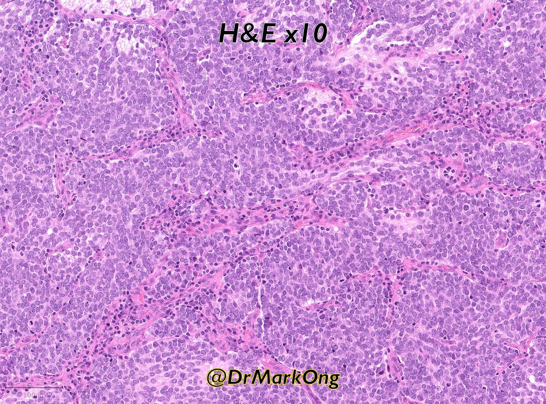
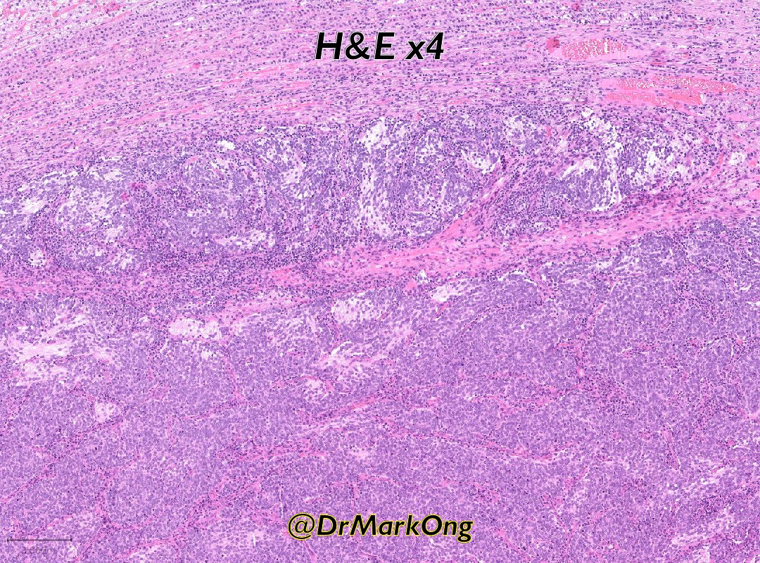
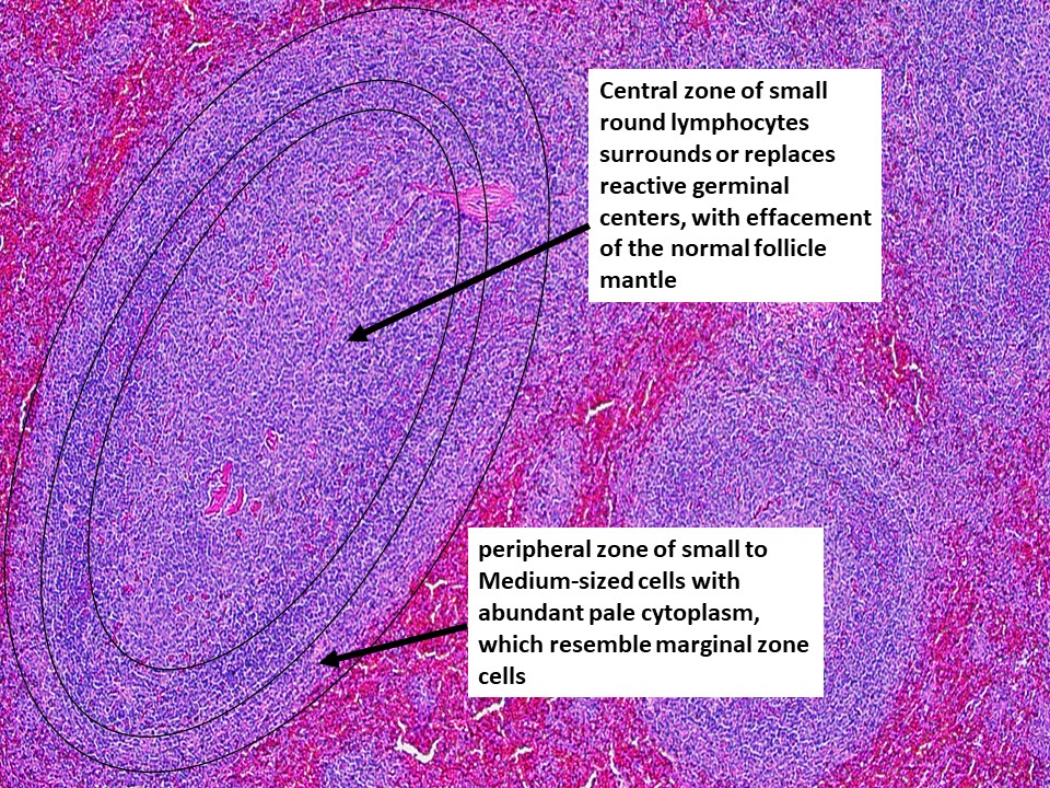
Splenic marginal zone lymphoma➡️look for these 2 zones:
🩸Central zone of small lymphocytes replacing reactive germinal centers
🩸Peripheral zone of small/medium-sized cells with abundant pale cytoplasm, resembling marginal zone cells
#hemepath#lymsm#PathTwitter#MedTwitter
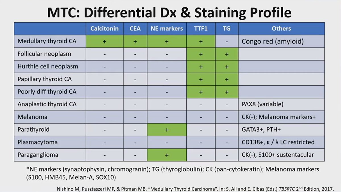
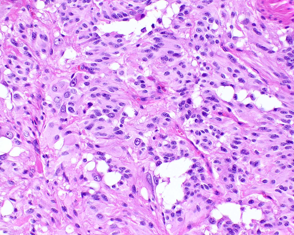
Do we have to grade medullary thyroid carcinomas? Yes.
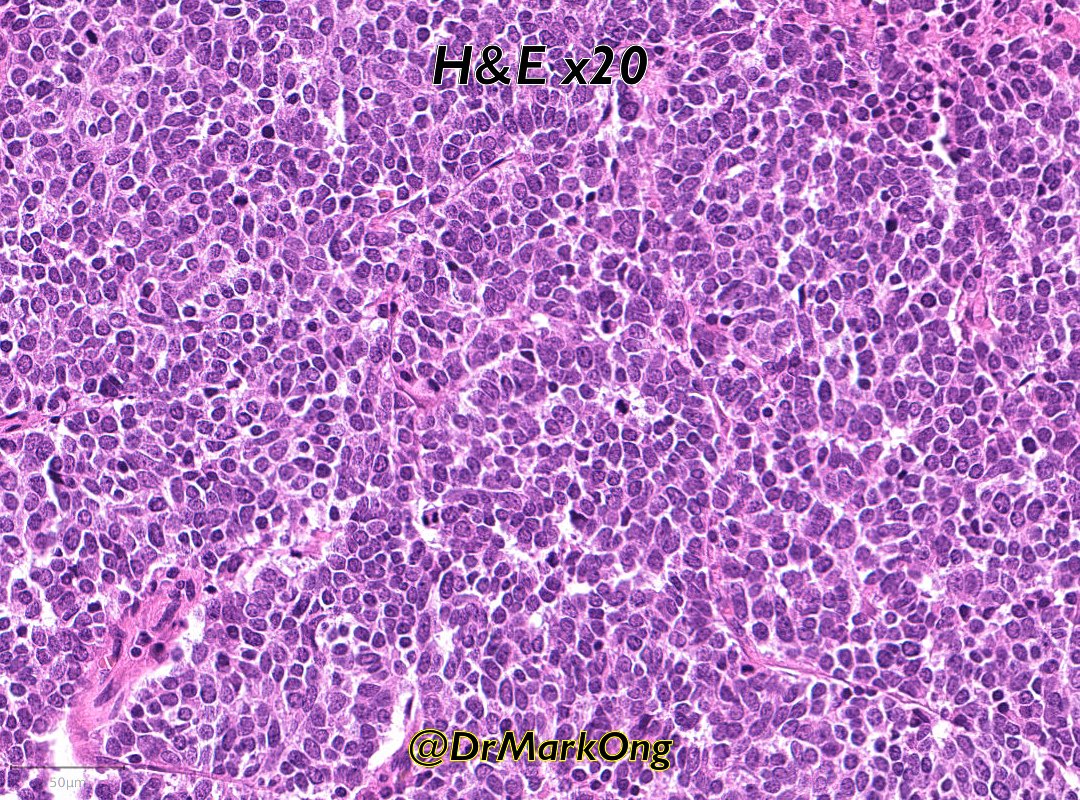
If there is tumor necrosis, ≥5 mitoses per 2 mm², or ≥5% Ki-67 labeling index, classify the tumor as high-grade MTC.
What constitutes necrosis? See pic 2
Dr. Nishino - 2025 Diagnostic Pathology Update #USCAP#pathology #PathX #PathTwitter
#liverpath The #nutmeg liver Macroscopic demonstration of Budd–Chiari syndrome due to IVC leiomyosarcoma and a great teaching image for liver pathology
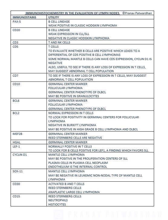
How do we use and interpret immunohistochemical stains in lymph node evaluations in hematopathology? Here is a summary table I prepared which I hope will be useful, especially for beginners! #PathEd#MedEd
Another papillary thyroid carcinoma (PTC) subtype:
Columnar cell PTC (0.2% of PTCs) is characterized by columnar cells with prominent nuclear pseudostratification. The pseudostratification and lack of abundant eosinophilic cytoplasm can help distinguish from the tall cell variant (see our last post).
#EndoPath #ENTPath #HNPath
Ovary-Endometrioid carcinoma
Diverse morphologic features (pic 1)
Endometrioid carcinoma with sex-cord pattern: Usually present presents with adenofibroma-like morphology
Not infrequently they have and aberrant IHC pattern like PAX-8 negativity and CDX2 positivity
Dr. Pilkington- Masters of Disguise: A Selection of Morphological Mimics in Gynae Tumour Pathology #ISGyP #Gynpath #pathology #PathX
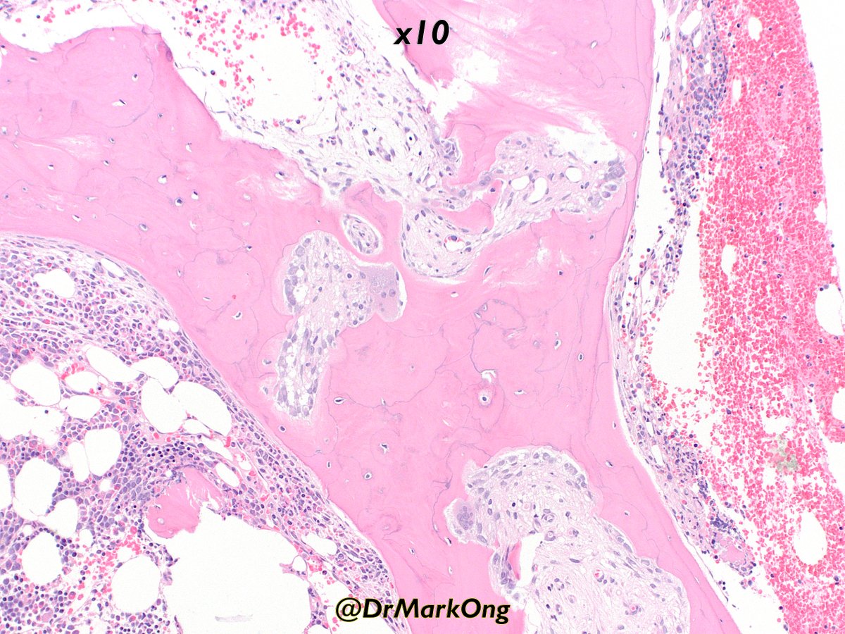
Bone marrow from elderly adult with end-stage renal failure.
Peri-trabecular fibrosis. Numerous osteoblasts, occasional osteoclasts. Dips in the bony trabeculae indicate bone resoprtion, but at the same time, abundant new bone formation (= woven rather than lamellar bone).
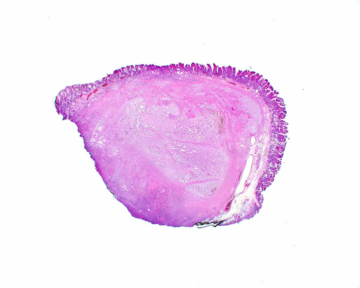
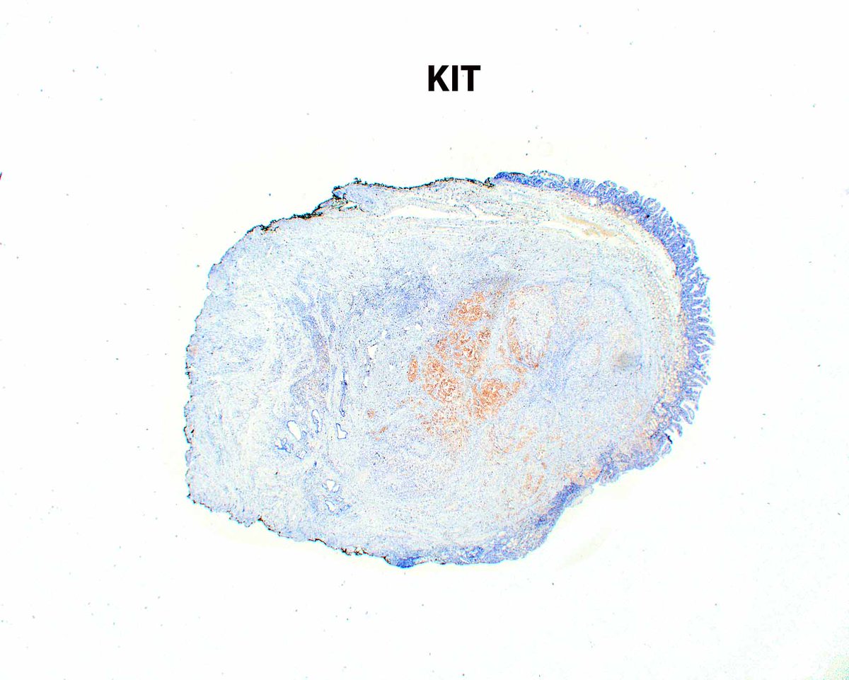
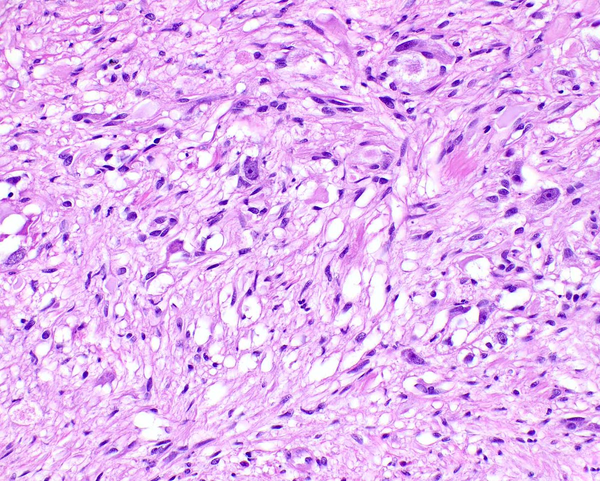
This week, I learned not only that gangliocytic paragangliomas were reclassified by the WHO as composite gangliocytoma/neuroma and neuroendocrine tumor (CoGNET) - thank you Rhonda Yantiss - but also that they can express CD117/KIT to result in wrong diagnoses on small biopsies.
FNA from a 75-year-old's 3.2 cm peripheral lung mass. What is your impression?
A) Benign
B) Malignant
C) I need a higher power 🖼️
#CytoPath#PathTwitter#Pathology
Reserve cells are immature epithelial cells residing underneath the columnar pulmonary cells as depicted in this sketch by @SaeedAsiry. Reserve cell hyperplasia can mimick small cell carcinoma (hyperchromatic nuclei with molding), but the cells are very small with no necrosis.
This is a crazy case of bladder cytitis cystica et glandularis with extensive intestinal metaplasia and low-grade dysplasia. It resembles a colorectal adenoma! There is lots to learn in Dr. Alex Kryvenko's new book. Get yours at https://t.co/11aBMJXEmQ #UMiamiPath
Fallopian tubes with nodular mesothelial/histiocytic hyperplasia - a benign, incidental finding
Good to know about all these incidentalomas so you don't mistake them for something else (also to not waste resources on unnecessary IHC)
#pathtwitter#gynpath#generalpath#pathoutpic