We’re excited to welcome Dr. Roberto Ochoa Jimenez back to Mount Sinai Morningside as an Advanced Cardiac Imaging Attending!
Dr. Ochoa completed his cardiology fellowship at Mount Sinai Morningside, followed by advanced imaging at UCSF. His clinical and academic interests encompass advanced echocardiography, structural heart imaging, cardiac CT, and cardiac MRI, reflecting a commitment to cutting-edge cardiovascular diagnostics and patient care.
Thrilled to have him rejoin our team! @RobertoC8a 👏👏 @MountSinaiHeart
#cardiotwitter
In Atrial Functional MR can get “Hamstringing”of posterior MV leaflet due to massive LA dilation;this “atriogenic” leaflet tethering is from displaced posterior annulus onto crest of LV inlet resulting in⬆️in annulopapillary distance restricting leaflet motion #echofirst
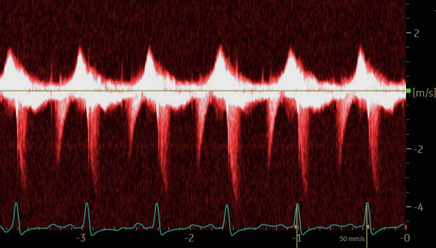
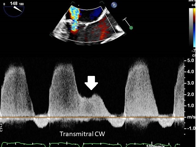
A patient for TEE prior to cardioversion. CW interrogation across the mitral valve in the long axis view. What is the low velocity signal indicated by the arrow?
**CASE OF THE MONTH**
Female, mid 20s
Fit & healthy
No CVS risk factors
Only 💊 is COC pill
Experiences central chest pain during sport (exertion), not happened before
Recently has been well, no coryzal or other symptoms
Here is admission ECG
Spectral Doppler tracings provide important hemodynamic insights. This book is dedicated to interpretation of spectral Doppler recordings, similar to EKG books focused on EKG interpretation. Examples of some spectral Doppler patterns which may not be widely known:
Notched aortic regurgitation
Interrupted aortic regurgitation
Phantom systole
Double peak LVOT signal
Notched tricuspid regurgitation
Giant L wave
Intermittent systolic flow reversal in hepatic veins
Merged tricuspid regurgitation
@EchoSoliman
Recognize the cute device bouncing in the LAA? Two years after transcatheter mitral paravalvular leak closure — Amplatzer device found embolized to the left atrial appendage. A reminder: closure success is more than the immediate result #CardioTwitter#StructuralHeart@PCRonline
💡 A rare very interesting case: persistent left superior vena cava (PLSVC) + right superior vena cava (RSVC) atresia --> challenging CRT-D implantation
⭐ Echo:
- Dilated coronary sinus
- Bubble test (agitated saline solution) via the left antecubital vein: bubbles first in the coronary sinus and then in the right atrium --> PLSVC
⭐ CT-scan + venography:
- Right superior vena cava (RSVC) atresia with the right brachiocephalic vein drained directly into the persistent left superior vena cava (PLSVC)
- PLSVC drained directly into the coronary sinus
⭐ Very challenging CRT-D implantation via left subclavian vein.
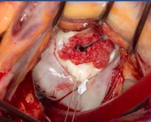
NBTE nonbacterial thrombotic endocarditis - look for sterile “vegetations” (platelet fibrin deposits) along the valve leaflet’s closing edges that appear to "kiss" in this characteristic pattern
#echofirst
🔎 Pneumopericardium can occur during pericardiocentesis even with slow drainage if >1L is removed. Sudden pressure shifts allow air entry. Air can be drained if the catheter is still in place. #Pericardiocentesis#CardioTwitter