In commotio cordis, which arrhythmia is most frequently triggered?
A. Atrial fibrillation
B. Ventricular fibrillation
C. Ventricular tachycardia
D. Complete heart block
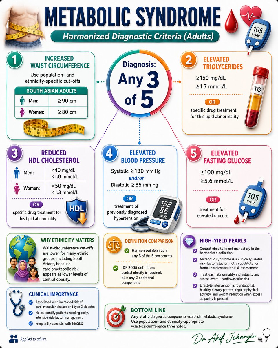
Metabolic syndrome is diagnosed when any 3 of 5 criteria are present. Early recognition helps guide targeted risk-factor management and cardiovascular risk assessment.
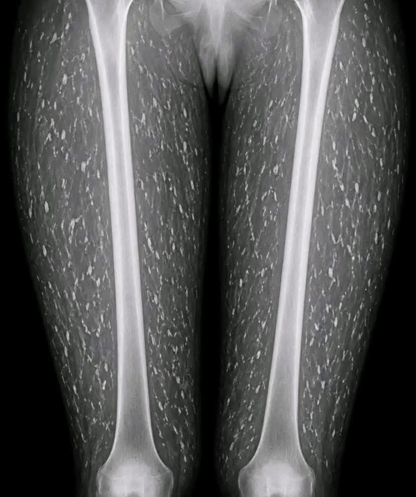
A patient presents with vague muscle aches and multiple firm lumps beneath the skin.
The X-ray reveals a striking finding.
What is the most likely diagnosis?
Understanding Diastolic Dysfunction, Made Simple with Echo
This visual shows how the heart’s diastolic function (its ability to relax and fill) changes across different stages of dysfunction , from normal to severely restrictive (Grade 3–4).
Here’s what’s being analyzed:
1️⃣ ECG: for timing
2️⃣ Mitral inflow: the E and A waves show how blood enters the left ventricle
3️⃣ Pulmonary vein flow: tells us about left atrial pressure
4️⃣ Tissue Doppler: checks the movement of the mitral annulus
5️⃣ Color M-mode: shows how quickly blood fills the ventricle (Vp = propagation velocity)
🟢 Normal:
E wave > A wave, good tissue movement, healthy filling
🟡 Grade 1 (Abnormal relaxation):
E < A, slowed filling often in older adults
🟠 Grade 2 (Pseudonormal):
Looks normal, but it’s not E > A again, but due to raised LA pressure
🔴 Grade 3–4 (Restrictive filling):
E ≫ A, very poor ventricular compliance and bad prognosis
Ventricular free-wall rupture, ventricular pseudoaneurysm, and papillary muscle rupture complicating acute myocardial infarction: A clinical consensus statement just published in #EHJ
👉 https://t.co/twtZtfY1f9
@RoccoMontone@ehj_ed