Inflammation is not one signal.
It is a biological conversation, and each marker lets us listen to a different voice.
By the way, this infographic is the perfect companion to my previous one on CRP, ESR and procalcitonin.
Tissue Doppler Imaging (TDI): A quick clinical refresher
TDI uses PW Doppler to capture low-frequency, high-amplitude signals from myocardial tissue, allowing us to evaluate longitudinal LV shortening and lengthening with far greater precision than standard Doppler.
Spectral TDI:
- The S wave represents systolic motion toward the transducer (positive).
- The E′ and A′ waves reflect early and late diastolic motion away from the transducer (negative), capturing the biphasic pattern of LV lengthening.
Color TDI:
🔴Red indicates myocardial motion toward the probe during systole.
🔵 Blue indicates motion away from the probe during diastole.
TDI helps identify subtle systolic and diastolic abnormalities long before changes appear on conventional measurements such as ejection fraction.
📸: BE Belwer MD
Echocardiographic views used in imaging the interventricular septum.
Adapted from Bulwer BE, Rivero JM, eds. Echocardiography Pocket Guide: The Transthoracic Examination
#POCUS transducer movement terminology: having a standard set of terms just makes teaching and learning smoother, especially when the instructor is remote.
#Nephpearls#FOAMed
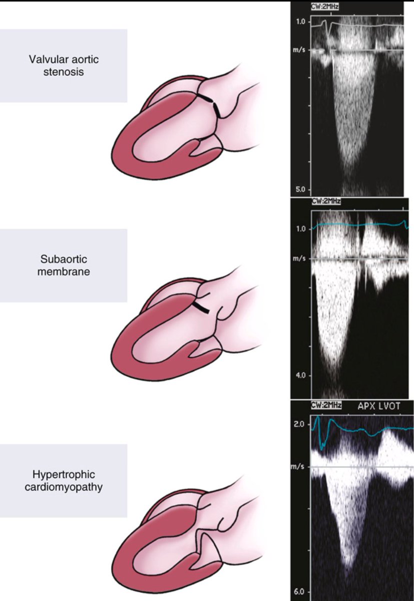
Not All LVOT Obstruction Is the Same.
Left ventricular outflow tract (LVOT) obstruction is not a single entity. Different pathologies produce similar pressure gradients, but their CW Doppler profiles reveal important clues.
⚫Valvular aortic stenosis produces a fixed obstruction at the valve level, resulting in a smooth, symmetric systolic velocity curve.
🟢A subaortic membrane causes a fixed subvalvular obstruction. The CW Doppler profile may look similar to valvular AS, but the systolic envelope often appears "rough" due to coarse fluttering of the aortic valve.
🔵Hypertrophic cardiomyopathy creates a dynamic obstruction within the LVOT. This produces a distinctly different Doppler pattern, with a late systolic peak and the classic dagger-shaped envelope.
Take-home message: high velocity alone is not enough. The shape of the CW Doppler signal, combined with 2D imaging and color flow, helps identify the exact level and mechanism of obstruction.
Ref: Catherine M Otto
https://t.co/wthq81gDJf
Detailed 3D rendering of coronary artery anatomy
(right-dominant circulation) highlighting major branches:
LMCA → LAD + LCx, RCA with PDA/PL branches, diagonals, OM, and LIMA (gold-standard CABG conduit). ❤️🫀"
#CoronaryArtery#CardiacAnatomy#Cardiology
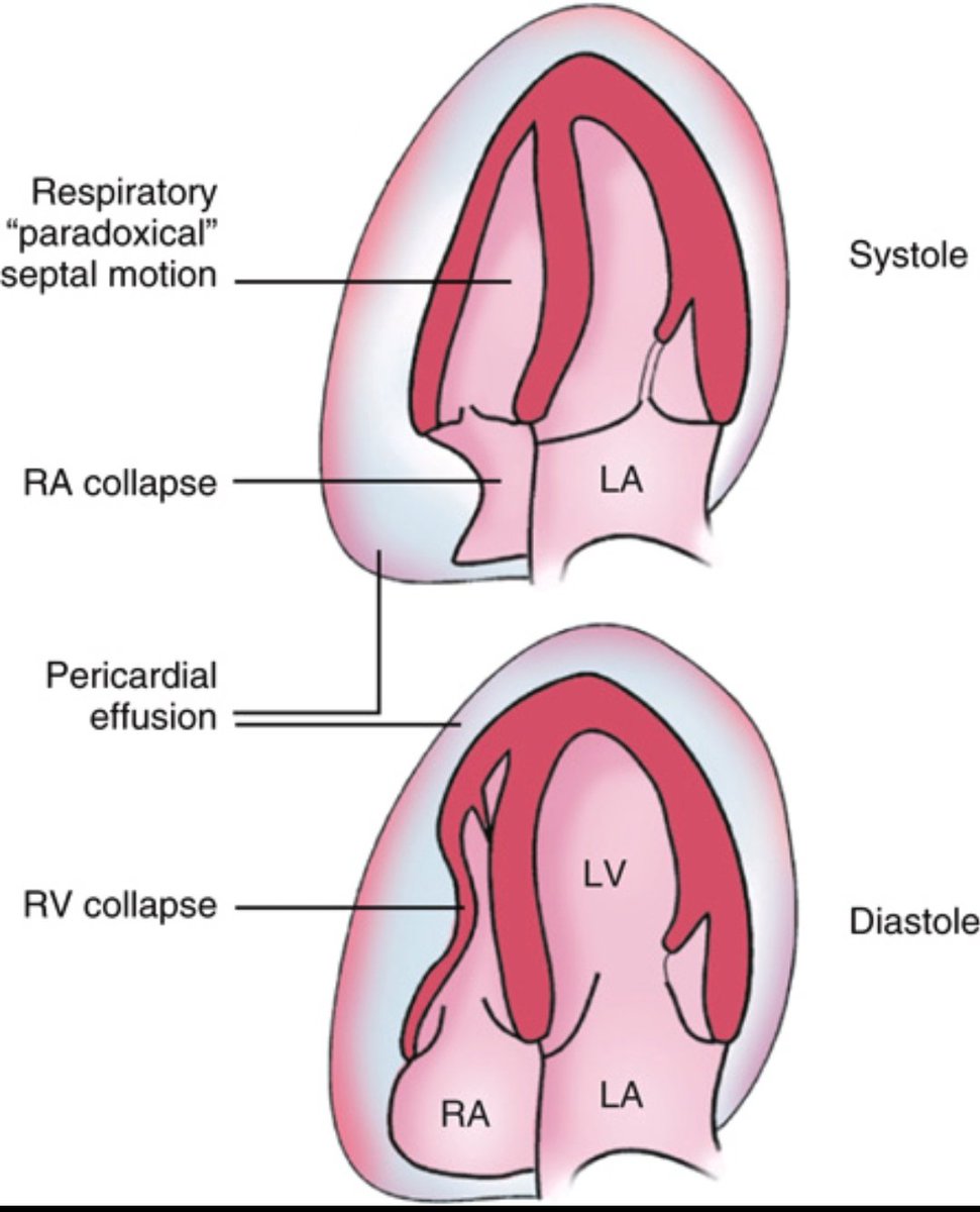
Cardiac Tamponade on 2D Echo
Key findings in tamponade physiology:
🫀 RA collapse in systole
🫀 RV collapse in diastole
🫀 Paradoxical septal motion due to ventricular interdependence
🫀 Pericardial effusion compressing the heart