We often take circulation for granted, yet it is rarely as simple as it seems. Grateful to these experts for providing fresh insights into hemodynamic management to help us better care for our patients. 🎩 tip to the authors, including @DrMCecconi
https://t.co/Sfr6vCn6le
Step up your management of patients with vasoplegia after cardiac surgery. Learn from the greats, including @pwierusz and @SXC71. 🎩 tip to the authors.
https://t.co/Sfr6vCn6le
#CardioNugget™
Echo-derived PVR on TTE
This is an empiric Doppler formula validated against cath data — the “×10 + 0.16” converts the Doppler ratio into estimated WU.
Think physiologically:
↑ TR velocity = ↑ pressure
↓ RVOT VTI = ↓ forward flow
So:
high pressure / low flow = high resistance.
#CardioNuggets #MedEd #EchoFirst
RF vs Anti-CCP vs ANA
Quick recall:
🔹 RF → Associated with RA, but not specific
🔹 Anti-CCP → Most specific antibody for RA
🔹 ANA → Screening test, not a diagnosis
Key pearls:
❌ RF ≠ RA
❌ ANA ≠ Lupus
✅ Anti-CCP predicts erosive RA
Positive autoantibodies must always be interpreted in the clinical context.
#Rheumatology #MedTwitter #RheumatoidArthritis #Lupus #ANA #AntiCCP #MedicalEducation #ClinicalPearls #FOAMed
Ever seen ESR 100 with a normal CRP? Or CRP 150 with ESR 15?
ESR and CRP measure different aspects of inflammation and often tell different stories.
Understanding when they disagree can prevent diagnostic errors and improve clinical decision-making.
What is the biggest ESR–CRP mismatch you’ve encountered in practice?
#Rheumatology #MedTwitter #InternalMedicine @docakx@IhabFathiSulima #MedicalEducation #ClinicalPearls #FOAMed #ESR #CRP #Inflammation #RheumatologyPearls
Understanding Diastolic Dysfunction, Made Simple with Echo
This visual shows how the heart’s diastolic function (its ability to relax and fill) changes across different stages of dysfunction , from normal to severely restrictive (Grade 3–4).
Here’s what’s being analyzed:
1️⃣ ECG: for timing
2️⃣ Mitral inflow: the E and A waves show how blood enters the left ventricle
3️⃣ Pulmonary vein flow: tells us about left atrial pressure
4️⃣ Tissue Doppler: checks the movement of the mitral annulus
5️⃣ Color M-mode: shows how quickly blood fills the ventricle (Vp = propagation velocity)
🟢 Normal:
E wave > A wave, good tissue movement, healthy filling
🟡 Grade 1 (Abnormal relaxation):
E < A, slowed filling often in older adults
🟠 Grade 2 (Pseudonormal):
Looks normal, but it’s not E > A again, but due to raised LA pressure
🔴 Grade 3–4 (Restrictive filling):
E ≫ A, very poor ventricular compliance and bad prognosis
Dr Nishimura 🫀✨This slide never gets old.
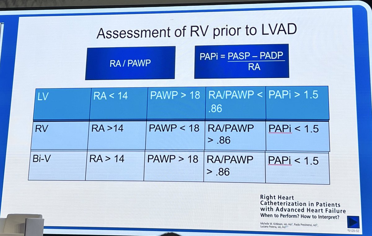
RHC hemodynamics — RA, PAWP, RA/PAWP & PAPi — remain the backbone for LV/RV/BiV failure assessment before LVAD.
Elegant physiology, real consequences.
#ICSC2026#CardioTwitter
🚨10 electrifying Hot Line sessions and 40+ pivotal trials debuting at #ESCCongress 2025 in Madrid!
‼️Trials targeting long‑held theories related to beta‑blockers post MI, digoxin for #HFrEF, antiplatelets & anticoagulants, novel treatments for Chagas, HCM, hypertension interventions.
Unmissable practice‑changing data!
Have we ever had so many important studies released at one meeting? Does anyone know the answer to this?
#LBCT #Cardiotwitter #CVPrev
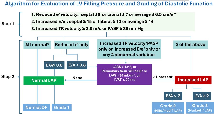
📢New Algorithm for Estimating LV Filling Pressure by Echo:
🔴increase in the number of patients in whom LV filling
pressure can be estimated
🟢only 2 cases of indeterminate LV filling pressure and the majority had a definitive LV filling pattern
https://t.co/4yL751yFFo