🌍 Proud to be a co-author of the recently published article: “Criteria for Hidradenitis Suppurativa Competence Centers” This international Delphi consensus study brings together global experts, patient representatives, and multidisciplinary leaders in hidradenitis suppurativa (HS) to establish the first evidence-based criteria for HS Competence Centers (HSCC). The publication highlights the importance of: ✔️ Multidisciplinary care ✔️ Standardized protocols and evidence-based management ✔️ Early diagnosis and timely referral ✔️ Specialized surgical and imaging support ✔️ Continuous medical education and patient-centered care ✔️ International collaboration networks I am especially pleased to contribute to the discussions regarding imaging and the role of ultrasound within specialized HS multidisciplinary teams, reinforcing the growing importance of imaging techniques in optimizing diagnosis, severity assessment, treatment planning, and monitoring in HS. This work represents an important step toward improving the quality and standardization of care for patients living with HS worldwide.
Reference: Marzano AV, Bechara FG, Shi VY, Martorell A, Hsiao JL, Van der Zee HH, Villani AP, Alavi A, Ingram JR, Sayed CJ, Tzellos T, Chandran NS, Ju Q, Kurokawa I, Frew JW, Gulliver WPF, Rivitti-Machado MC, Wortsman X, McGrath BM, Pintori G, Jordan V, Thomas N, Alarcon I, Zouboulis CC. Criteria for Hidradenitis Suppurativa Competence Centers. Dermatol Ther (Heidelb). 2026 May 23. doi: 10.1007/s13555-026-01783-1. Epub ahead of print. PMID: 42176118.
Link: https://t.co/DG3gNbBk0K #HidradenitisSuppurativa #HS #Dermatology #Ultrasound #DermatologicUltrasound #MedicalImaging #InflammatorySkinDiseases #PatientCare @sonoskin
✨ Excited to share our new publication in the Journal of Ultrasound in Medicine! ✨ “Dynamic Color Doppler Ultrasound Features of Cutaneous Mastocytoma at High and Ultra-High Frequency.” This study explored dynamic ultrasound changes incutaneous mastocytoma before and after a gentle friction maneuver, demonstrating that high- and ultra-high-frequency color Doppler ultrasound can reveal increased dermal thickening and hypervascularity that may support the diagnosis of these lesions. Among the highlights of the study: 🔹 Dynamic increase in dermal vascularity after friction maneuver 🔹 Detection of hypoechoic dermal thickening at high and ultra-high frequency 🔹 Potential role of ultrasound as a non-invasive support tool for early diagnosis and management 🔹 Largest case series to date evaluated with high and ultra-high frequency ultrasound Very proud to collaborate with this outstanding international team and continue contributing to the advancement of dermatologic ultrasound and innovative imaging applications in skin diseases. Congratulations to all coauthors for this important contribution!
Reference: Sechi A, Tagliati C, Marzano AV, Dini V, Oranges T, Wortsman X. Dynamic Color Doppler Ultrasound Features of Cutaneous Mastocytoma at High and Ultra-High Frequency. J Ultrasound Med. 2026 May 22. doi: 10.1002/jum.70309.
Link: https://t.co/hzIgF5bYCg #DermatologicUltrasound #Ultrasound #ColorDoppler #Mastocytoma #CutaneousMastocytosis #SkinImaging #Radiology #Dermatology #HighFrequencyUltrasound #UltraHighFrequencyUltrasound #Innovation #MedicalImaging @xworts@sonoskin
✨ Paris · January 2026 🇫🇷 ✨
I’m very happy to share my participation in the Ultrasound in Aesthetics IMCAS Course (January 28–31) and the ThinkIn Course (January 26–27) held in Paris.
It was a privilege to be part of these high-level educational courses during IMCAS, where ultrasound was highlighted as a key tool for safer, more precise, and evidence-based aesthetic practice.
These courses reinforced how essential ultrasound is to see before we treat: understanding anatomy in real time, managing complex cases and complications, and ultimately improving patient safety and outcomes in aesthetic medicine.
Grateful for the inspiring discussions, outstanding faculty, and the opportunity to share and learn alongside colleagues from all over the world. Paris was, as always, the perfect setting for science and innovation. 🖤🔍
#IMCAS2026 #UltrasoundInAesthetics #ThinkInCourse
#AestheticUltrasound #DermatologicUltrasound
#ImageGuidedAesthetics #PatientSafety
#Paris #MedicalEducation #Innovation @xworts@sonoskin
✨ New Publication | Aesthetic Ultrasound & Fillers ✨
I’m pleased to share our recently published literature review on the ultrasonographic evaluation of cosmetic fillers and their most frequent complications, now available in Clinical Imaging 🩺📡
🔍 Why is this important? With the growing use of cosmetic fillers, complications can occur — sometimes immediately, sometimes years later. High-frequency ultrasound, combined with color Doppler, allows us to:
✔️ Identify the type of filler (HA, CaHA, PLLA, PMMA, PCL, silicone, fat, and others) ✔️ Determine depth, location, and distribution ✔️ Detect early and late complications such as vascular occlusion, granulomas, migration, abscesses, and fat necrosis ✔️ Guide treatment safely, including ultrasound-guided hyaluronidase injections and drainage procedures
📌 Key message: 👉 Ultrasound is not optional in modern aesthetic medicine — it is a first-line, safety-driven imaging tool that improves diagnostic accuracy, guides management, and protects patients.
Honored to collaborate with an excellent multidisciplinary team and to contribute to advancing evidence-based, image-guided aesthetic practice.
Reference: Khorasanizadeh F, Momeni A, Daneshvar A, Ghannadzadeh R, Etesami I, Wortsman X. Ultrasonographic evaluation of cosmetic fillers: patterns and frequent complications - A literature review. Clin Imaging. 2026 Jan 2;131:110708. doi: 10.1016/j.clinimag.2025.110708. Epub ahead of print. PMID: 41505983.
#AestheticUltrasound #DermatologicUltrasound #CosmeticFillers #PatientSafety #HighFrequencyUltrasound #ColorDoppler #ImageGuidedAesthetics #UltrasoundFirst #ClinicalImaging #Radiology #Dermatology
#aesthetics
I’m very pleased to share our recently published case demonstrating the first reported ultrasound image ofa shagreen patch in Tuberous SclerosisComplex 🙌
Using high-frequency dermatologic ultrasound, wewere able to visualize characteristic dermal thickening and decreased echogenicity, providingnew insight into this important cutaneous sign. Thisreport reinforces how ultrasound can enhancedermatologic diagnosis and deepen our understandingof rare genodermatoses.
🧠 Why it matters: Shagreen patch is one of the major diagnostic criteriaof Tuberous Sclerosis Complex. Until now, itscharacteristics had never been documented withultrasound. This opens new possibilities for non-invasive assessment and improved clinicalcharacterization.
📄 Read the article here: Diagnostics (2025) — Shagreen Patch in Tuberous Sclerosis Complex at High-Frequency Ultrasound Authors: Corrado Tagliati & Ximena Wortsman
🔍 Advancing dermatologic ultrasound, one step at a time!
Reference: Tagliati C, Wortsman X. Shagreen Patch in Tuberous Sclerosis Complex at High-FrequencyUltrasound. Diagnostics (Basel). 2025 Dec12;15(24):3168. doi: 10.3390/diagnostics15243168. PMID: 41464169; PMCID: PMC12731642.
@xworts@sonoskin #dermatologicultrasound
✨ Honored to be featured in the newspaper El Mercurio (Chile) for our international project on Artificial Intelligence for the discriminating cosmetic fillers on ultrasound. Together with colleagues from Spain and an interdisciplinary and international team of radiologists, dermatologists, computer scientists, and imaging experts, we developed an AI-based system capable of automatically distinguishing the most common types of cosmetic fillers on ultrasound.
🔍 Why is this important? As aesthetic procedures rise worldwide, many patients present with multiple substances—often without knowing exactly what was injected. This complicates diagnosis and treatment, especially when managing adverse events.
Our AI tool seeks to support physicians by: • Automatically identifying the most common filler materials • Improving the diagnostic workflow for complications • Helping guide safer interventions and personalized treatment plans • Standardizing evaluations across different centers and ultrasound systems
🧠 This is the first initiative to combine high-frequency ultrasound with artificial intelligence deep learning to improve safety for users of cosmetic fillers. The project analyzed thousands of images from multiple countries and across several ultrasound platforms, ensuring broad clinical applicability.
This work reflects years of research on imaging of fillers and is a step toward creating robust, reliable tools that assist specialists in everyday practice—and ultimately improve patient care. We are grateful to the entire international team and excited about the next phase, as we continue to refine the algorithm and expand its capabilities.
📄 Full article available in the section Innovation of El Mercurio, Chile.
Article Published in the Journal of Ultrasound in Medicine
Reference: Wortsman X, Lozano M, Rodriguez FJ, Valderrama Y, Ortiz-Orellana G, Zattar L, de Cabo F, Ducati E, Sigrist R, Fontan C, Rezende J, Gonzalez C, Schelke L, Zavariz J, Barrera P, Velthuis P. Artificial Intelligence Deep Learning Ultrasound Discrimination of Cosmetic Fillers: A Multicenter Study. J Ultrasound Med. 2025 Sep 29. doi: 10.1002/jum.70079.
Link to Article :https://t.co/L7EDkIPVBJ @xworts@sonoskin@elmercurioec@AIUMultrasound
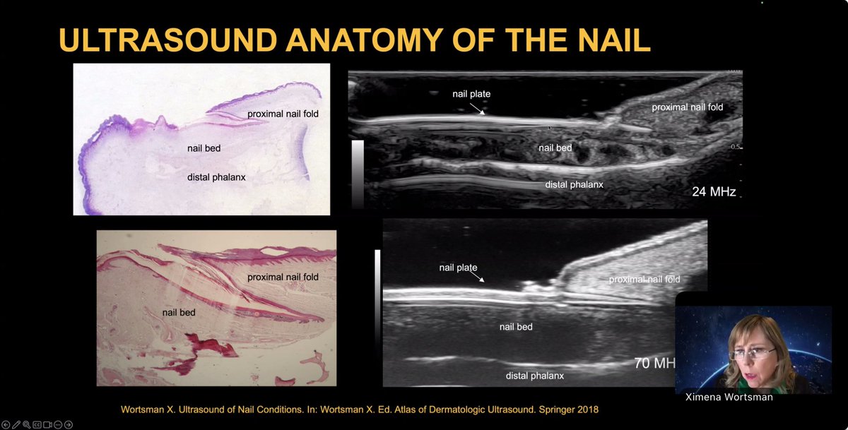
Dermatologic ultrasound is evolving fast! Evidence-based guidelines & standardized protocols are improving accuracy, consistency, and outcomes across skin, nail & aesthetic imaging.
Explore Dr. Ximena Wortsman’s new insights⬇️
https://t.co/dsxZL0R0VI
🌍 The Tremendous Importance and Impact of Guidelines and Standardized Protocols in Dermatologic Ultrasound
💡📖 I´m glad to share this article, recently published in the Journal of Ultrasound in Medicine, that emphasizes the following points:
- Dermatologic ultrasound differs from conventional soft tissue ultrasound—integrating clinical evaluation, color/power Doppler, spectral analysis, and high-to ultra-high-frequency technology to visualize skin structures in unparalleled detail.
-Evidence-based guidelines and standardized examination protocols are transforming dermatologic ultrasound into a mature and structured imaging subspecialty.
🖋️ From inflammatory diseases to oncology, nail disorders, and aesthetics, standardized protocols ensure:
✅ Higher diagnostic accuracy and reproducibility ✅ Improved therapeutic decisions through subclinical detection ✅ Enhanced collaboration between dermatologists and radiologists ✅ Precise cost estimation and time allocation of the ultrasound examinations for real-world practice
🔗 Read more in the Journal of Ultrasound in Medicine
Reference: Wortsman, X. (2025), The Tremendous Importance and Impact of Guidelines and Standardized Protocols in Dermatologic Ultrasound. J Ultrasound Med. https://t.co/npWDcmTndE
#DermatologicUltrasound #Guidelines #Protocols #Standardization #HighFrequencyUltrasound #UHFUS #SkinImaging #Dermatology #UltrasoundEducation #ImagingStandards @xworts@sonoskin@AIUMultrasound
Thank you Dr Wortsman @xworts for excellent overview of how using ultrasound in cutaneous lupus as well as HS and Morphea in diagnosis and assessing disease activity. You can also access the nails with US. Thank you @AstraZeneca for sponsoring
#MSUS#Rheum#Derm#Lupus#USSONAR
Join us next week Monday, October 6 at 8:30 PM EST as we continue our Lupus Webinar Series on! CME available
Join us for Cutaneous Manifestations of Lupus and Dermatologic Ultrasound, presented by:
Dr. Matt Lewis, MD, MPH
Dr. Ximena Wortsman, MD, FAIUM
https://t.co/HQNr5Z89WA
Just Published!!! 🌍✨ International Multicenter Study Published on Artificial Intelligence Deep Learning Ultrasound Discrimination of Cosmetic Fillers
We are proud to share the results of an international collaborative study exploring the use of artificial intelligence (AI) and deep learning (YOLOv11) to discriminate cosmetic fillers on dermatologic ultrasound (US) in the Journal of Ultrasound in Medicine.
🔹 Study Highlights:
Conducted by 14 physicians across 6 countries.
Dataset: 1,432 ultrasound images of hyaluronic acid (HA), polymethylmethacrylate (PMMA), calcium hydroxyapatite (CaHA), and silicone oil (SO).
AI achieved an average accuracy of 92%, with outstanding performance for HA (F1 score 0.96) and SO (F1 score 0.94).
Further work is needed to refine AI recognition of CaHA and PMMA.
🔬 This is the first study to demonstrate AI’s role in supporting the reliable discrimination of cosmetic fillers on ultrasound under real-world conditions.
🤝 Special thanks to all international collaborators and institutions who made this research possible.
More info at:
Wortsman X, Lozano M, Rodriguez FJ, Valderrama Y, Ortiz-Orellana G, Zattar L, de Cabo F, Ducati E, Sigrist R, Fontan C, Rezende J, Gonzalez C, Schelke L, Zavariz J, Barrera P, Velthuis P. Artificial Intelligence Deep Learning Ultrasound Discrimination of Cosmetic Fillers: A Multicenter Study. J Ultrasound Med. 2025 Sep 29. doi: 10.1002/jum.70079. Epub ahead of print. PMID: 41024593.
Journal Link: https://t.co/L7EDkIPVBJ
Pubmed link is:https://t.co/09OkEdnwYe
@xworts@sonoskin@AIUMultrasound
#artificialintelligence #dermatologicultrasound #ultrasound #dermatology #fillers #aesthetic
I am truly honored and grateful for the invitation to Sonoderm 2025 in Bucharest, Romania. It was a wonderful experience to share my knowledge in dermatologic ultrasound with outstanding colleagues, while also discovering Bucharest for the first time—a fantastic city with beautiful parks and elegant architecture. We were fortunate to enjoy exceptional guided tours led by Prof. Olga Simionescu.
My heartfelt thanks to Prof. Dr. Maria Crișan, Prof. Dr. Radu Badea, Prof. Dr. Diana Crișan, and Prof. Dr. Olga Simionescu for their kind hospitality.
It was also a joy to reconnect and share with dear colleagues and friends: Prof. Dr. Fernando Alfageme, Dr. Claudia Gonzalez, Dr. Orlando Catalano, and Dr. Valentina Dini.
@xworts@sonoskin
We are proud to share the WFUMB Consensus on Best Practice in Aesthetic Dermatologic Ultrasound, now available in Ultrasound in Medicine & Biology.
🔹 Why is this important?
Dermatologic ultrasound has become an essential tool in aesthetics, supporting filler identification, vascular mapping, complication management, and safe guided procedures.
🔹 What’s new?
This global consensus—developed by experts from multiple specialties and countries—establishes 24 key recommendations on:
✅Equipment requirements
✅ Safety protocols & infection control
✅Standardized scanning techniques
✅Reporting & documentation models
✅Training & accreditation pathways
✅Ultrasound guidance in managing complications
🔹 Impact:
These guidelines aim to standardize practice worldwide, ensuring safer, evidence-based, and more precise patient care.
📖 Read the full article: Chammas MC, Sigrist R, Alfageme F, Gonzalez C, Cavallieri F, Desyatnikova S, Crisan M, Velthuis P, Schelke L, Catalano O, Cartier H, Mandava A, Weiner S, Master M, Wortsman X. WFUMB Position Paper: Consensus on Best Practice in Aesthetic Dermatologic Ultrasound. Ultrasound Med Biol. 2025 Aug 26:S0301-5629(25)00231-5. doi: 10.1016/j.ultrasmedbio.2025.07.003.
#Dermatology #AestheticMedicine #Ultrasound #DermatologicUltrasound #WFUMB #Consensus #MedicalGuidelines #SkinImaging @sonoskin@xworts
🩺✨ New Publication Alert: Ultrasound for Detecting Tumor in Vivo and Ex Vivo Margins inSkin Cancer Surgery!
A recent study investigated the in vivo and ex vivo use of high-frequency ultrasound (HFUS) to evaluate tumor margins during micrographic-controlled surgery (MCS) for non-melanoma skin cancer in 136 tumors ( Basal Cell Carcinoma and Squamous Cell Carcinoma). 🕵️♀️💡
✅ Key findings:
• HFUS showed >98% specificity for basal cell and squamous cell carcinomas.
• Correctly identified tumor-free margins in 89% of lesions after first resection.
• Detected tumor residues in incomplete excisionsin 6%, allowing immediate re-excision.
🏥 Why is this important? Using HFUS during surgery could reduce surgical steps and hospitalization time, improving patient care and healthcare efficiency.
🖊️Citation: Crisan D, Wortsman X, Alfageme F, Scharffetter-Kochanek K, Crisan M, Bernhard L, Tarnowietzki E, Schneider LA, Schmid-Wendtner MH. In vivo and ex vivo sonographic evaluation of tumor margins during micrographic-controlled surgery: a promising new tool in dermato-oncology? J DtschDermatol Ges. 2025 Jul 2. doi: 10.1111/ddg.15827. Epub ahead of print. PMID: 40599105.
📖 Journal: Journal der Deutschen DermatologischenGesellschaft (2025).
🔗Link to Article: https://t.co/k3a1ZRkfN5
#Dermatology #Ultrasound #SkinCancer #TumorMargins #NonMelanomaSkinCancer #HFUS #MicrographicSurgery #Research #Innovation #Dermatosurgery #dermatologicultrasound
@xworts@sonoskin
🧠✨ New Study Alert! High-Frequency Ultrasound (HFUS) in Frontal Fibrosing Alopecia (FFA)
📌 HFUS reveals structural changes in the scalp and helps detect subclinical inflammation in FFA—even when clinical signs are absent!
🖼️ Key findings: 🔹 Dermal thinning and follicular miniaturization in affected areas 🔹 Shallower and fewer hair follicles at the frontal hairline 🔹 Power Doppler ultrasound detected inflammation in 33.3% of patients with no visible signs of disease activity
🧪 This non-invasive imaging technique improves the diagnosis, activity monitoring, and potentially the treatment follow-up in FFA.
📖 Published in the Journal of Ultrasound in Medicine 👩⚕️ Authors: Giavedoni P, Vera-Morandini F, Fuertes de Vega I, Combalia A, Fernández-Quiroga C, Podlipnik S, Wortsman X
#FFA #FrontalFibrosingAlopecia #UltrasoundDermatology #HFUS #SkinImaging #ScarringAlopecia #Trichoscopy #HairLossDiagnosis #UltrasoundInDermatology #SubclinicalInflammation
@xworts@sonoskin
🔬📈 New Scoring System for Hidradenitis Suppurativa (HS) Inflammation
I'm excited to share my publication in the Journal of Ultrasound in Medicinepresenting the Modified Ultrasound Hidradenitis Suppurativa Activity Scoring (mUS-HSA).
This simplified and enhanced scoring tool helps radiologists and dermatologists assess the degree of inflammatory activity in HS using color Doppler ultrasound. It includes updates on lesion size, vascularity, and corporal distribution—offering a more practical and standardized way to guide clinical management and research.
🟡 Basal + Control Phases
🟢 Max score: 18 points
🧭 Designed for real-life practice and clinical trials
Reference: Wortsman, X. (2025), Modified Ultrasound Hidradenitis Suppurativa Activity Scoring (mUS-HSA). J Ultrasound Med. https://t.co/D8D8Wdz2KK
🔗 Read more: https://t.co/u5RUCho6yf
Thank you to the Journal of Ultrasound in Medicine for publishing this contribution.
#HidradenitisSuppurativa #UltrasoundDermatology #HSUltrasound #ColorDoppler #mUSHSA #InflammatorySkinDisease #SkinUltrasound #JUM #XimenaWortsman
#HidradenitisSuppurativaultrasound
@xworts@sonoskin
🇧🇬 I had the incredible honor of being invited to teachdermatologic ultrasound at the Hidradenitis Suppurativa Masterclass in Stara Zagora, Bulgaria. This was my first visit to Bulgaria, and I was truly moved by the warm hospitality and professional excellence of the Bulgarian dermatology team, led by the remarkable Professor Dr. Evgeniya Hristakieva. The experience far exceeded my expectations and will remain unforgettable. I was invited and guided to explore historic cities, museums, and monuments, gaining insight into Bulgaria’s rich culture and meeting outstanding dermatologists whose passion and dedication were inspiring. My heartfelt thanks to Prof. Evgeniya Hristakieva, Prof. Nicolay Tsankov, Dr. Gavrail Poterov, Dr. Karen Manuelyan, and Dr. StoyanMurdjev for this wonderful opportunity.
#DermatologicUltrasound #HidradenitisSuppurativa #MedicalEducation #Dermatology #UltrasoundInDermatology #ultrasounddermatology @xworts@sonoskin
Great news just published: A Fusion of Imaging Technologies: Dermoscopy and High Frequency Ultrasound! “Dermoscopy-guided high-frequency ultrasound for preoperative assessment of basal cell carcinoma lateral margins” in the British Journal of Dermatology.
📌 This pilot study evaluates a portable device that combines dermoscopy and high-frequency ultrasound (DG-HFUS) to map the lateral margins of basal cell carcinoma (BCC) prior to surgery.
📊 Key findings: ✔️ Sensitivity: 94.4% ✔️ Specificity: 93% ✔️ Overall diagnostic accuracy: 93.4%
👨⚕️👩⚕️ Authors from the Hungary, USA, Chile, Italy, Belgium, Switzerland, and Poland.
📲 DG-HFUS may offer a valuable alternative in settings where Mohs surgery is unavailable, potentially improving surgical precision in BCC treatment.
🔗 Read the full article: https://t.co/ne07rHCPdw
#SkinCancer #BasalCellCarcinoma #Ultrasound #DermatologyResearch #Dermatoscopy #NonInvasiveImaging #DermatologicUltrasound #BCC #UltrasoundInDermatology #SkinCancerAwareness #AcademicDermatology #dermatologicultrasound #ultrasound #skincancerultrasound
@xworts@sonoskin
Glad to share the news of the Faculty of Medicine ofUniversity of Chile.
🌟 Chilean Professor in Elite Publication of HidradenitisSuppurativa at the The Lancet 🌟
Dr. Ximena Wortsman, a professor at the Faculty ofMedicine of the University of Chile and an internationalleader in dermatologic ultrasound imaging, is a co-author ofan article published in The Lancet, one of the world’s mostprestigious medical journals.
🧬 The study provides a comprehensive reviewof hidradenitis suppurativa (HS)—a chronic, painful, and stigmatizing inflammatory skin disease that primarily affectsyoung individuals.
🔬 This publication highlights the commitment and excellence of Chilean science on the global stage.
🔗 Read the full articlehere: https://t.co/ccKEyovdMq
#ChileanScience #TheLancet #HidradenitisSuppurativa #Dermatology #MedicalResearch#dermatologicultrasound #ultrasound #dermatology
@sonoskin@TheLancet@xworts@udechile