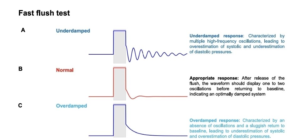
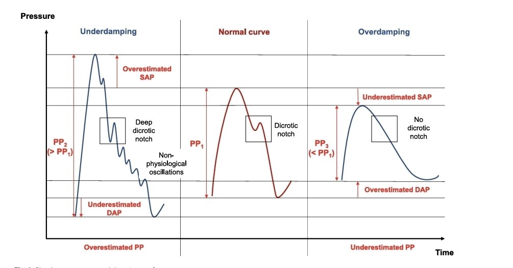
🫀Fisiología utilizando la presión arterial invasiva como herramienta de monitorización 6️⃣ puntos relevantes para la práctica clínica.🫀
📜Notas cafeteras ☕️
🫀El uso de la presión arterial invasiva (PAI) como una herramienta fisiológica dinámica, más allá de un simple valor numérico (PAM), integrándola en la evaluación del estado cardiovascular, acoplamiento ventrículo-arterial (V-A) y perfusión tisular.
☝🏻🤓La verdadera utilidad de la línea arterial no es alcanzar un MAP objetivo, sino entender si el sistema cardiovascular está acoplado, eficiente y capaz de entregar oxígeno (DO₂) a los tejidos.
☝🏻🤓La línea arterial permite evaluar si un paciente es respondedor a volumen (es decir, si aumentará su gasto cardíaco tras líquidos) mediante el análisis de interacciones corazón-pulmón reflejadas en la onda arterial.
💎No mide volumen directamente, sino la dependencia del gasto🫀 cardíaco a la precarga.
🫀 PAI como ventana fisiológica🩺
📚La señal arterial invasiva permite analizar en tiempo real la interacción entre:
▪️Corazón (Ees: elastancia ventricular)
▪️Sistema arterial (Ea: carga arterial)
▪️Retorno venoso y volemia efectiva
👉 La onda arterial se convierte en un biomarcador continuo de la eficiencia hemodinámica.☝🏻🤓
📈📉 Componentes de la onda arterial y su significado fisiológico✨️
🫀●Interpretación ●
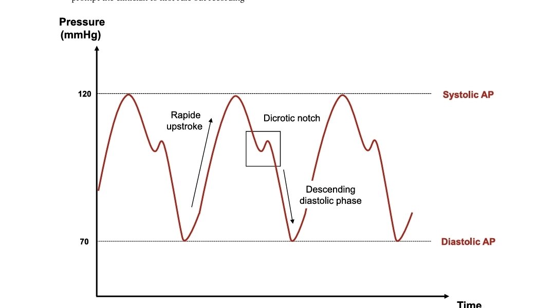
1️⃣ Fase sistólica. ( Pendiente ascendente sistólica)" Upstroke"
▪️Contractilidad ventricular izquierda
▪️Precarga efectiva
▪️Sincronía ventrículo-arterial
⏩️El upstroke es la fase inicial ascendente de la onda de presión arterial invasiva, que ocurre justo después de la apertura de la válvula aórtica.🫀
👉 Representa el momento en que el ventrículo izquierdo eyecta sangre hacia la aorta, generando un aumento rápido de presión.
☕️¿Porqué la importancia de Ascenso sistólico de la onda arterial?🤔
🫀El upstroke es un reflejo dinámico de la interacción entre:
⏩️Contractilidad ventricular izquierda (Ees)
⏩️Precarga efectiva
⏩️Poscarga (resistencia arterial)
⏩️Acoplamiento ventrículo-arterial (Ees/Ea)
👉 Es uno de los indicadores más sensibles de función sistólica en tiempo real.
📚Características del Upstroke
☆ 1. Pendiente (rapidez de ascenso)
▪︎Normal: ascenso rápido y definido
Representa una eyección eficiente.
☆ 2. Amplitud inicial.
▪︎Relacionada con el volumen sistólico.☝🏻🤓
☕️El upstroke no mide presión… mide cómo el corazón empuja la sangre contra el sistema arterial
🔴 " Upstroke" hiperdinámico (rápido y alto) Sugiere:
📈🫀Estado hiperdinámico ( sepsis temprana)
▪️↓ Resistencia vascular sistémica
▪️↑ Volumen sistólico
🔴 "Upstroke" lento:
▪️ ↓ contractilidad (shock cardiogénico)
▪️. ↑ poscarga (vasoconstricción extrema)
▪️↓ acoplamiento V-A.
🫀El "upstroke"es contractilidad en tiempo real.
💎 Un upstroke lento con MAP normal puede ocultar un bajo gasto cardíaco.
💎 Cambios en el upstroke durante ventilación mecánica ayudan a detectar:
●Respuesta a volumen
●Interacción corazón-pulmón
💎 Integrarlo siempre con:
●Presión de pulso
●Lactato
●Ecocardiografía
2️⃣ Pico sistólico⏩️Influido por: Volumen sistólico, Rigidez arterial
Pico bajo → bajo gasto cardíaco o hipovolemia 🩸
🔹 Fase diastólica
○
▪︎Determinada por:Tono vascular (resistencias sistémicas)
▪︎Tiempo de relajación arterial
▪︎Presión diastólica baja → vasoplejía ( sepsis)
🔹 Incisura dicrótica
▪︎Marca el cierre valvular aórtico
●Alteraciones●
▪︎Difusa o ausente → ↓ tono vascular
▪︎Muy prominente → ↑ resistencia periférica
3️⃣Interpretación avanzada: integración fisiológica
📌 Más allá de la PAM🚀
▪️La PAM no refleja adecuadamente:Flujo (CO) Distribución de perfusión,Coherencia hemodinámica
📌 Integración con acoplamiento V-A
📈Onda arterial permite inferir:
▪️Relación entre Ees/Ea
🔴Patrón ideal:
📈Upstroke rápido + presión sistólica adecuada + diastólica conservada
→ Acoplamiento eficiente → máximo rendimiento energético☝🏻🤓
🔴Desacoplamiento:
-🩸Hipovolemia → ↓ Ea funcional.👇🏽 continua...
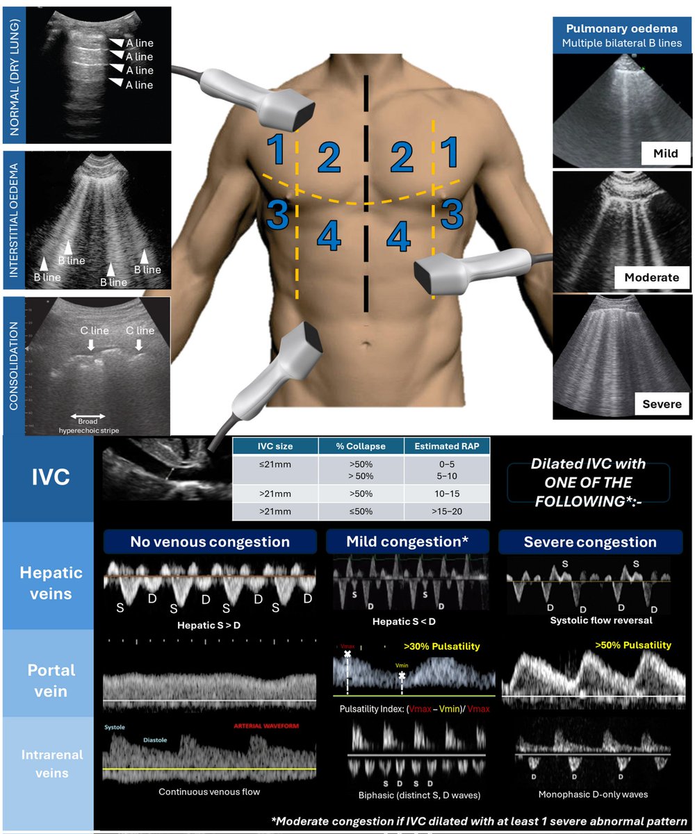
Ultrasound Assessment of Congestion.
Thoracic and venous excess ultrasound (VeXUS)
(Top) Thoracic ultrasound to determine the degree of pulmonary oedema
(mild: 2–3 B lines; moderate: ≥4 B lines; severe: confluent B lines);
C lines: short hyperechoic lines with broad hyperechoic stripe below suggest consolidation.
(Bottom) Venous excess ultrasound based on plethoric IVC with abnormal venous waveform in at least one venous system
(original figure created by author PT).
https://t.co/gXQKYj4CX7
1/9
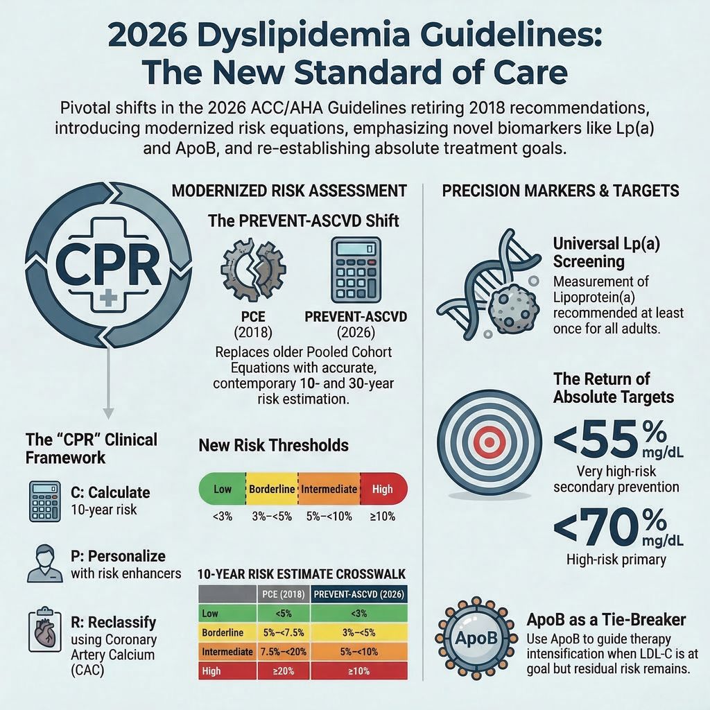
The 2026 ACC/AHA Dyslipidemia Guidelines are officially here!
Replacing the 2018 guidelines, these new updates bring major shifts in risk assessment, novel lipid markers, and cholesterol targets.
Here are the most salient features you need to know. 🧵👇
#Cardiology #MedTwitter
1/10
Coronary artery calcium (CAC) scoring is a quick CT scan that detects calcified plaque in your heart arteries — often called a powerful tool for predicting heart attack risk.
But large studies show the added value is more limited than the hype suggests.
Here's what the evidence (MESA, DANCAVAS, CONFIRM & more) actually says 🧵 #HeartHealth #CACScore
🦵💧 THE 300-mL MANEUVER: WHY RAISING THE LEGS IS BETTER THAN OPENING THE IV FLUIDS
ICU patient. Hypotensive, oliguric, rising lactate.
The automatic phrase during many handovers is still:
“Give a 500-mL saline bolus and see if he responds.” 🌊
🛑 THE DANGER OF “LET’S SEE IF HE RESPONDS”
The problem with traditional “fluid challenges” is that fluid goes in—but it doesn’t come out. If the patient is not fluid responsive (the heart is already on the flat part of the Frank–Starling curve), those 500 mL will only flood the lungs, worsen renal edema, and increase mortality.
❌ THE CVP (CENTRAL VENOUS PRESSURE) MYTH
For decades we were taught to look at CVP:
“If CVP is 4, give fluids. If it’s 15, the patient is full.”
Today we know (thanks to meta-analyses by Marik and others) that CVP predicts fluid responsiveness about as well as flipping a coin (50%). It’s a static pressure, not a dynamic indicator.
💡 THE SOLUTION: REVERSIBLE AUTO-TRANSFUSION (PASSIVE LEG RAISING – PLR)
Instead of giving real fluids, lay the patient flat and raise the legs to 45° for 1–2 minutes.
What does this achieve?
You shift about 300 mL of venous blood from the legs and the splanchnic circulation toward the right heart. It’s an endogenous fluid challenge.
🔬 HOW TO INTERPRET IT (THE MASTER TOUCH WITH POCUS)
While raising the legs, measure the aortic VTI (Velocity–Time Integral) with ultrasound.
✅ If VTI increases >10–15%:
The patient is fluid responsive. The heart benefits from the extra preload. You can give fluids safely.
🚫 If VTI does not change:
The patient does not need fluids—they need inotropes or vasopressors. When you lower the legs, the 300 mL returns to where it came from, causing no harm and no edema.
🩺 On-call lesson:
Never give water to a heart that doesn’t know what to do with it. Be dynamic, use ultrasound, and do the leg-raising test.
💬 Discussion:
In your units, do you still use CVP as a resuscitation target, or have you fully transitioned to dynamic measures (PLR, VTI, Pulse Pressure Variation)?
#MedTwitter #FOAMed #MedX
Mastering the Suprasternal View in Echocardiography
The suprasternal view is a crucial but often underutilized echocardiographic window that provides key insights into aortic pathology and great vessel abnormalities.
📌 How to Obtain:
- Place the probe in the suprasternal notch with the marker directed towards the chin.
- Apply gentle downward pressure while angling slightly posterior to visualize the aortic arch.
- Optimize gain & depth to enhance image clarity.
- Use Color Doppler to assess flow abnormalities in the great vessels.
Patient Positioning:
- Supine with neck extended (placing a small pillow under the shoulders helps).
Structures Visualized:
✅ Aortic Arch & its branches (Brachiocephalic, Left Common Carotid, Left Subclavian)
✅ Descending Thoracic Aorta
✅ Right & Left Pulmonary Arteries
✅ Superior Vena Cava (SVC)
✅ Left Atrium
Key Pathologies Detected:
📌 Aortic Coarctation – Look for narrowing of the aorta with post-stenotic turbulence on Doppler.
📌 Aortic Dissection – Identify an intimal flap separating true & false lumens.
📌 Patent Ductus Arteriosus (PDA) – Color Doppler reveals continuous left-to-right shunting.
📌 Aortic Aneurysms – Assess dilation & wall abnormalities to detect aneurysmal changes.
📌 Aortic Thrombi & Emboli – Visualizing thrombi can help assess embolic risk.
Why is the Suprasternal View Important?
This view is critical in pediatric & adult echocardiography, especially in suspected congenital heart disease, unexplained hypertension, and aortic syndromes. Its role in screening for aortic dissection, PDA, and coarctation makes it invaluable in emergency and routine cardiac assessments.
Do you routinely use the suprasternal view in your echo practice?
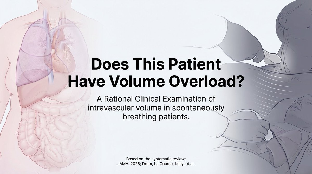
🆕 HOT OFF THE PRESS in JAMA (Feb 23, 2026)
“Does This Patient Have Volume Overload?”
The 2026 Rational Clinical Examination (40 studies, 11,490 patients).
In an obese patient with mixed wet/dry signs… how do you actually decide?
Thread with the key LRs + real-world case 👇
#MedTwitter #POCUS #JAMA #HeartFailure
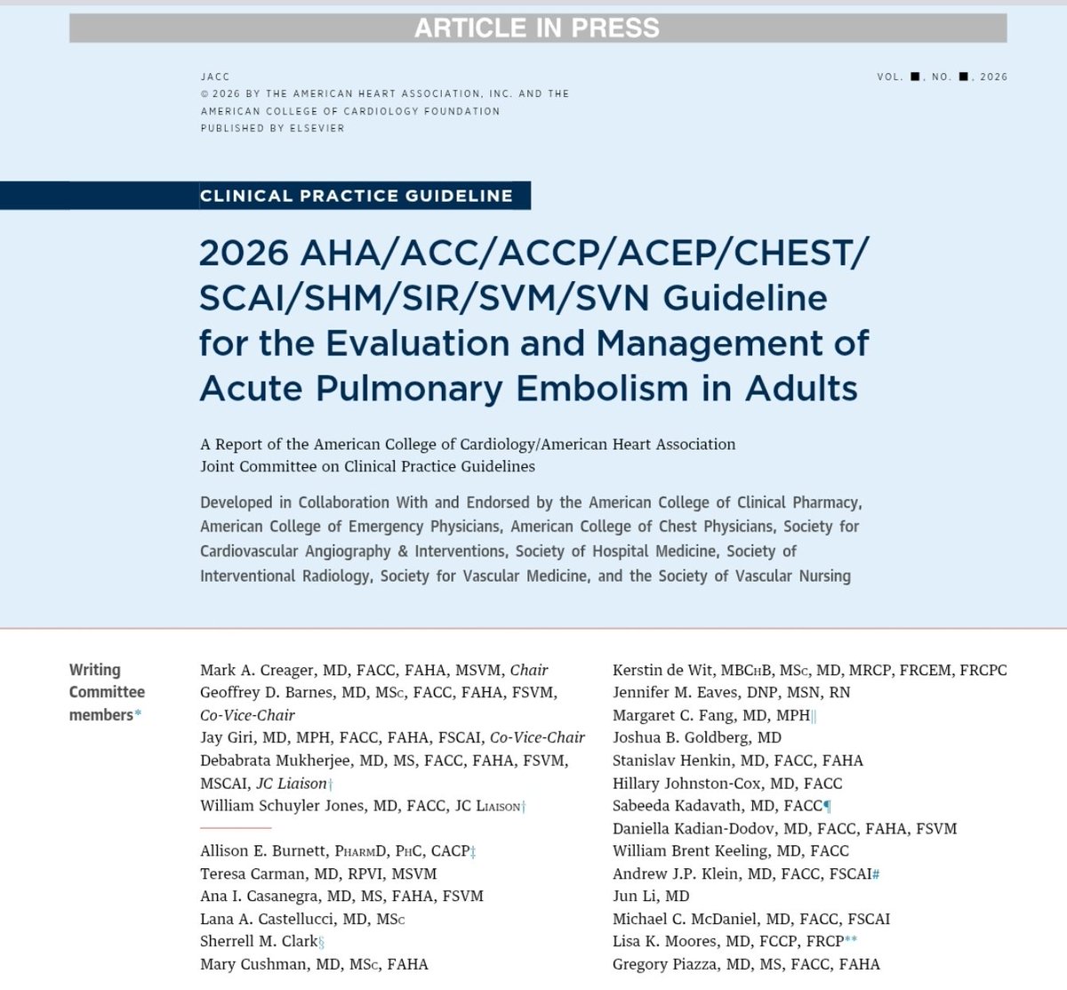
🚨 The 2026 AHA/ACC PE guidelines changed how we think about pulmonary embolism.
Not just new treatments — a new clinical framework.
Say goodbye to “massive vs submassive.”
Meet A–E PE Clinical Categories 🧵👇
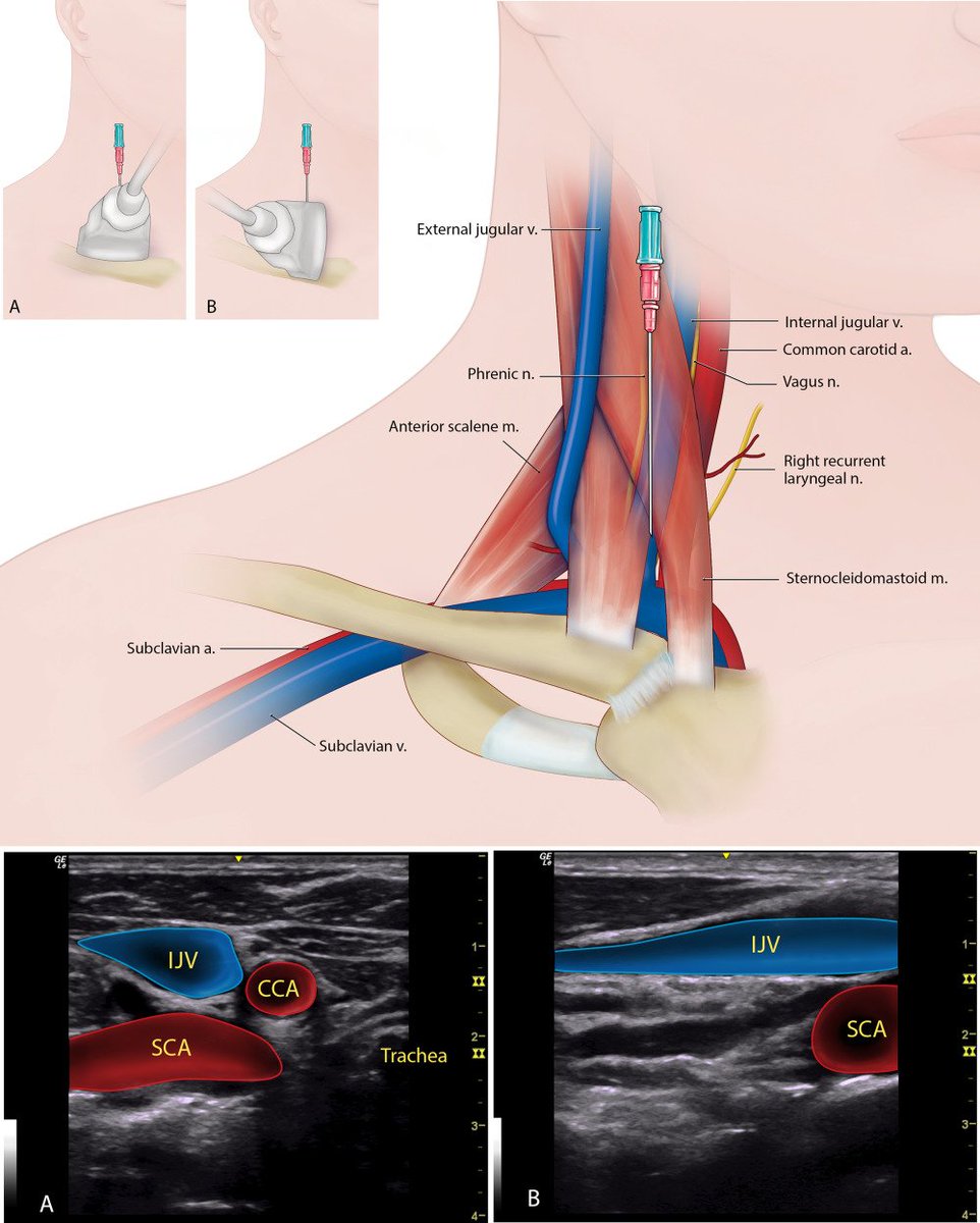
#POCUS - central line illustrations
#FOAMed#Nephpearls
From 🔗Clin Anat. 2017;30(2):237-250. doi: 10.1002/ca.22768. PMID: 27521991.
1. Internal jugular vein (A. Transducer placement in short axis, B. long axis)
So, according to IRCTC call centre supervisor guys, I can't buy e-ticket for my sister & her minor child in my IRCTC account. They consider it as commercial tranx not personal.
My account is blocked for more than 2 months due to this .
Is this a valid explanation @IRCTCofficial
@IRCTCofficial Already raised 2 months back , no response from back end . Today while raising issue again I came to know that this is the cause of deactivating my account.
Irony is I'd put money in my wallet just 1 wk prior .
@IRCTCofficial Incredible service from @IRCTCofficial , even after 1.5 months no response, no quarry from your department.
still my account is disabled.
In meantime how can I book tickets ?
@IRCTCofficial , why my IRCTC account is blocked without any intimation.
Some money is kept in my IRCTC wallet.
I've raised complaint 15-16 days back, but still no response.
My account was Adhaar linked .
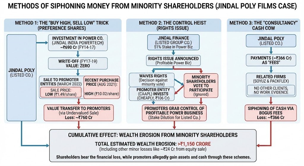
In 2022, Jindal Poly Films decided to transfer ₹690 Cr of "worthless" investments directly to its Promoters.
The price? Pennies.
Then, Ankit J (shareholder) & ors (others) sued the company for "fraudulent scheme" to divert ~₹760 Cr (& more) from public shareholders to promoters.
Write-offs, cheap buybacks, and "consultancy" fees. 🧵
Here's a quick primer on Jindal Poly's "siphoning" methods & how you can spot & Avoid it in your PF companies👇