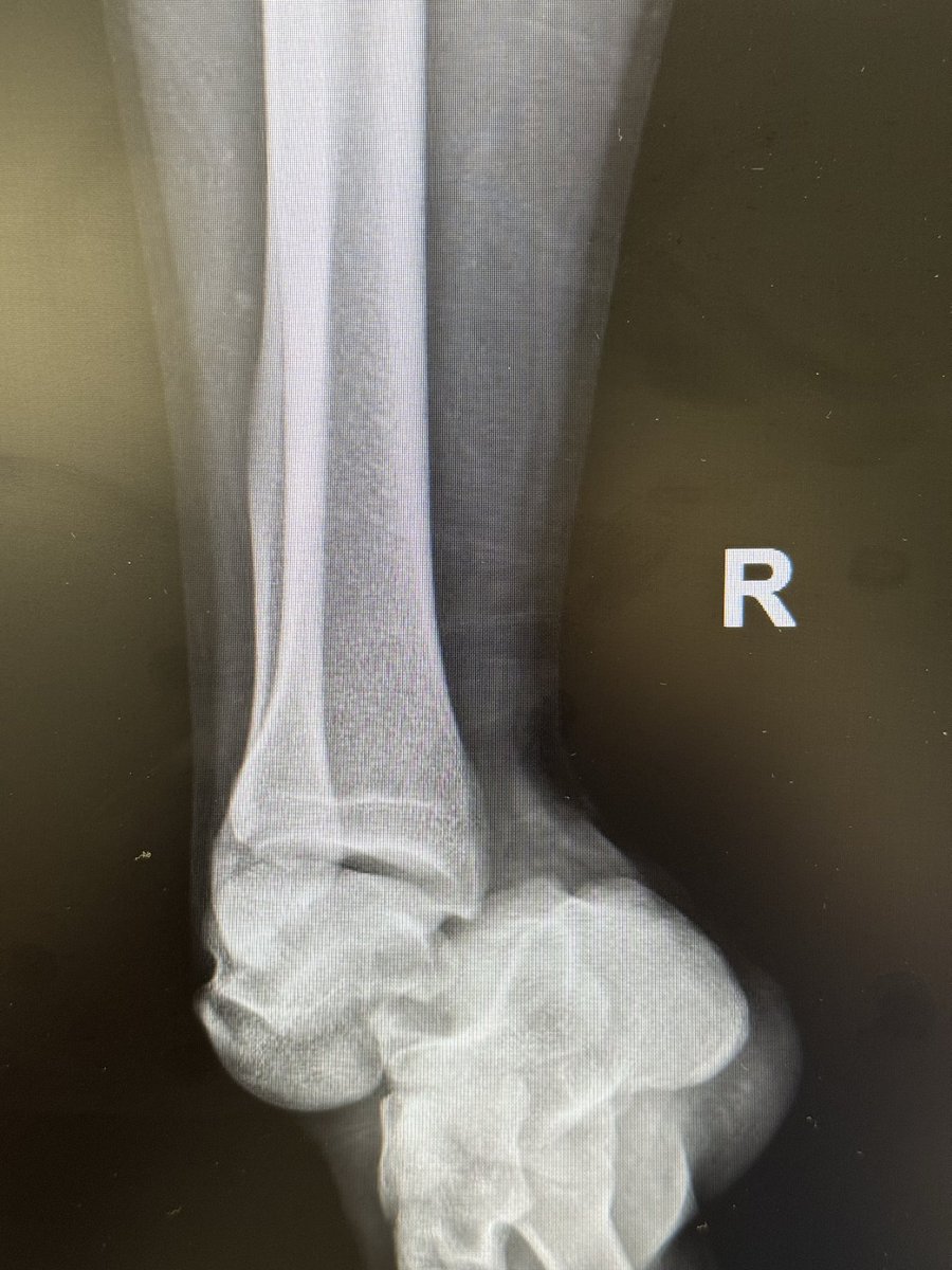
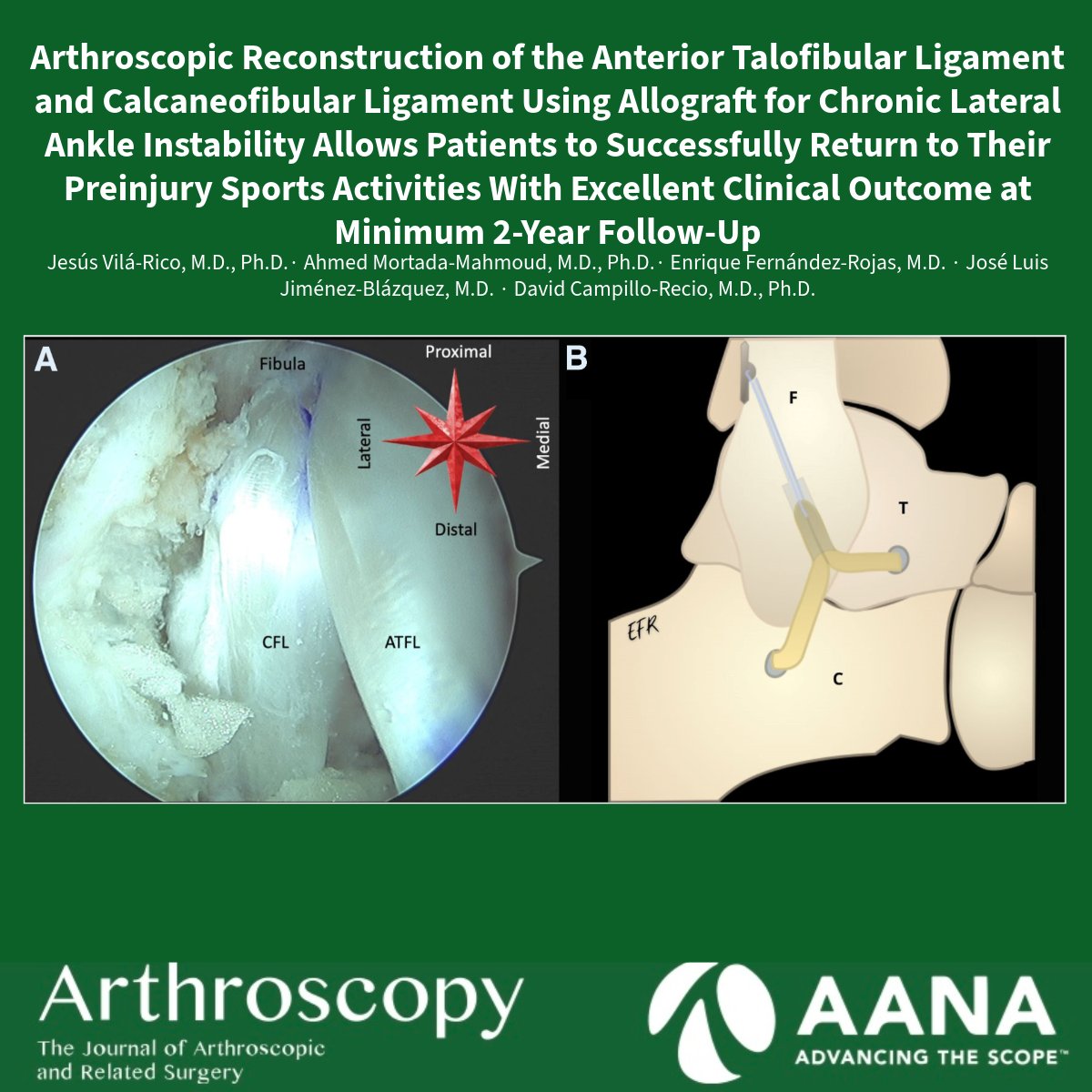
Can a complete Achilles rupture heal without surgery—even in a high-risk patient? The case underscores the importance of individualized care pathways and evaluation of nonoperative approaches.
Read the full report here: https://t.co/bY29McYAkT
Tratamiento con fisioterapia sin mejoría. Plantillas para mejora biomecánica. Siguen los síntomas y la rodilla continúa con un dolor severo. Cojera cada vez más marcada. En estos casos NUNCA debe faltar una radiografía de las caderas (AP y AXIAL)
MECANISMOS FISIOLÓGICOS DE MEJORA DEL RENDIMIENTO CON EL ENTRENAMIENTO CON RESTRICCIÓN DE FLUJO SANGUÍNEO El entrenamiento con restricción del flujo sanguíneo mejora el rendimiento y reduce la fatiga neuromuscular en mayor medida que el entrenamiento convencional. Estas mejoras se explican principalmente por adaptaciones periféricas integradas, especialmente a nivel vascular y neuromuscular, más que por cambios en la capacidad respiratoria mitocondrial. (lee el artículo completo en BLOG JL Chicharro en https://t.co/ghlyA0rsYU) https://t.co/DmVfVlDr0k
🦴 ¿Eres hiperlaxo/a y no lo sabes?
La hiperlaxitud es la capacidad de mover las articulaciones más de lo normal… pero no siempre es un problema.
Para saberlo, usamos la escala de Beighton, que va de 0 a 9 puntos.
Aquí os explico cómo saberlo ⤵️
Over-pronators are prone to suffering from PTTD (Posterior Tibial Tendon Dysfunction)
1. Ankle Pronation
2. Collapse of the Arch
3. Lack of Big Toe Control
4. Excessive Tibial External Rotation
5. Uncontrolled Hip Internal Rotation
All common biomechanical deficiencies I see within people who tend to over-pronate
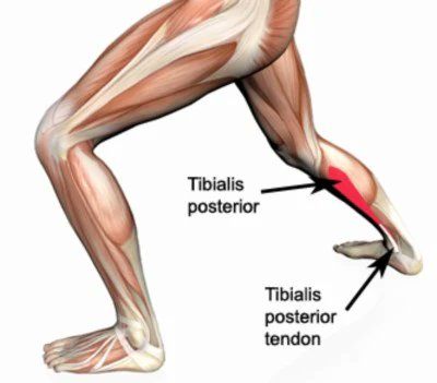
A main contributor to these patterns is due to a weakness within the Posterior Tibialis
The Posterior Tibialis is one of the primary 'supporting' muscles of the arch of the foot
Strengthening the Posterior Tib is a critical for recovery from PTTD (example exercise in post below)
But as I said, PTTD isn't just a Posterior Tibialis issue..
Over-Pronation creates a kinetic chain disaster & it's vitally important to the address the issue through a collective "From the Ground Up" approach
The Tibialis Posterior is one of the most underrated muscles for fixing knee, ankle, and foot pain.
Weakness here shows up in countless injuries - yet most rehab overlooks it. Let’s break down why it matters & how to train it 👇
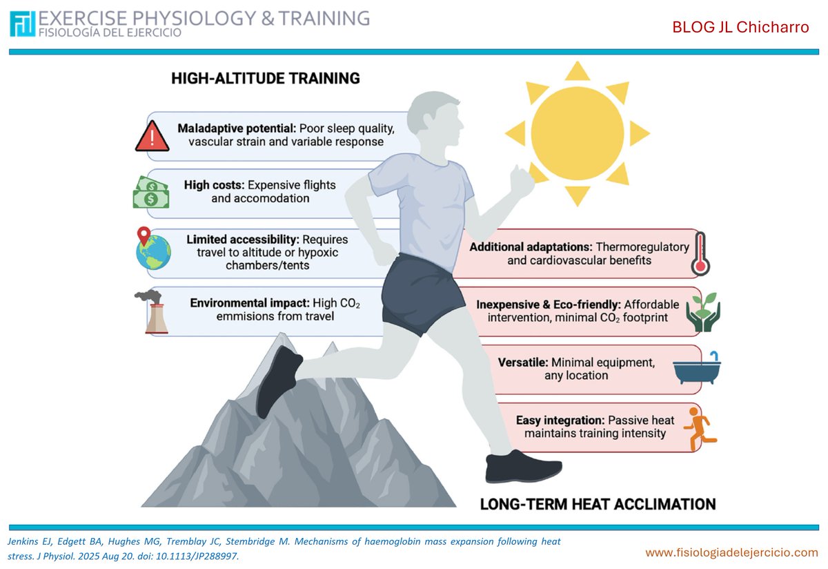
Aumento de la masa de hemoglobina con el calor
La aclimatación prolongada al calor aumenta la masa de hemoglobina y el VO₂max, ofreciendo beneficios comparables a la altitud pero con menor coste y riesgo. Los mecanismos incluyen isquemia renal, mayor consumo de oxígeno y la teoría del critmeter como reguladores de la EPO. (lee el artículo completo en BLOG JL Chicharro en https://t.co/ghlyA0rsYU) https://t.co/yyqL8rAY71
UFC Hall of Famer @GeorgesStPierre posted this on his IG
“Building power is like building a house-start with a solid foundation. That's why I train my feet!”
The elites know the importance of foundational health
From the Ground Up!
This is the most important part of ankle mobility & function you’ve never heard of:
The Cuboid-Locking Mechanism.
This is essential to train if you want to help improve how your foot & ankle moves, but also potentially to fix pain like Plantar Fasciitis and Achilles issues.
Los #tendones son tejidos conectivos fibrosos organizados JERÁRQUICAMENTE. Compuestos principalmente de colágeno tipo I (70%), elastina, proteoglicanos y glicoproteínas.
¿Qué tipo de ejercicio combate mejor la sarcopenia?
Un nuevo metaanálisis lo deja claro: el entrenamiento de fuerza es clave para mejorar movilidad y fuerza muscular en mayores.
Beneficios mayores entre 65–70 años y más marcados en mujeres.
Lee más 👉
https://t.co/KSv3AxDx9m
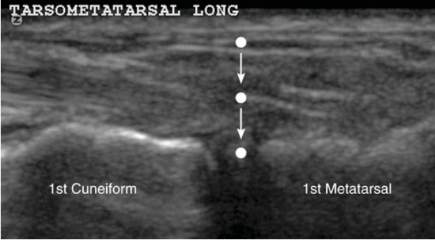
A lisfranc injection can be performed under ultrasound guidance in patients with certain types of lisfranc injuries or refractory pain in the midfoot/ forefoot! Click/ follow to learn more!
Read: https://t.co/pxQGcwkxXa
#sportsmedicine#physicaltherapy#physiotherapy #orthopedics #rehab #sportsinjury #injuryprevention #rehabilitation #athletictraining #meded
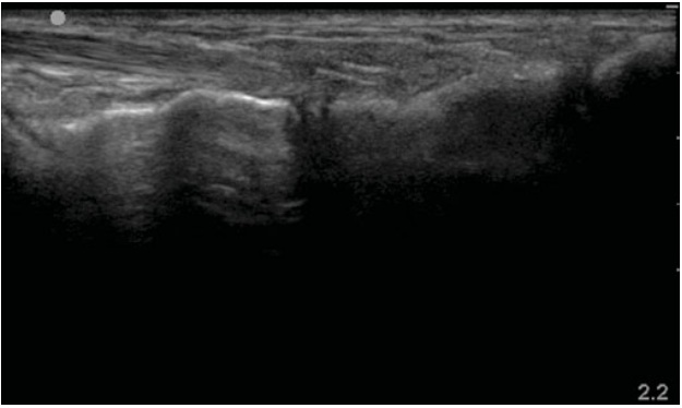
Posterior tibial tendon instability due to retinacular injury
The flexor retinaculum is detached from its medial malleolar insertion, creating a retinaculo-periosteal pocket into which the posterior tibial tendon can dynamically dislocate. This instability may lead to chronic friction, degenerative changes, and eventual tendon rupture.
The typical injury mechanism involves forced hindfoot inversion with reflex contraction of the posterior tibial muscle.
Dynamic ultrasound is essential for diagnosis, as static imaging may appear less apparent. Sometimes, just like in life (”and this case”), a small push is all it takes to move forward.
#mskrad