Having trouble remembering what to look for on scans for Alzheimer’s dementia?
Want the key findings to stay in your memory?
Medial temporal atrophy (MTA) score quantifies hippocampal volume loss
It rates the amount of CSF around the hippocampus as a marker of volume loss
CSF surrounds the hippocampus like a snake surrounding its prey—how far the snake has gone in getting its prey determines the MTA score:
MTA-1: Choroidal fissure opens, looks like the snake has started to slither in
Remember: the number one looks flat like a slightly opened choroidal fissure
MTA-2: Temporal horn widens, looks like the snake has surround the hippocampus
Remember: the number two look like the CSF curving around as the temporal horn opens
MTA-3: Moderate hippocampal atrophy, looks like the snake has begun to squeeze the hippocampus
Remember: the number three has a tight curve, so it looks like it is crushing the hippocampus & it looks small (atrophied)
MTA-4: Severe hippocampal atrophy, looks like the snake has crushed the hippocampus
Remember: Number four has only a tiny hole in it, so the hippocampus has to be super tiny & atrophied to fit inside!
Hopefully, this will help you commit this rating system to memory so you’ll never forget what you need to say for Alzheimer’s!
Reactive Arthritis
Think beyond the classic triad. A recent GU or GI infection followed by acute asymmetric lower-limb oligoarthritis, enthesitis, or dactylitis should raise suspicion—while septic arthritis and anterior uveitis must never be missed.
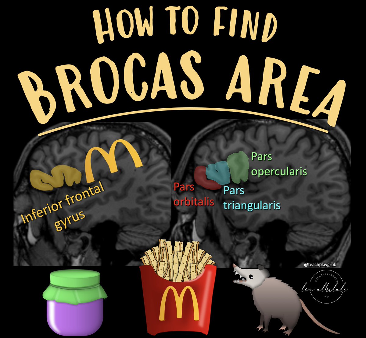
1/Do you get a Broca’s aphasia trying remember the location of Broca's area?
Does trying to remember inferior frontal gyrus anatomy leave you speechless?
Don't be at a loss for words when it comes to Broca's area
Here how to remember the anatomy of this key region!
1/I always say you can tell a bad read on a spine MR if it doesn’t talk about lateral recesses.
What will I think when I see your read? Do you rate lateral recess stenosis?
Here’s a thread on lateral recess anatomy & a grading system for lateral recess stenosis
1/Time is brain! But what time is it?
If you don’t know the time of stroke onset, are you able to deduce it from imaging?
Here’s a thread to help you date a stroke on MRI!