@Doctors_GUILD The correct answer is A. Duodenum.
After abdominal surgery, gastrointestinal motility returns in this general order:
Small intestine (especially duodenum) → within a few hours
Stomach → about 24–48 hrs
Colon → about 48–72 hrs
Duodenum regains motility earliest after surgery.
Pattern recognition: Testicular Sonography.
I have spent the past few weeks perfecting my knowledge of testicular sonography, and I'll be sharing some of the most important patterns you should know in three parts.
Here's part 1. 👇👇
Contributions and corrections are welcome.
Quiz: What's the difference between an epididymal cyst and spermatocele on ultrasound?
A clinical algorithm and medication table for the diagnosis and management of heart failure (HF). It categorizes patients based on left ventricular ejection fraction (LVEF) and outlines recommended drug therapies and dosages.
https://t.co/L6fjzE1VxQ
“Tell me where it hurts.”
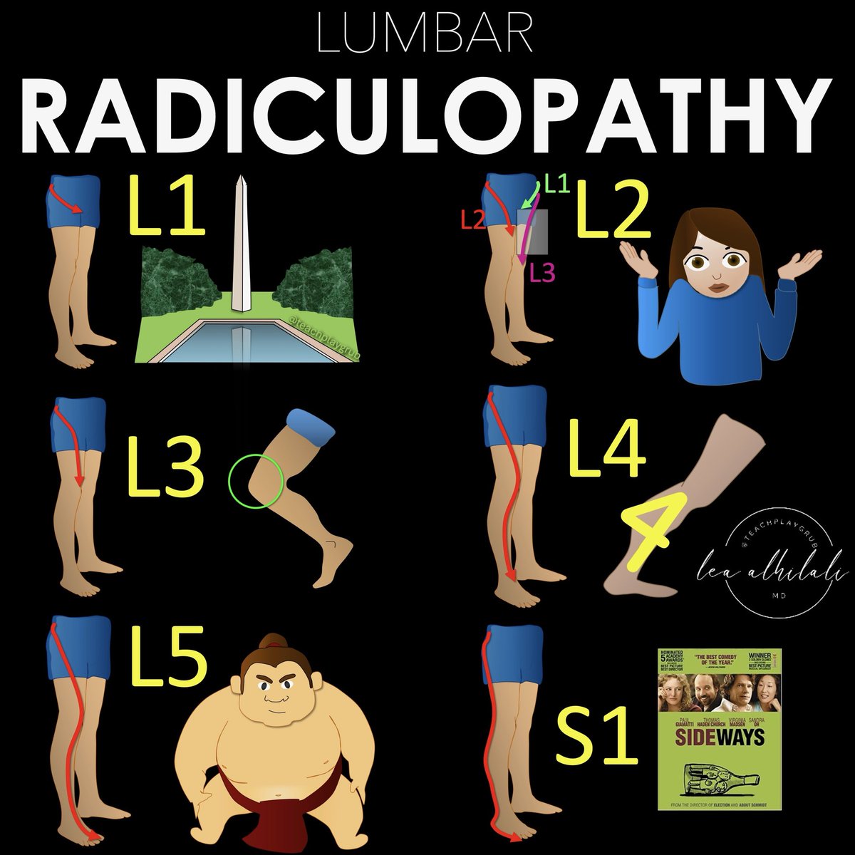
How back pain radiates can tell you where the lesion is—if you know where to look!
Do YOU know where to look?
Here’s how to remember the lumbar radicular pain distributions!
Keep this cheat sheet as a BACKUP for when you are dealing with BACK pain!
➡️L1
🔸L1 radiates to the groin
🔸Remember that b/c the number 1 is, well, um…phallic. So phallic number 1 radiates to the groin.
➡️L2
🔸L2 radiates to thigh
🔸Two is the number between 1 and 3, so the distribution of L2 is between the distributions of L1 and L3—and between the groin (L1) and knee (L3) is the thigh.
➡️L3
🔸L3 radiates to the knee
🔸Remember L3 is to the knee—easy, it rhymes!
➡️L4
🔸L4 radiates to the calf.
🔸Remember this bc the number 4 looks like the calf, 🔸Top part of the 4 looking like a bulging gastroc & the bottom part of the four is the rest of the calf connecting to the ankle.
➡️L5
🔸L5 radiates to the big toe.
🔸Remember the little rhyme “Five is to the big guy!”
🔸L5 is also foot drop. Remember big guys are heavy, and heavy gravity = drop.
➡️S1
🔸S1 radiates to the side of the foot.
🔸Remember this because both S1 and Side start w/S.
So now you know where in the lumbar spine to a look when a patient tells you the pain radiates down their leg!
🚨Remember, there are many variations & this is just a starting guide🚨
But hopefully now remembering the lumbar radicular distributions won’t be a pain in the backside!
ECG Interpretation: Breaking Down Every Wave and Interval.
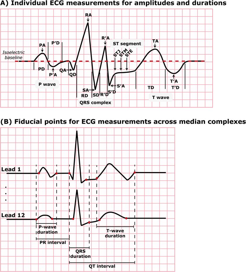
Your ECG is more than waves, it’s a precise timeline of cardiac electrical activity.
🟢 P wave: atrial depolarization initiated by the SA node
🟣 PR interval: conduction through the AV node (physiologic delay)
⚪ QRS complex: ventricular depolarization and onset of systole
🔴 ST segment: electrically neutral phase between depolarization and repolarization
⚫ T wave: ventricular repolarization
🟡 QT interval: total duration of ventricular depolarization + repolarization
Waveform nuances matter:
P and T waves may be positive, negative, or biphasic depending on lead orientation
Q, S, and S′ are defined as negative deflections, though their amplitudes are expressed as positive values
ST segment precision:
J point (STJ): level at QRS offset
STM: ST level at QRS offset + 1/16 RR interval
STE: ST level at QRS offset + 1/8 RR interval.
Modern ECG analysis (e.g., Marquette 12SL) measures global intervals by detecting the earliest onset and latest offset across all 12 leads, not just a single lead.
Every millisecond on ECG reflects a real electrical and mechanical cardiac event.
A simple way to see the 12-lead ECG
Color-coding helps pattern recognition:
🟢 Inferior: II, III, aVF
🟡 Lateral: I, aVL, V5–V6
🔵 Anterior/septal: V1–V4
🔴 Right: aVR (± V1)
Instead of memorizing 12 leads individually, think in territories.
This makes STEMI localization faster, cleaner, and harder to miss.
FOOT DROP
Fibular (peroneal) neuropathy vs L5 radiculopathy!
You have a patient come in with a foot drop. There is no pain, but they have a history of low back pain. How do you differentiate between an L5 radiculopathy and fibular neuropathy in the clinic?