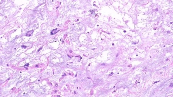
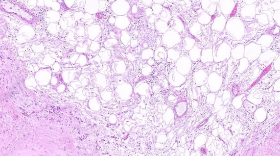
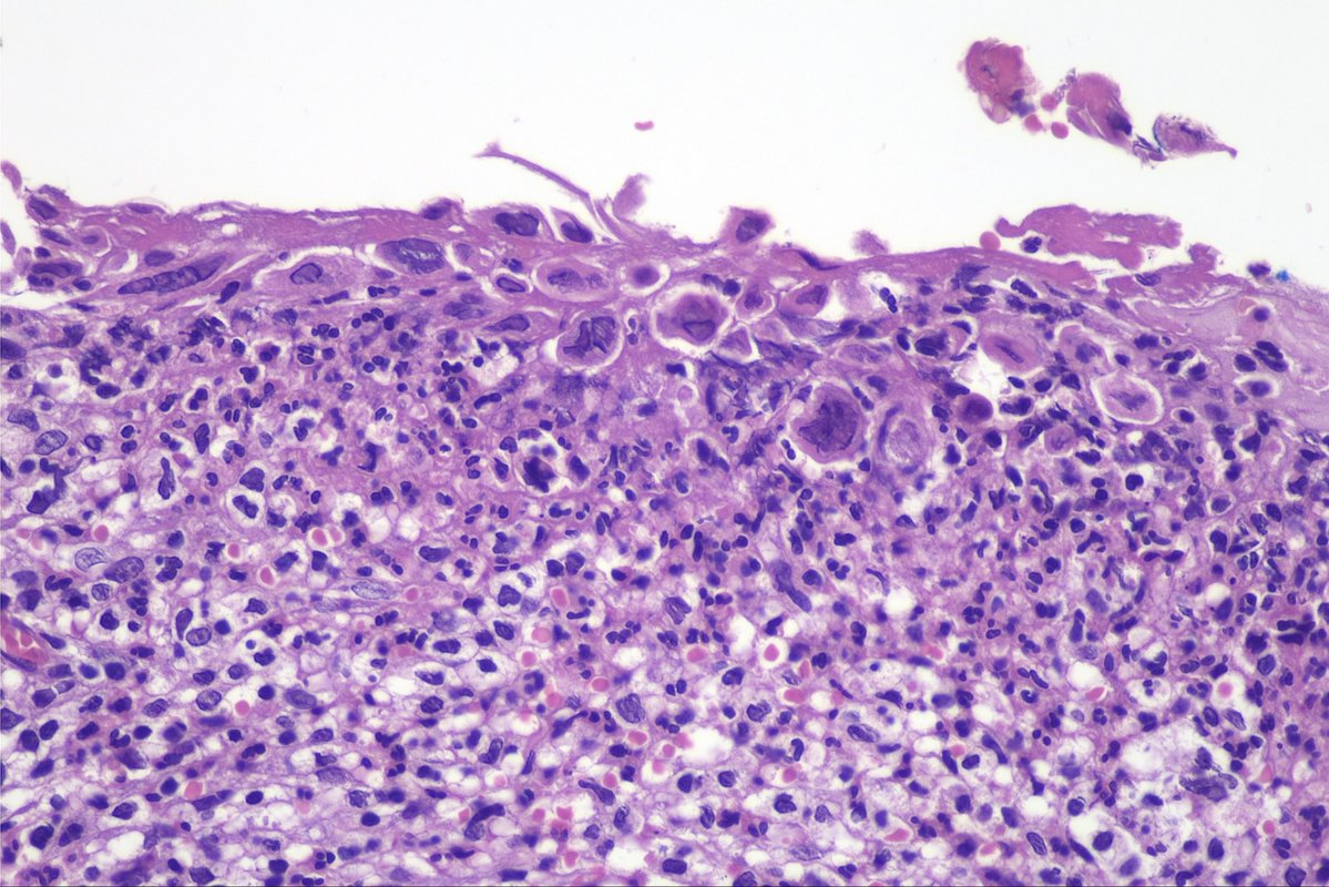
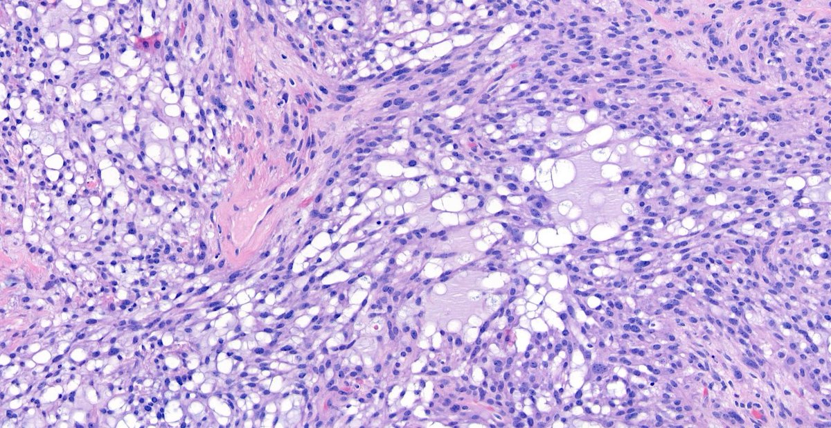
Masterclass on bone #pathology w special guest Dr. Andrew Rosenberg!
Video: https://t.co/Z3rVipx4y0
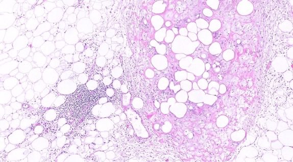
He discusses normal bone #Histology, #Embryology, & non-neoplastic pathology, reactive changes, woven bone, fracture callus, & Paget disease. Amazing #meded!
What drives local recurrence risk after breast-conserving surgery? The DCIS component—especially when an extensive intraductal component (EIC) is present—is a major driver of ipsilateral breast recurrence
What drives systemic recurrence risk? Primarily the invasive component: size (T stage), histologic grade, lymphovascular invasion, nodal status, and molecular subtype/biomarkers
Dr. Bloom. Microinvasive carcinoma. CAP on demand https://t.co/CFLLpZmOyB #pathology #PathX #breastpath
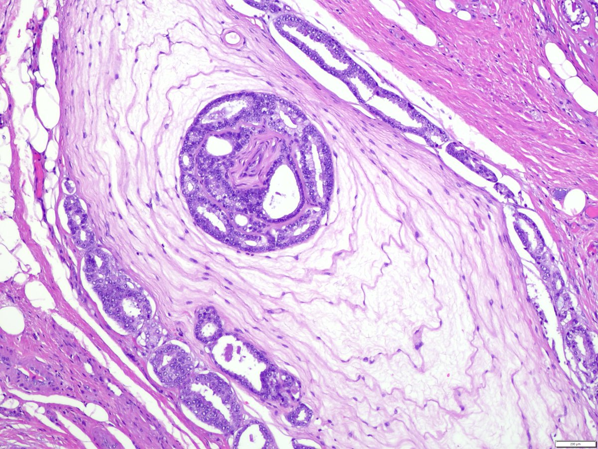
How to handle cases with microinvasive carcinoma?
-Obtain deeper levels and pay attention to lymphoid inflammation areas around DCIS
- In cases with multiple foci, the sum of the sizes should not be used for determining the pT stage
- Report biomarkers if possible, if exhausted report ER/PR from adjacent DCIS
Dr. Huina Zhang. Microinvasive carcinoma CAP on demand https://t.co/CFLLpZmOyB #pathology #PathX #Breastpath
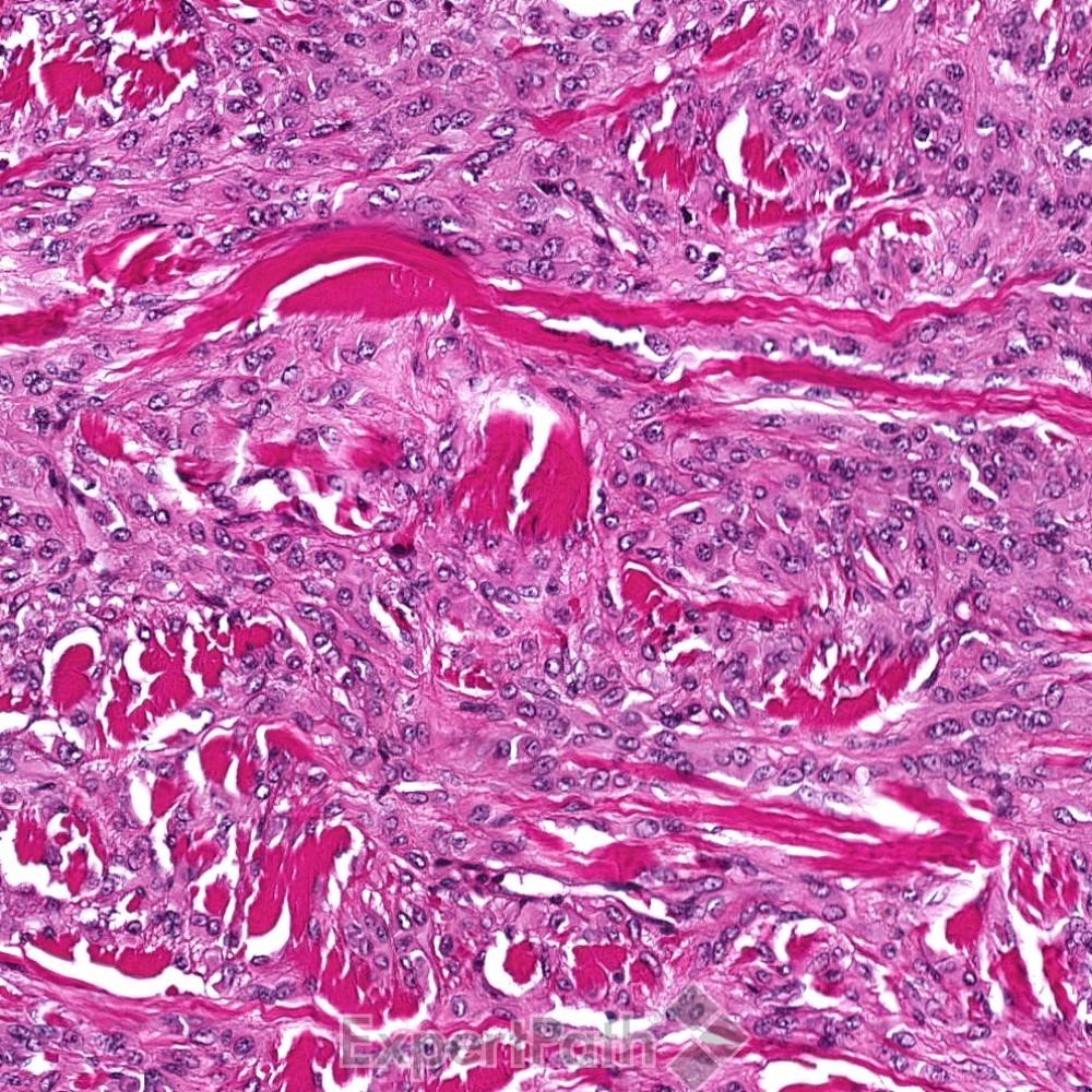
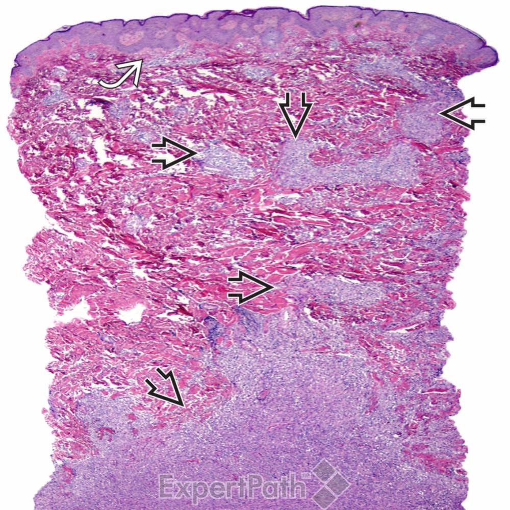
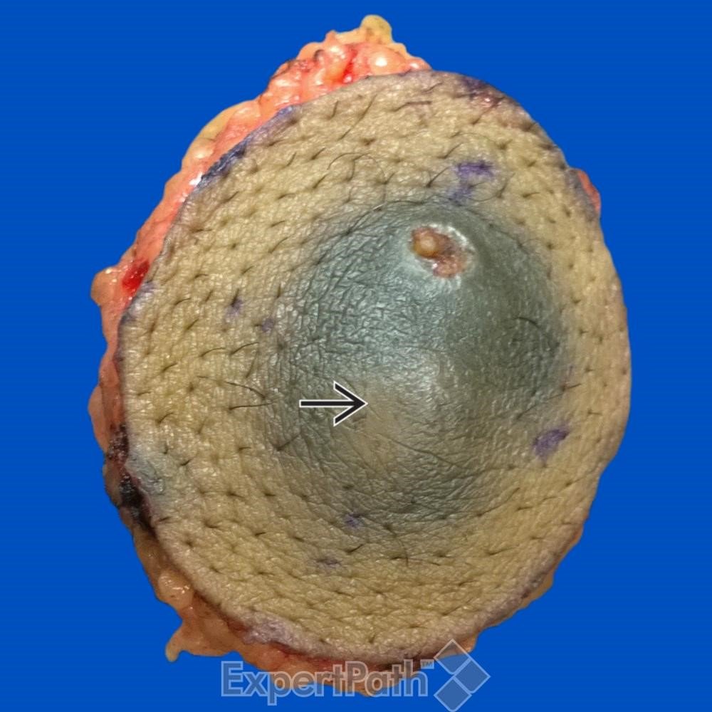
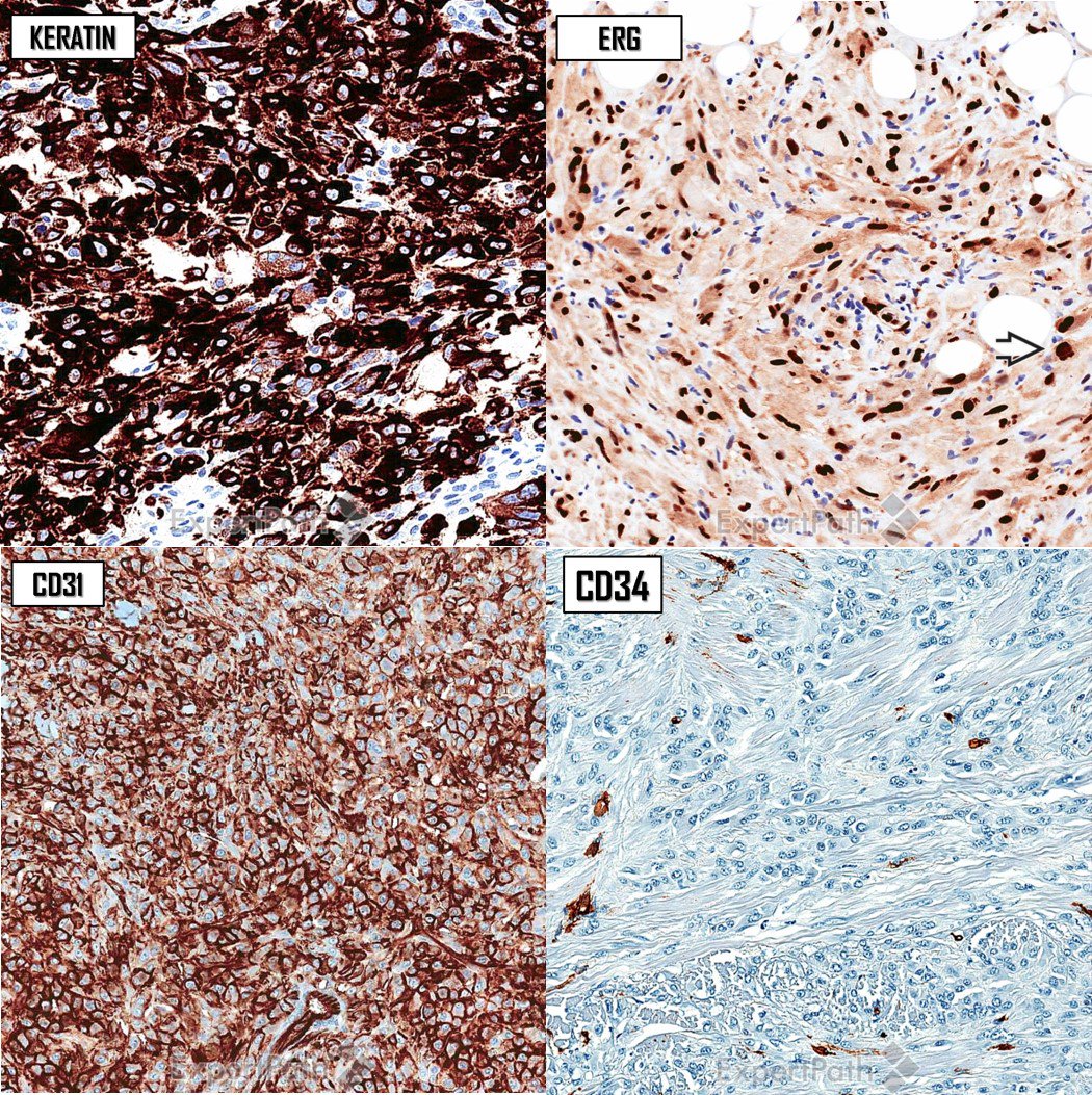
Now THAT's Cherry-Red Nucleoli! (but it isn't melanoma). Large thigh mass in an adult
Answer ✅ https://t.co/vN6bODcYyI
More pics: https://t.co/Hqc8QkCAXA.
How I work up pleomorphic spindle cell tumors: https://t.co/SSY2q2lEhW
#BSTpath#pathologists#pathology#pathTwitter #dermpath #dermatology #dermtwitter
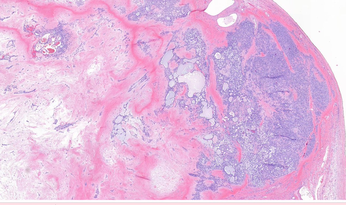
Solid Papillary Carcinoma of the Breast
(with emphasis on in situ × invasive disease)
🔬 A rare and generally indolent neoplasm, but with one of the most interesting diagnostic pitfalls in breast pathology: absence of myoepithelial cells does NOT, by itself, indicate invasion.🧵
📌 Definition
Solid papillary carcinoma (SPC) is characterized by expansile solid nodules traversed by delicate fibrovascular cores, frequently showing neuroendocrine differentiation.
👥 Epidemiology
Predominantly affects postmenopausal women, usually in the 7th decade of life or later.
📍 Sites
It may occur in any region of the breast, but there is a predilection for the central/subareolar region.
🧬 Pathogenesis
A tumor with a luminal phenotype, generally ER+/HER2−. Reported alterations include 1q/16p gains, 16q loss, and PIK3CA mutations in a subset of cases.
🩺 Clinical Features
It may present as:
• palpable mass;
• mammographic abnormality;
• bloody nipple discharge.
🧪 Laboratory Diagnosis / Core biopsy
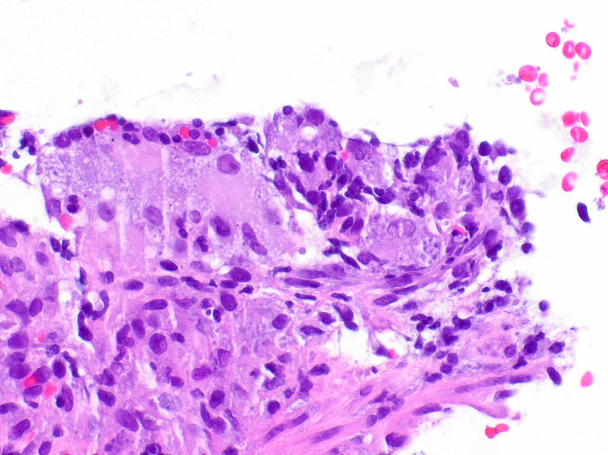
Core biopsy may recognize the pattern, but frequently does not adequately sample the tumor periphery, limiting assessment of invasion. Post-biopsy changes may also mimic infiltration.
🔬 Histopathology
• Solid, rounded, expansile nodules.
• Delicate fibrovascular cores, sometimes inconspicuous.
• Monotonous round/oval or spindle-shaped cells.
• Granular or plasmacytoid cytoplasm.
• Nuclear palisading around vessels.
• Intracellular or extracellular mucin may be present.
⚠️ The main pitfall
SPC in situ:
→ smooth, rounded, circumscribed nodules.
→ a peripheral myoepithelial cell layer may NOT be demonstrable.
Invasive SPC:
→ irregular/angulated contours;
→ geographic “jigsaw puzzle” pattern;
→ small infiltrative nests;
→ desmoplasia and/or adipose tissue infiltration.
👉 Therefore: negative p63 ≠ automatic invasion.
In SPC, the morphology of the tumor–stroma interface is fundamental.
🧫 Immunohistochemistry
• ER: usually strong and diffuse.
• PR: frequently positive.
• HER2: usually negative.
• Ki-67: low/intermediate.
• Synaptophysin / Chromogranin / INSM1: frequently positive.
Neuroendocrine differentiation is common, but SPC should not simply be reclassified as a neuroendocrine tumor of the breast.
🔎 Differential Diagnosis
• Papilloma + florid UDH → mosaic CK5/6, heterogeneous ER, preserved myoepithelium.
• Encapsulated papillary carcinoma → cystic space/capsule and more evident true papillae.
• Mucinous carcinoma → carcinoma truly infiltrating mucin pools.
• Invasive lobular carcinoma → evaluate E-cadherin/p120.
• Metastatic NET → absent/weak ER should raise this possibility.
📈 Prognosis
Excellent in the pure/in situ form. Lymph node and distant metastases are essentially related to the presence of an invasive component.
💊 Treatment
SPC in situ is managed as in situ disease. When invasion is present, management follows the principles applicable to the corresponding invasive breast carcinoma.
🎯 Take-Home Messages @Notas_Patologia
• Look for delicate fibrovascular cores within an apparently solid proliferation.
• Absence of myoepithelium does not define invasion in SPC.
• Circumscription favors in situ; irregularity + desmoplasia/adipose infiltration favor invasion.
• Neuroendocrine differentiation is frequent, but does not change the entity.
• For staging, use the size of the invasive component, not the total size of the SPC.
📚 Selected References
• WHO Classification of Breast Tumours, 5th ed.
• Saremian J, Rosa M. Arch Pathol Lab Med. 2012;136:1308–1311.
• Otsuki Y et al. Virchows Arch. 2023;482:687–695.
• Hoda RS, Wen HY. Human Pathology. 2025;162:105759.
• PathologyOutlines �� Solid papillary carcinoma.
⚠️Disclaimer:⚠️
This text is an educational summary for healthcare professionals and students. It does not replace full pathology reports, local guidelines, or individualized clinical decision-making.
#MedicalEducation #NotasDePatologia #pathology #PathTwitter #BreastPathology #BreastCancer
🖋️ Case and slide coloration courtesy of Dr. Ivison Xavier Duarte - Laboratório Patologika (https://t.co/AmtxJzycUs), as part of an academic partnership project.