

Top Tweets for #Pathbugs

The answer to #Parasite case 821 is cystic echinococcosis ! Read the full post here👇
https://t.co/vBSaRiCxbi
Many thanks to Loranne Lambregts and @ITMantwerp for this beautiful case.
#Parasitology #Pathology #Microbiology #mayopath #PathBugs #CrittersOnTwitter
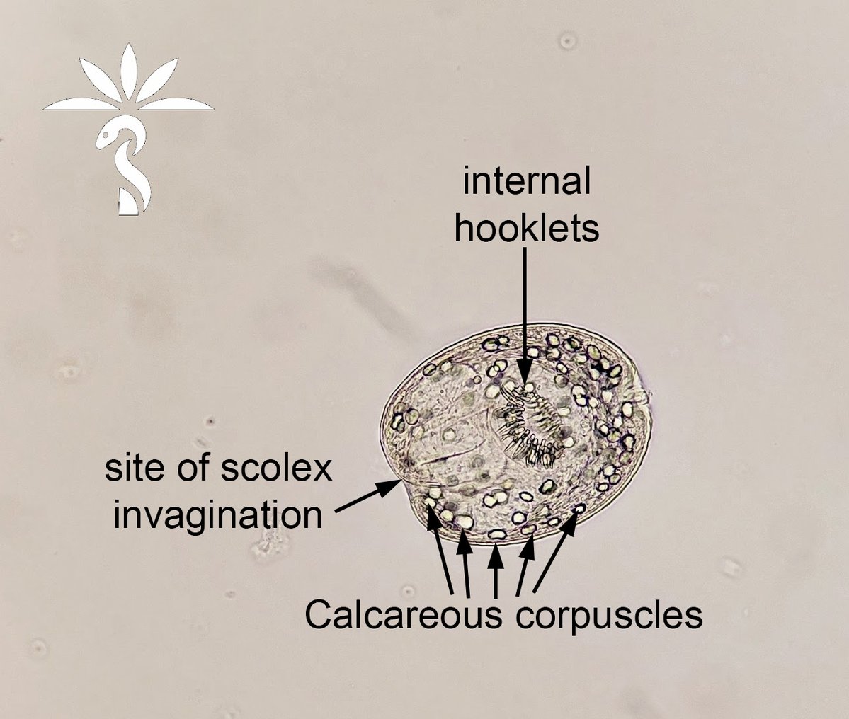
Answer to #Parasite case 821 is cystic echinococcosis! Check out full discussion here:
https://t.co/vBSaRiBZlK
Many thanks to Loranne Lambregts and @ITMantwerp!
#Parasitology #Pathology #Microbiology #mayopath #PathBugs #CrittersOnTwitter
#Parasite Case 821 is up, thanks to Loranne Lambregts and @ITMantwerp! This one comes from liver cyst fluid. 😯 What’s your identification? Post your guess below or in the blog comments: https://t.co/wyYZS5K4mu later this week!
#mayopath #pathology #PathBugs #CrittersOnTwitter
@MayoClinicINFD
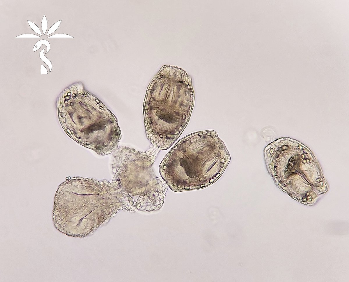


Happy Halloween from these creepy critters and their scary counterparts 🎃. #Halloween #pathart #pathology #pathbugs #happyhalloween


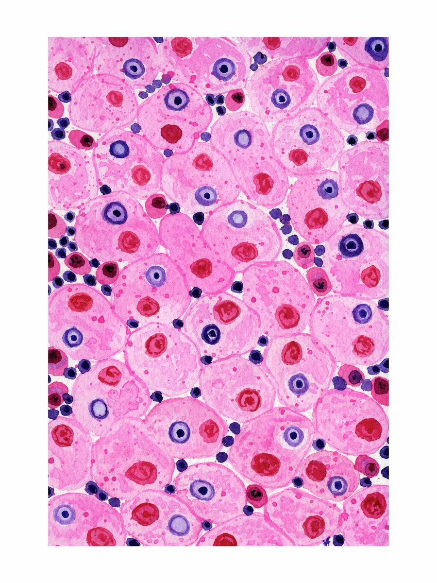

Döhle-like body inclusions in MHA and Trypanosoma are my next spooky cells. 🥹 #pathology #pathx #pathbugs #hemepath #pathart


Skin biopsy. What is the bug?

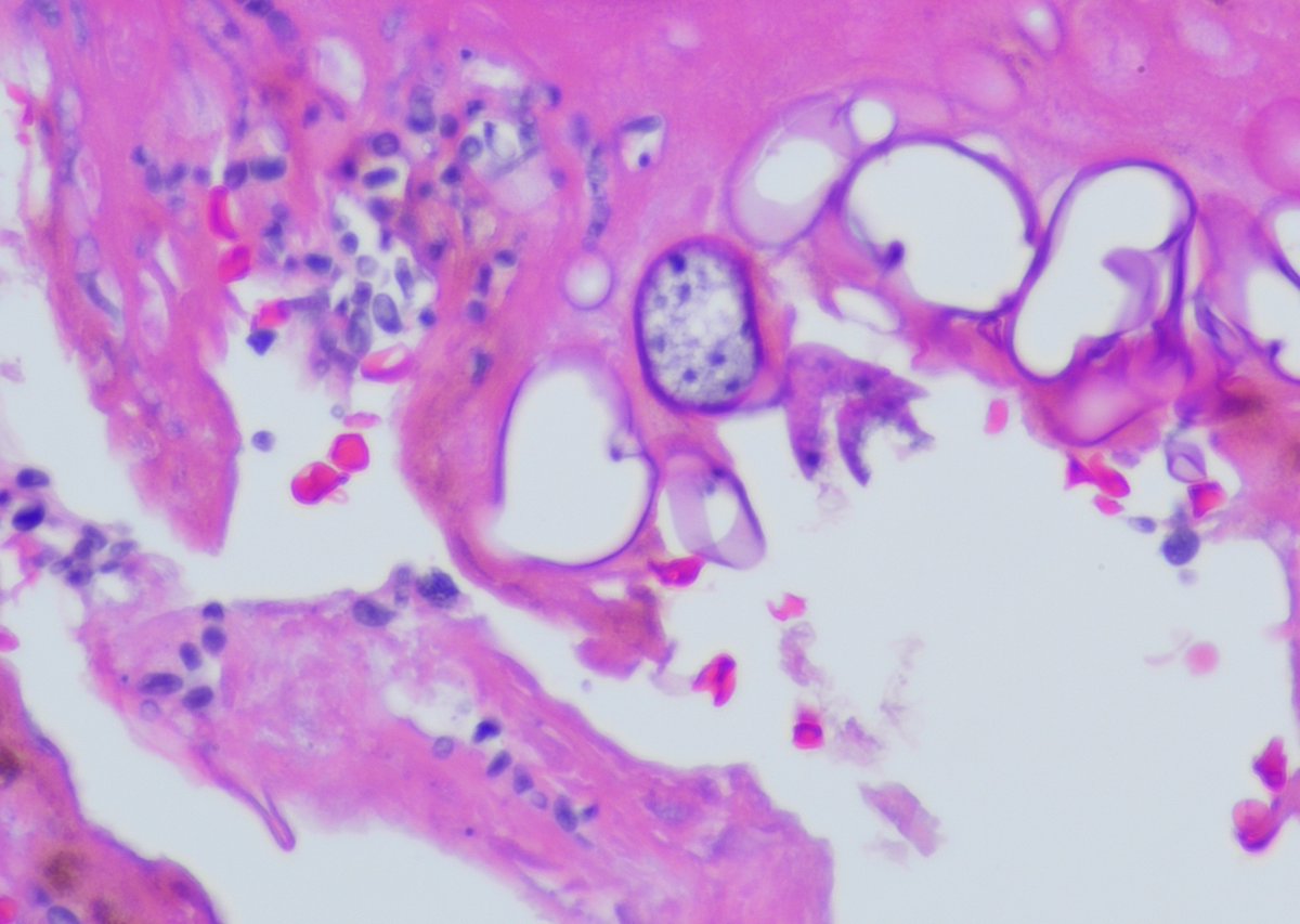
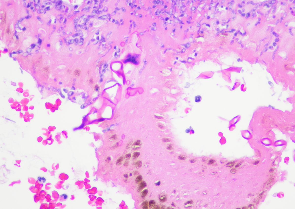


Always enjoy your fascinating parasite cases, @ParasiteGal! Such a wonderful way to challenge our diagnostic skills and keep us learning. Thanks for sharing! 🔬🪱
#PathBugs #Pathresidents #Pathologists #Infectiousdisease
The #Parasite Case 820 is up, a subcutaneous nodule in a man from Liberia. Check out the whole slide image to make your diagnosis! https://t.co/q6pG8UBlnF 🪱
Answer later this week. #mayopath #pathology #PathBugs #CrittersOnTwitter


The #Parasite Case 820 is up, a subcutaneous nodule in a man from Liberia. Check out the whole slide image to make your diagnosis! https://t.co/q6pG8UBlnF 🪱
Answer later this week. #mayopath #pathology #PathBugs #CrittersOnTwitter

Bug day! Tunga penetrans!!!


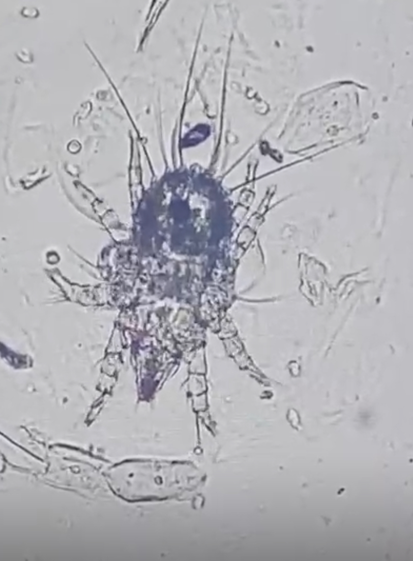
The answer to #Parasite Case 819 is up: Mite, not a human pathogen! A mighty interesting incidental finding 🕷️🔬 Read the full blog post for the key morphologic features and more info: https://t.co/2AvVMODTzS #mayopath #pathology #PathBugs #parasitology
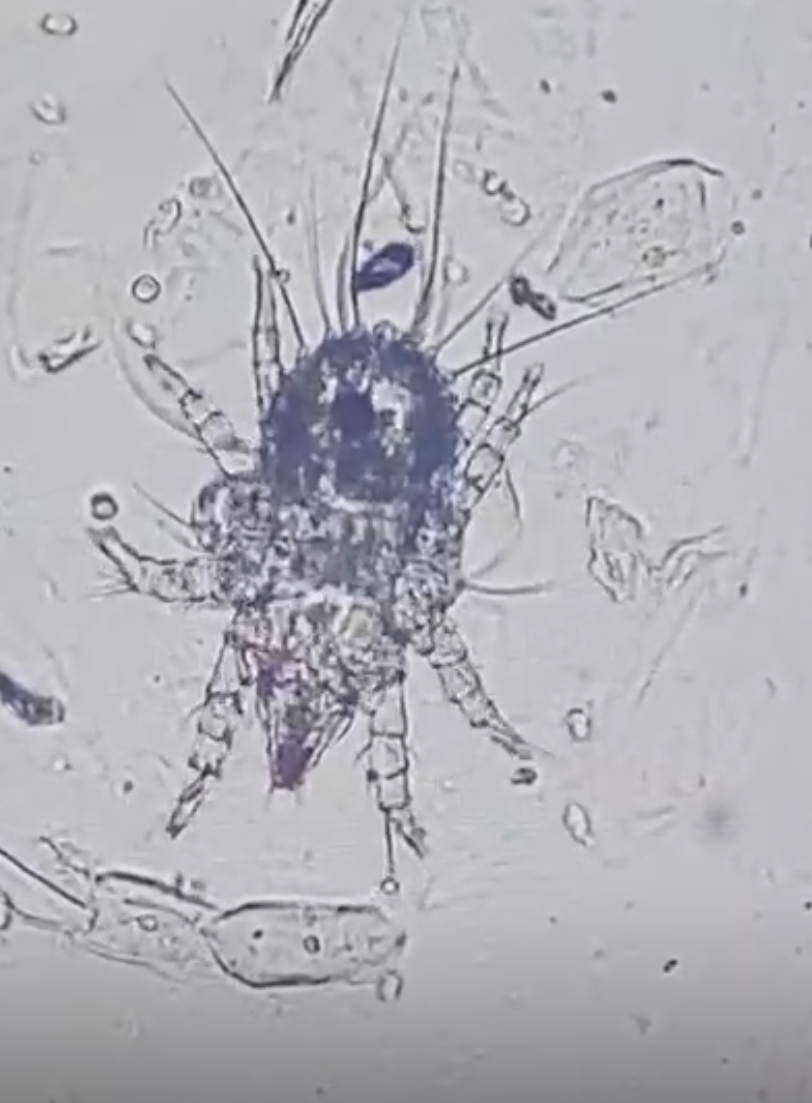

Woah, what do we have here? How did these get into the eye?
#Pathbugs #pathology #Eyepathology #Ocularpathology #Ophthalmicpathology #Pathtwitter #Pathx
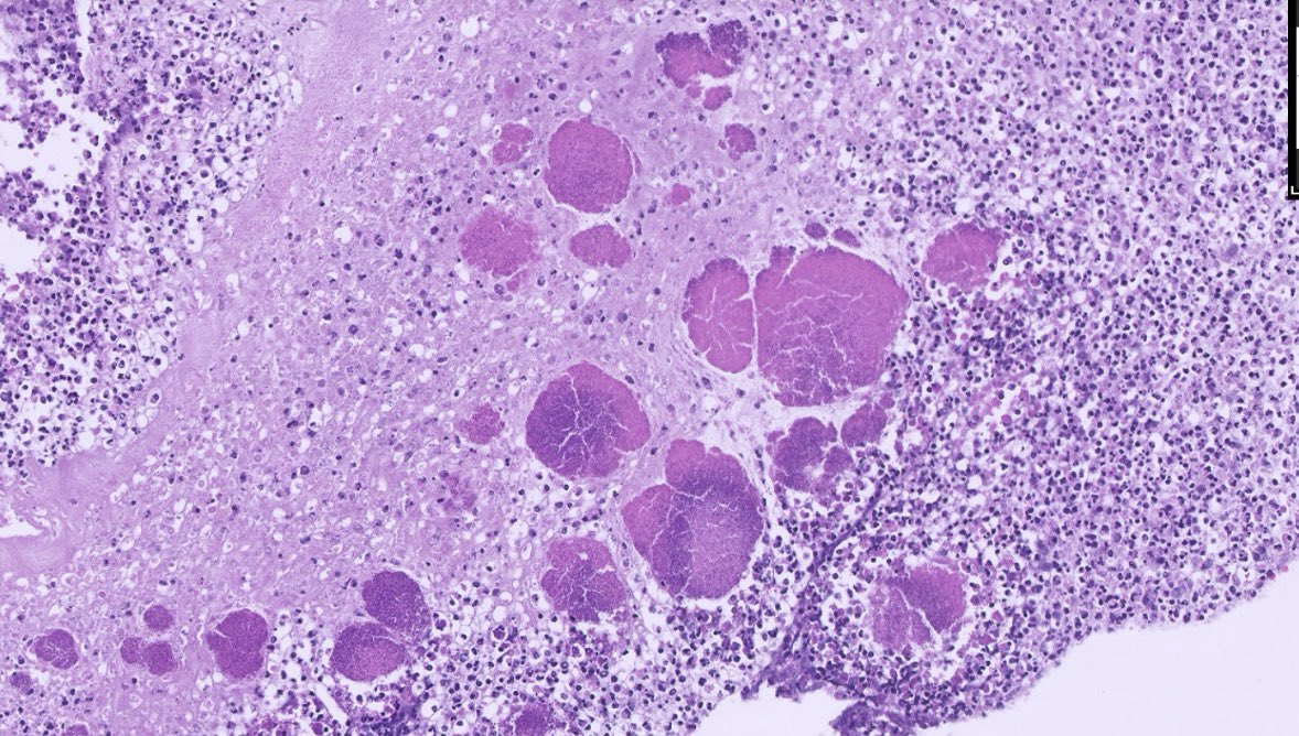
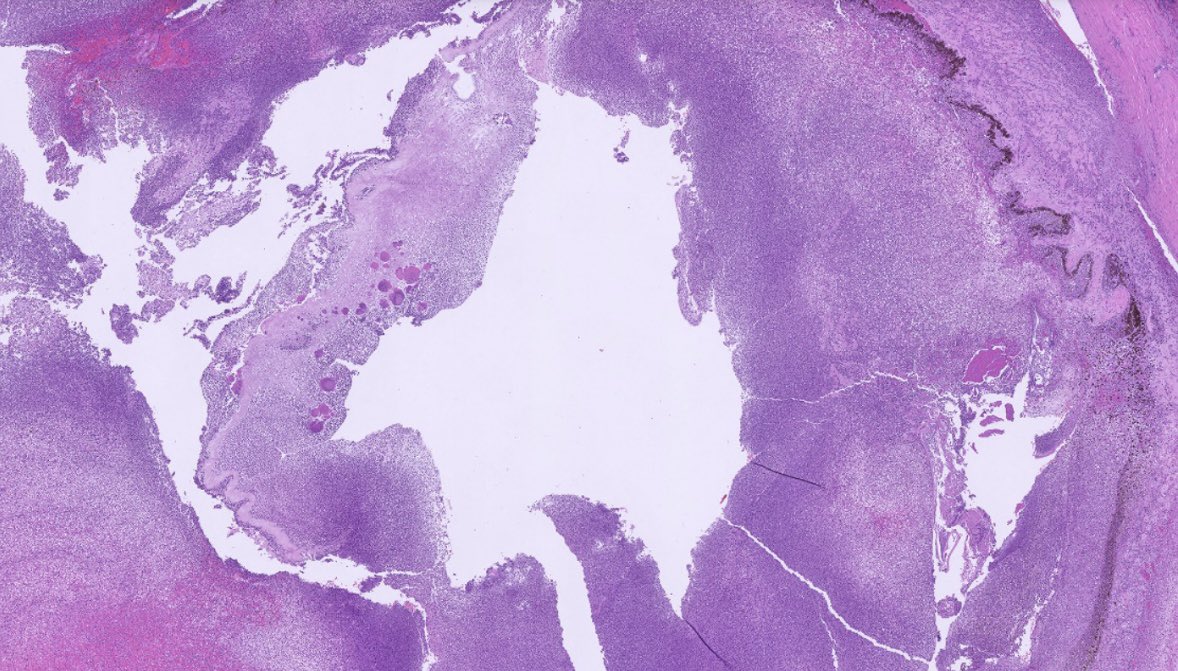
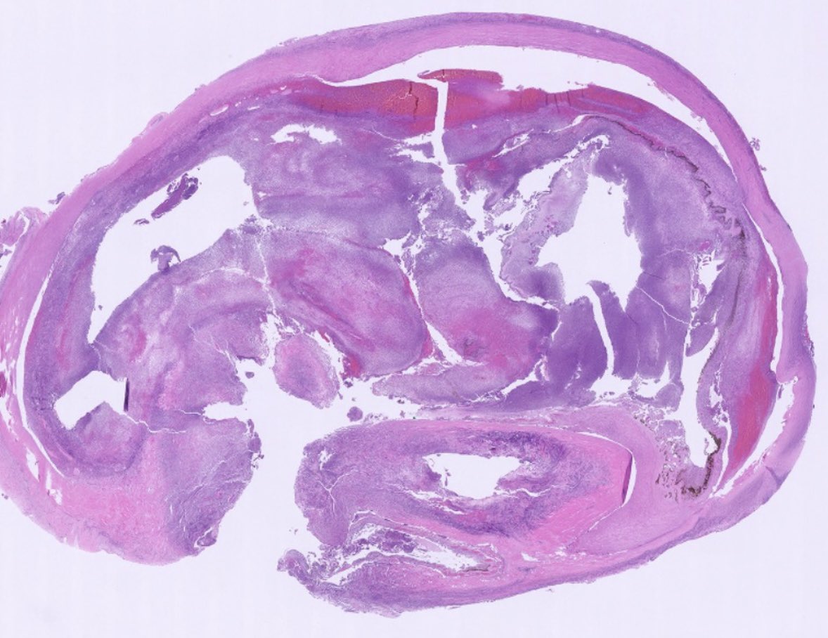

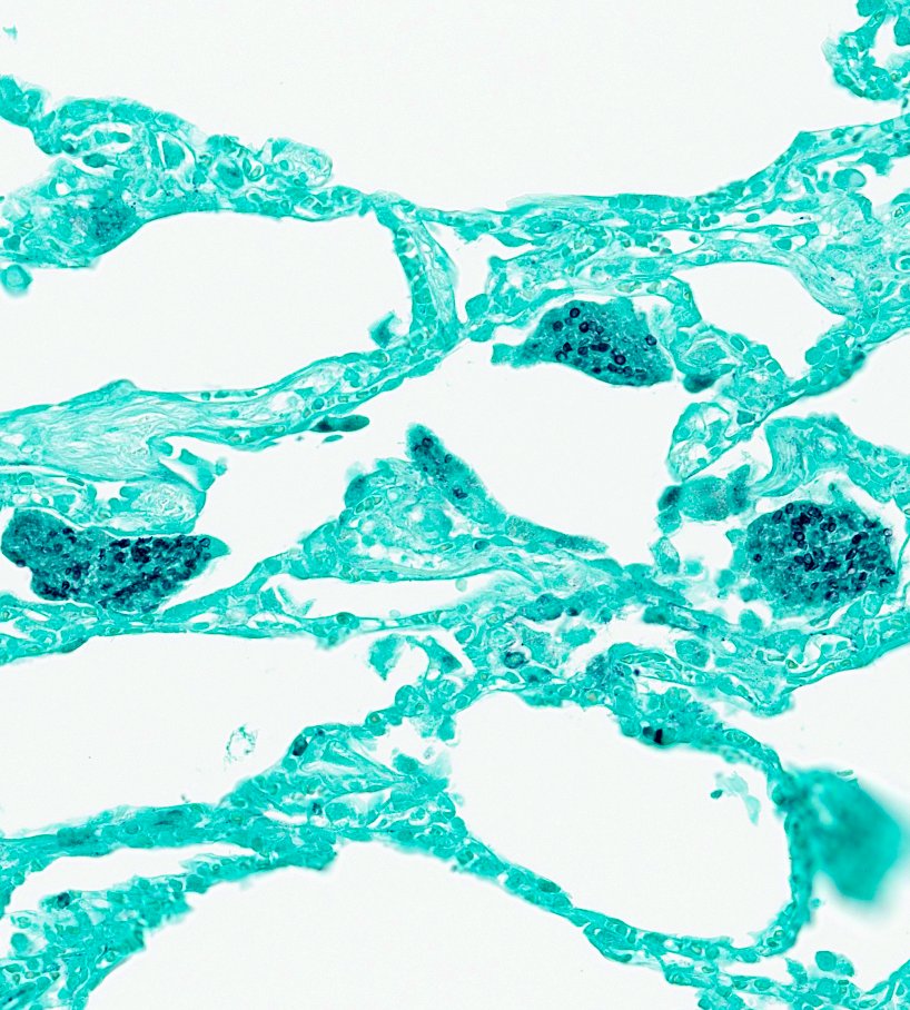
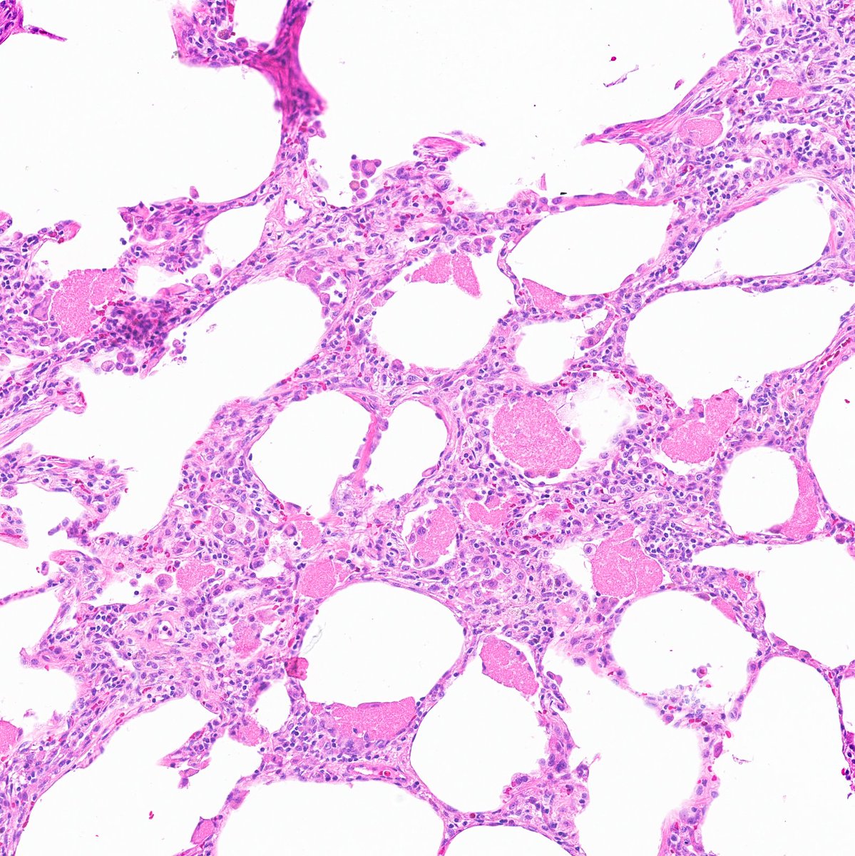
🔬 Lung wedge resection: H&E and GMS.
What’s your diagnosis?
Clue: Pink froth+ ping-pong balls 🧼🏓
#PathTwitter #MedTwitter #PulmPath #LungPath #USMLE #PathFellow #PathResidents
The #Parasite Case 819 is up, donated by @VasuMicroCaucus: an arthropod found in a urine specimen from a woman living in rural India. What is your identification? https://t.co/PKzNN5nfSp
Answer later this week! #mayopath #pathology #PathBugs #CrittersOnTwitter

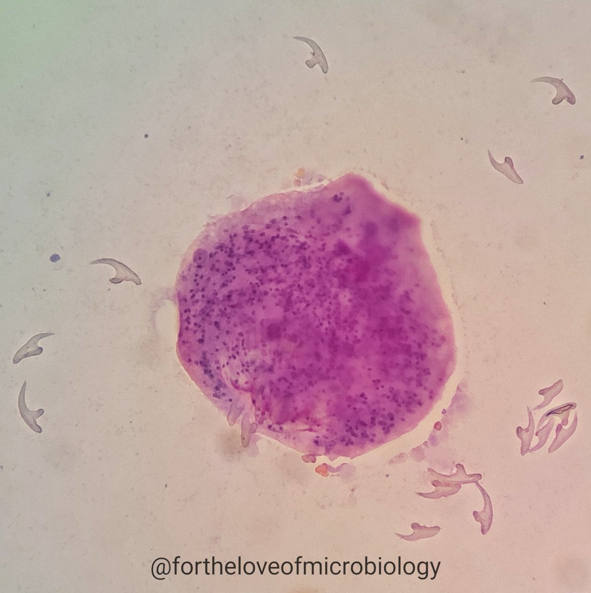
Gram stain of a renal cyst aspirate demonstrating many hooklets of Echinococcus.
Human echinococcosis (hydatidosis, or hydatid disease) is caused by the larval stages of cestodes of the genus Echinococcus. Echinococcus granulosus causes cystic echinococcosis and is the form most frequently encountered.
EG infections often remain asymptomatic for years before the cysts grow large enough to cause symptoms in the affected organs. The rate at which symptoms appear typically depends on the location of the cyst.
Hepatic and pulmonary signs/symptoms are the most common clinical manifestations, as these are the most common sites for cysts to develop In addition to the liver and lungs, other organs (spleen, kidneys, heart, bone, and central nervous system, including the brain and eyes) can also be involved, with resulting symptoms.
Rupture of the cysts can produce a host reaction manifesting as fever, urticaria, eosinophilia, and potentially anaphylactic shock; rupture of the cyst may also lead to cyst dissemination.
Hooklets are small, chitinous structures found on the rostellum of Echinococcus tapeworms, specifically Echinococcus granulosus and Echinococcus multilocularis.
Hooklets are a key diagnostic feature of Echinococcus hydatid cysts. They are found in the protoscolices, which are the larval heads of the tapeworm that develop inside the cyst.
These hooklets, numbering 25 to 50, assist the parasite in adhering to the intestinal mucosa of the definitive host, typically canids.
The presence of hooklets is crucial for the identification of these parasites, as they are key morphological features in diagnosing echinococcosis.
They may be the only identifiable Echinococcus structure present in some cases, especially if the cyst has ruptured.
Visualization of hooklets, either directly or with special stains, helps confirm the diagnosis of hydatid cyst disease caused by Echinococcus tapeworms.
#Fortheloveofmicrobiology #clinicalmicrobiology #microrounds #IDpath #ASMClinmicro #STEM #womeninmicrobiology #microbiologypakistan #pathbugs
Enterobius vermicularis ova were identified on stool microscopy from a 9-year-old child.
Microscopic appearance: The eggs are colorless, smooth and ovoid, with a moderately thick, double-layered shell. One side is characteristically flattened, giving the egg its classic “D-shaped” appearance. They measure approximately 50–60 × 20–30 µm.
Unlike the eggs of many other intestinal nematodes, E. vermicularis eggs are passed partially embryonated, and a developing larva may sometimes be visible within the egg.
About the parasite: E. vermicularis is a nematode (roundworm) with a worldwide distribution. Infection is particularly common in children.
Adult worms inhabit the cecum and adjacent colon. At night, gravid female worms migrate to the perianal region to deposit their eggs. This explains the characteristic symptom of nocturnal pruritus ani (night-time anal itching). In heavier infections, sleep disturbance, irritability and reduced appetite may occur.
Although E. vermicularis eggs may occasionally be detected in stool—as in this case—stool microscopy is not the optimal diagnostic method. The preferred method is the cellophane/cellulose adhesive tape test, performed on the perianal skin, ideally in the morning before bathing or defecation.
A beautiful example of how a characteristic microscopic morphology can point directly toward the diagnosis.
#Fortheloveofmicrobiology #clinicalmicrobiology #microrounds #IDpath #ASMClinmicro #STEM #womeninmicrobiology #microbiologypakistan #pathbugs


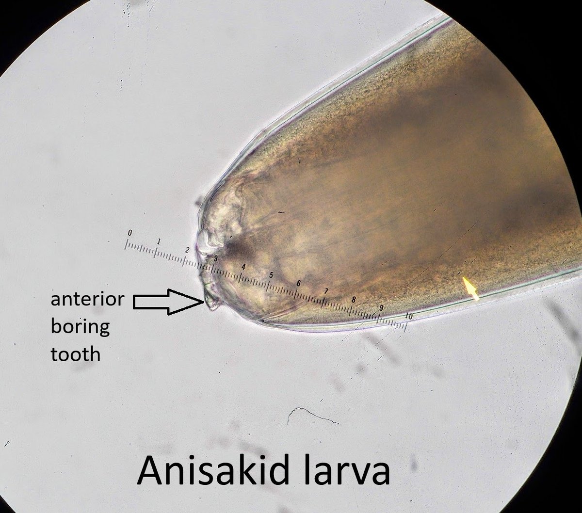
The answer to #ParasiteCase 818 is up: anisakid larva! 🪱🐟
Check out the full post for the key morphologic features—including the boring tooth and mucron—and learn how these fish-associated larvae are identified:
https://t.co/wtD59gnVY4
#PathBugs #Parasitology #Pathology #MayoPath
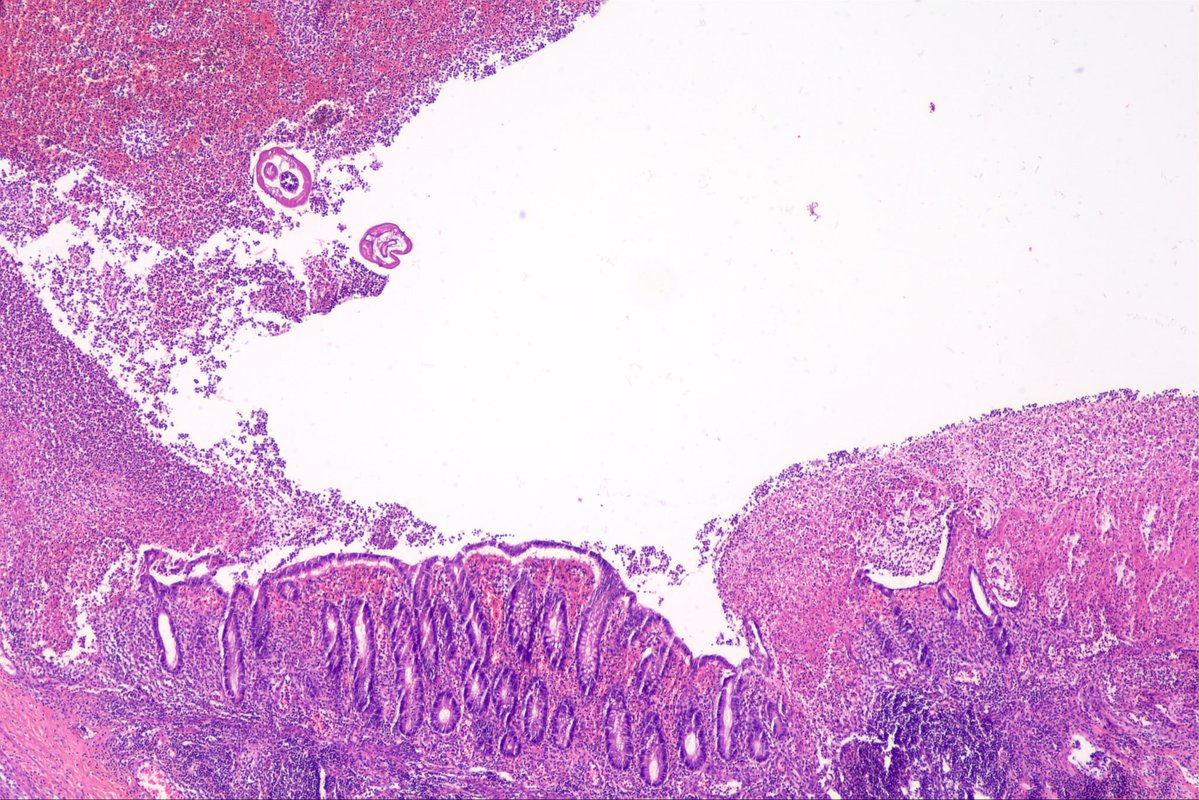
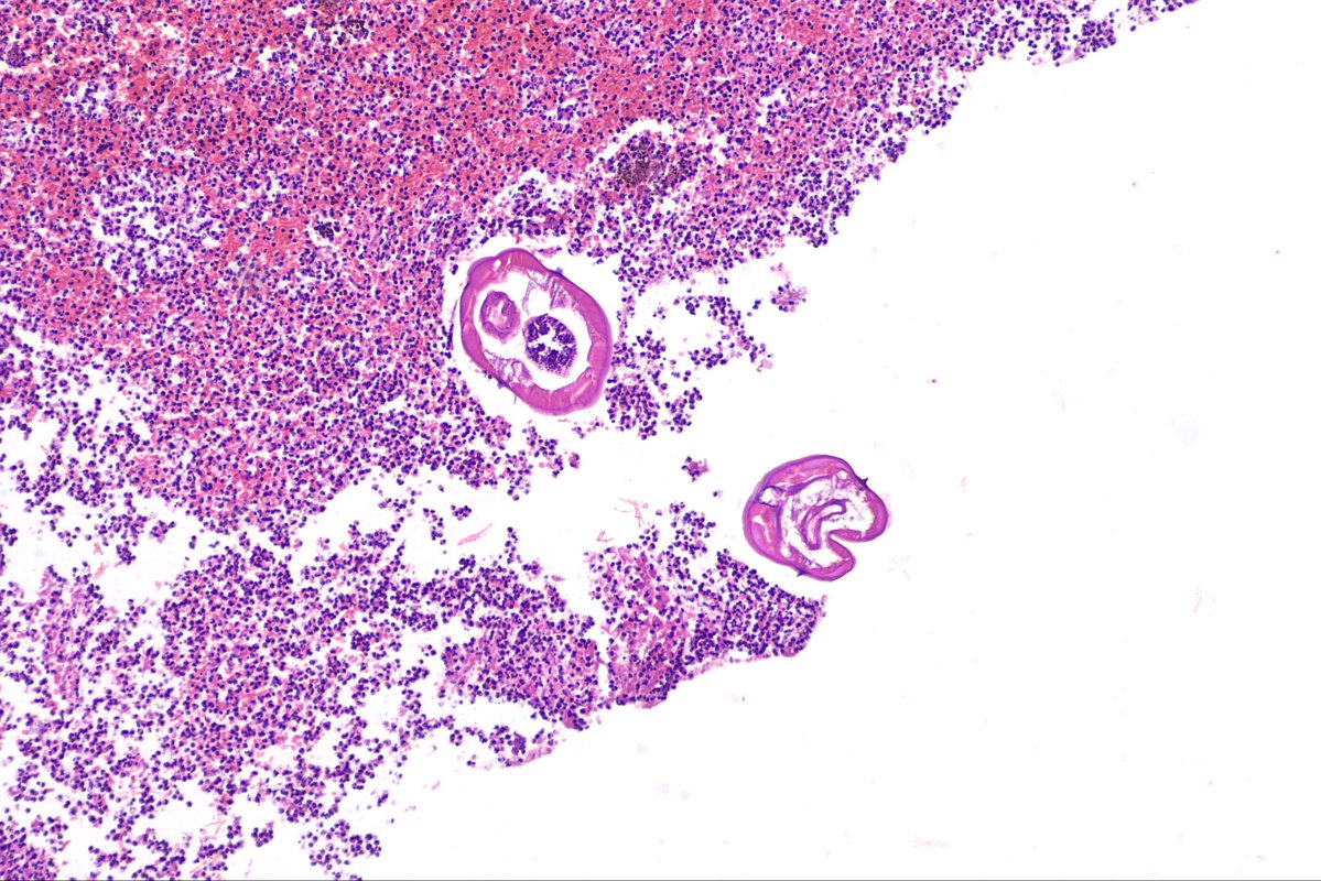
#LiverPath #Pathbugs #Infectiousdisease #GIPath
Interesting contrast with our region: PAHO currently reports no ongoing endemic transmission in Central America. In the Americas, transmission is concentrated mainly in Brazil and Venezuela, with focal transmission in Suriname.
I learned a new thing today: the country with most Schistosomiasis cases in the world is Nigeria
Pic: Schistosoma egg in liver
#pathbugs #crittersontwitter #pathology #liverpath #yalerosenslidecollection



Last Seen Hashtags on Sotwe
Trends for you
Most Popular Users

Elon Musk 
@elonmusk
241.3M followers

Barack Obama
@barackobama
118.8M followers

Cristiano Ronaldo
@cristiano
114.5M followers

Donald J. Trump
@realdonaldtrump
111.9M followers

Narendra Modi
@narendramodi
107M followers

Rihanna
@rihanna
98.7M followers

NASA
@nasa
92.3M followers

Justin Bieber
@justinbieber
91.8M followers

KATY PERRY
@katyperry
90M followers

Taylor Swift
@taylorswift13
84M followers

Lady Gaga
@ladygaga
75.5M followers

Virat Kohli
@imvkohli
73.4M followers

Kim Kardashian
@kimkardashian
70.8M followers

YouTube
@youtube
68.7M followers

Neymar Jr
@neymarjr
66.4M followers

Bill Gates
@billgates
65.2M followers

Selena Gomez
@selenagomez
63.1M followers

The Ellen Show
@theellenshow
62.2M followers

CNN
@cnn
61.7M followers

X
@x
60.7M followers