Patient in Rwanda: Pedestrian struck by vehicle presents to the ED
•Pt. is awake and alert but noted to have hypotension
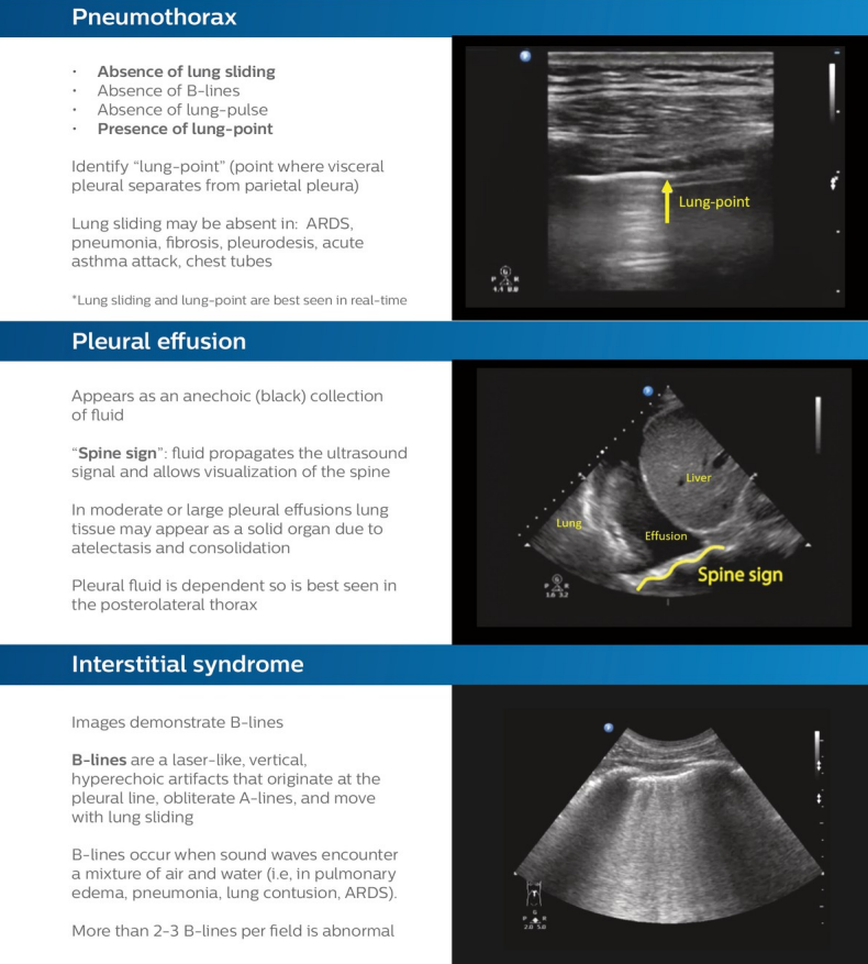
•No free fluid in the abd or pleural space on initial E-FAST
•Cardiac views show pericardial effusion w/ evidence of tamponade physiology
Great case showing how a paramedic used Lumify Reacts in a traumatic out-of-hospital cardiac arrest and was able to collaborate with a hospital based physician using tele-ultrasound. https://t.co/RkNFtvnc1Q
Optimizing the A4C view
• All 4 chambers are seen and the long-axis of the heart is vertical
• Don’t see the atria: transducer is aimed too posterior, tilt face of transducer upwards
• Heart is tilted to the right: move transducer laterally
#POCUS#FOAMed
Optimizing the PSAX view
• The LV should be round and in the center
• If the LV is pear-shaped, try scanning one intercostal space higher
• If the LV is asymmetric, rotate the transducer clockwise or counter-clockwise
#POCUA#FOAMed
Optimizing the PLAX view
• Septum & LV wall should be as parallel & horizontal as possible
• If septum & LV wall are vertical, move transducer up one intercostal space
• If the LV appears “closed,” rotate the transducer
#POCUS
Depth
• Depth increases or decreases the field of view
• Important to have the appropriate depth setting for each view
• Insufficient depth will not display all of the anatomy that is of interest
• Excess depth may not allow the visualization of details needed
#POCUS
Subcostal IVC view
• From subcostal 4-chamber view, rotate transducer 90 degrees counter-clockwise
• Orientation marker at ˜12 o’clock
• Depth 16-24 cm
• Tip: It is important to see the IVC merging into the RA. This will confirm that you are not visualizing the aorta.
#POCUS
Subcostal view
• Patient is supine
• Transducer 2-3 cm below the xyphoid process
• Orientation marker ~3 o’clock
• Direct transducer toward pts chin/left shoulder
• Depth: 16-24 cm
• Tip: Hold transducer palm down to facilitate cephalad angulation of ultrasound beam
Apical 4-chamber view (A4C)
• Transducer on apical impulse
• Orientation marker to pts left (˜3 o’clock)
• Tilt face of transducer up until the u/s beam cuts through long axis of the heart
• Depth: 14-20 cm
See more: https://t.co/jmk48DMVtc.
Parasternal short-axis view (PSAX)–LV level
• From PLAX view, rotate the transducer 90 degrees clockwise
• Orientation marker toward the pts left shoulder (˜2 o’clock)
• Depth: 12-16 cm
• Tilt transducer face slightly downward toward the pts left hip
https://t.co/eNzTZGId2Z.
Parasternal long-axis view (PLAX)
• Transducer in 3rd-4th intercostal space
• Orientation marker toward the patient’s right shoulder (˜10 o’clock)
• Depth 12-16 cm
• For assessment of pericardial and pleural effusions use a depth of 20-24 cm.
https://t.co/cGwuGhoVJg.
See how the use of Lumify Reacts used in the pre-hospital setting affected clinical decision-making for this patient in apparent PEA. https://t.co/W6y5MWjjwh
#POCUS#FOAMed
Teaching #POCUS to new students this fall? @RubbleEM created a great interactive “Intro to ultrasound for students” lecture. Check it out: https://t.co/QII6isixaP.
#FOAMed