This Friday: Kara Gordon, our Director of Neurobiology, will be speaking at an @elixirgensci webinar!
Attend to learn more about our work using hiPSC-derived models to model epilepsy in a dish 🔬🧠
https://t.co/rqcodMPXJA
Please contact us to learn more about how our hiPSC-based screening services can answer your toxicology and drug discovery questions!
https://t.co/e9O1xx3g1s
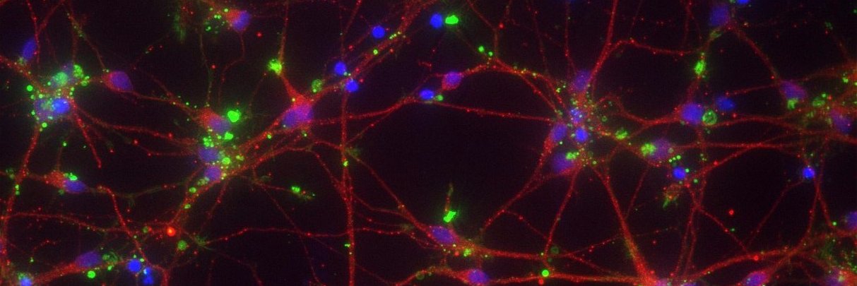
This image shows hiPSC-neural progenitor cells from
@elixirgensci, which we have induced towards neuronal/glial differentiation for 3 weeks. A newly differentiated astrocyte (red) is "hugging" a neuron (green).
CyteSeer, Vala's high-content image analysis platform, finds hundreds of cells in each image and then uses biomarkers like MAP2 and GFAP to discriminate between cell types and to measure cellular morphology and protein expression.
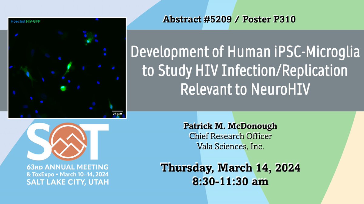
Stop by our poster today, Thursday, March 14 from 8:30-11:30 am at #SOT2024!
Our Chief Research Officer, Pat McDonough is presenting on how we differentiate and infect iPSC-derived microglia to study HIV replication and HIV-associated neurological symptoms.
Our CEO, Jeffrey Price, is at #SOT2024. Stop by our poster today, March 13 from 2:15-4:15 pm!
Learn more about the process of creating a literature-informed knowledgebase on adverse drug reactions and discuss best approaches to drug screens.
Abstract 4410 / Poster Board P197
Our liquid handler streamlines drug treatment and imaging for live cells cultured in 96-well plates. It has adjustable drug dispensing times and rates to accommodate various cell types and experimental designs.
Combined with CyteSeer®, our single-cell video analysis software, Vala's liquid handler can produce accurate, data-rich readouts of drug effects in high throughput. We continue to develop new liquid handler applications, especially for hiPSC-derived neurons and cardiomyocytes.
New video showing our automated liquid handler in action! 🔬
To capture single-cell drug responses in real time, we developed an automated liquid handler for our Kinetic Image Cytometer® high content screening platform.
https://t.co/SqzyTYF0he
CyteSeer® then analyzed the movies to detect single-cell calcium transients at millisecond resolution. Single-cell analysis allowed the authors to identify intermediate reprogrammed states in which calcium transients were rare and/or asynchronous.
Future work can build on this information to further improve reprogramming conditions and to produce cells that can effectively replace mature cardiomyocytes in patients.
The study authors also used KIC® to evaluate the electrophysiological phenotypes of reprogrammed cells by recording high-speed movies of a fluorescent intracellular calcium indicator at 50 frames per second for 60 seconds.
The high throughput of KIC® and CyteSeer® supported fast and accurate testing of reprogramming yield from 24 patients samples using thousands of combinations of overexpressed transcription factors, culture media supplements, and small molecule modulators of signaling pathways.
Using KIC® and CyteSeer®, Vala's automated cell image analysis software, the authors measured single-cell nuclear fluorescence intensity of the cardiomyocyte reporters MYH6 and TNNT2 and then calculated the percentage of cells positive for each reporter in each tested condition.