We are excited to share that our NanoX project has been featured in the Rhein-Neckar-Zeitung @RNZonline! Thank you to Amira Sanli for visiting our laboratories and showcasing our work in the newspaper!
#NanoX#HepatitisE#softxraytomography#cellbiology
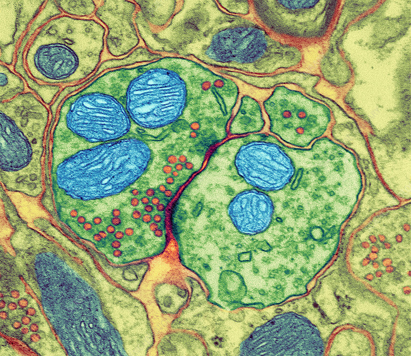
Brain mitochondria are gorgeous! 🥹
Beautiful MitoBrainMap v1.0 collaboration with neuroanatomist, neuropsychologist, and white matter expert @MichelTdS
The brain is full of mitochondria!
But how many, and where?
Different types of mitochondria specialize for energy transformation and other functions
In MitoBrainMap v1.0 we provide the first maps of mitochondrial content and OxPhos capacity across the human brain
Thrilled to announce our latest work on #SXT imaging of biofilms is now published in NPJ Biofilms and Microbiomes!
Read the full publication here: https://t.co/0qW7xCpkY9
A huge thank you to all incredible collaborators who made this research possible! 🙏
I am happy to share our latest preprint "Soft #Xray tomography reveals variations in B.subtilis biofilm structure upon tasA deletion". Amazing work by Anthoula and many thanks to our collaboration partners! Below is a small snapshot of our work, enjoy!
https://t.co/SBmHyB9qRW
We are recruiting a Full Professor in Molecular Biology with a focus on Biological Engineering. Details can be read up in the advertisement on Nature Careers. Please share with researchers who are active in this field.
https://t.co/gxdjgSrldD
Job Alert! Love #microscopy, including the #imaging analysis and data management side? Come join the growing Imaging Unit at the Core Facility Hohenheim, here in Stuttgart! Deadline 31 Dec. https://t.co/hZuj2EcAmK
The discovery of nitrogen-fixing organelles in eukaryotes is one of the breakthroughs of 2024, recognized by @ScienceMagazine. Soft X-ray tomography helps to reshape our understanding of cellular evolution. What a remarkable year for #SXT! https://t.co/q1j3aUIhwR
Tonight great talk for the general public by James Hudspeth of @RockefellerUniv at @DAIHDGermany on the biophysics of hearing. Second chance to hear him talk: coming Tue at 4 pm in the #BioQuant seminar
The recording of the #Virtual#I2K2024 workshop about our #BioImageAnalysis software #CellACDC is now available on YouTube https://t.co/kJEP5CtRt8
Huge thanks to the organisers, this was a very useful conference!
Course alert!
Do you want to learn light microscopy? Registration for the Pasteur course 'Principles and Applications of Fluorescence Microscopy' in March 2025 is open!
https://t.co/lXEwI13gjV
March – 21 March 2025
#PAFM
is back after a two-year break 🤩🌈🔬🪛 👇thread
🔊 Exciting AI Research Opportunity! my team at @czbiohub SF is hiring a Research Scientist to develop foundational AI models for BioImage analysis. Join us to work on cutting-edge ML/CV! 💻🔬🧪🧠 #AIJobs#ML
https://t.co/BIa3VHs9kn
Think of it as a "postdoc on steroids" in AI/ML/CV. You will have access to world-class computing: one of the largest nonprofit #AI clusters (1000x H100)! , unique datasets, and an incredibly supportive collaborative environment!
Do you use 3D imaging like CT or MRI for research, teaching, outreach, or art? Check out our new article in @iScience_CP featuring a detailed workflow for producing 3D cinematic renderings with stunning realism! https://t.co/dcsHTT8TjJ @BIOTECH_UH@HiLIFE_helsinki@helsinkiuni
If you still haven’t had a chance to read our latest publication, here’s a video summarizing our SmARTR pipeline for creating 3D cinematic renderings of imaging data with stunning realism: https://t.co/XOzXbQl2z8 @BIOTECH_UH@HiLIFE_helsinki@helsinkiuni
@LennartHilbert@Cluster3DMM2O@StefanBraese@KITKarlsruhe@UniHeidelberg Dear Lennart, Stefan Bräse, Christian Bräse, and co. are extremely efficient and already have a project website: https://t.co/ofIJ2zD1gh. We are not yet as far. You can read some insights on our project from press releases: https://t.co/iO8WdMx7k6
https://t.co/LEwCpHukAi
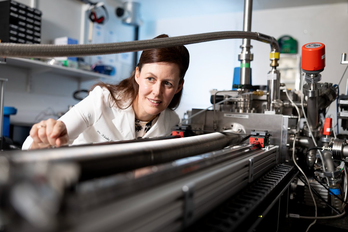
👩🔬🩸 Exciting news! Congratulations to infectious disease expert Assistant Professor Nicola Fletcher, one of only two Ireland-based recipients of the coveted ERC Synergy Grant this year. Her groundbreaking project, NanoX, will receive over €6 million in funding to better understand the pathophysiology of hepatitis E infectWell done, Nicola
🗣️ Professor Kate Robson Brown, UCD Vice President for Research, Innovation and Impact: "Her achievement exemplifies the excellence of our researchers who are building strong international collaborations to drive innovation and address global challenges.”
Soft X-ray microscopy is a new imaging technique that allows the visualisation of cells in great detail, but it has not yet been applied to imaging whole tissues.
NanoX seeks to merge the skillsets of international experts in infectious disease, physics and structural biology to develop new techniques that can image tissue micro-biopsies.
🗣️ Assistant Professor Nicola Fletcher, Ad Astra fellow at UCD School of Veterinary Medicine and Fellow of the UCD Conway Institute: "I am confident that this project will deliver new ways to diagnose and treat a wide range of diseases in humans and animals."
“I’m passionate about One Health, the idea that animal, human and environmental health are all linked, and we must consider all of them when trying to improve the health of anyone. This project fits perfectly within One Health and will benefit all species.”
"The UCD Conway Institute is home to the world’s first commercially available laboratory scale soft x-ray microscope, built and designed by Sirius XT. Together with the University of Heidelberg and the Diamond Light Source in Oxford, we have the state-of-the-art facilities we need to develop new ways to image tissue biopsies in exquisite detail. This will allow us to characterise diseases at the cellular level.”