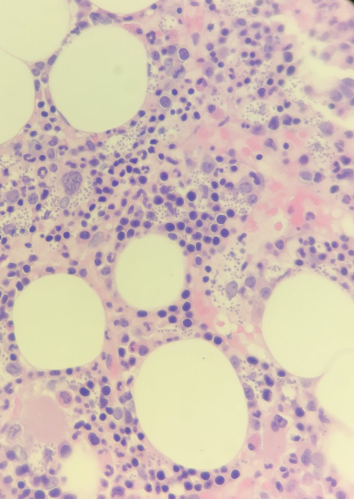
@MahrukhMumtaz9 I'll give it a try..
A- aspirate clot (?)
B- biopsy (H&E)
C- touch imprint
D- bone marrow aspirate
E- unstained bone marrow aspirate
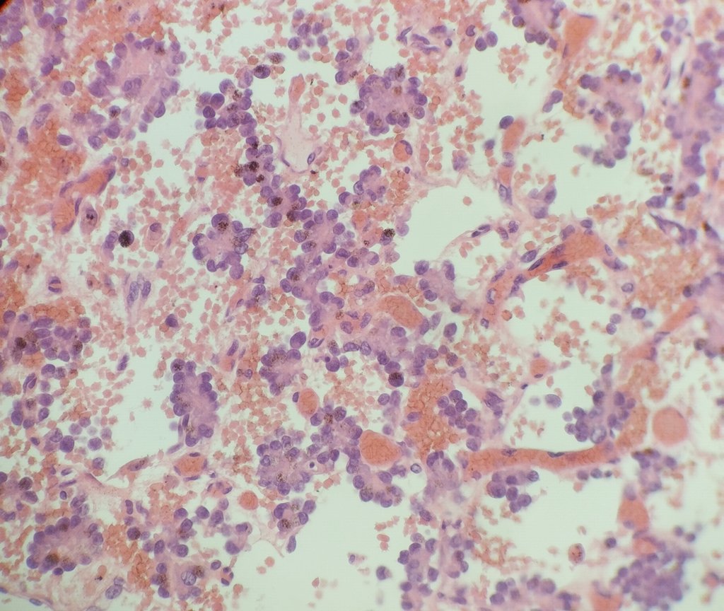
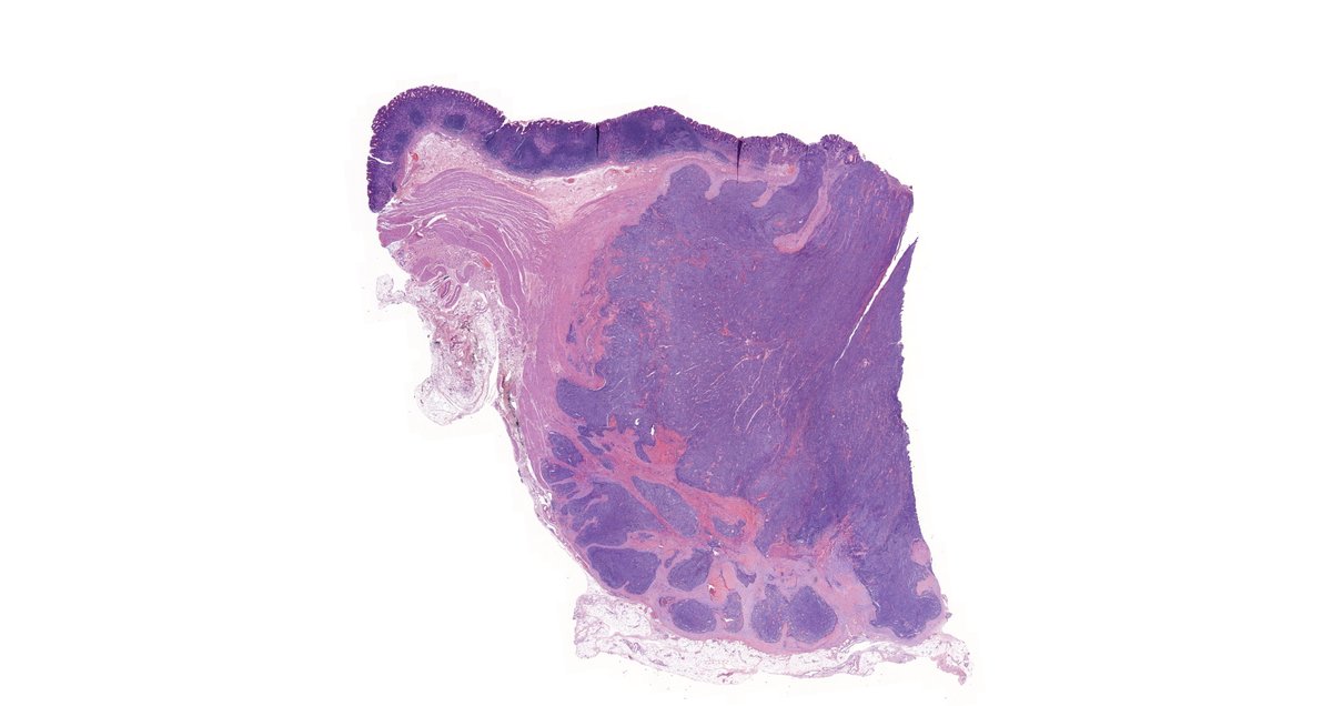
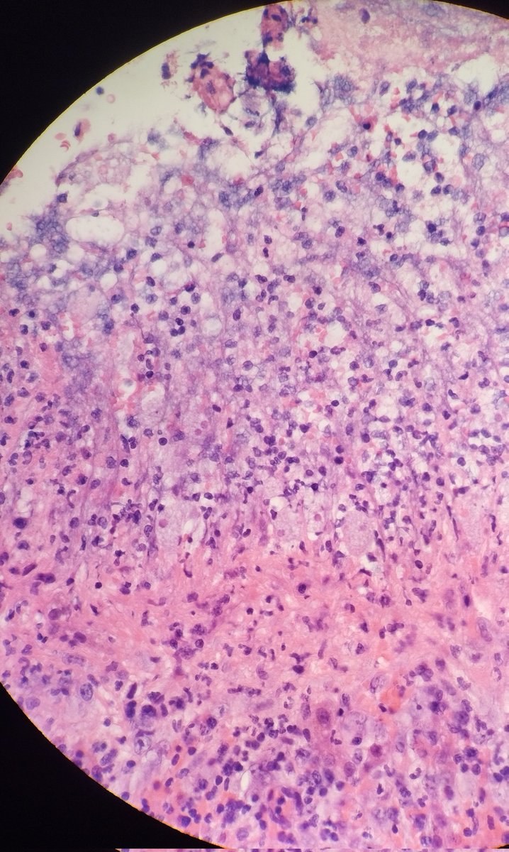
Is this a common pattern observed in treated Ewing sarcomas of the bone?
Viable tumor consisted exclusively of perivascular rosette-forming clusters!
@JMGardnerMD@JLHornick
Here's my first case of appendiceal goblet cell adenocarcinoma! Further sampling revealed cell clusters floating in extracellular mucin (high grade feature).
How many cases have you come across in your practice so far?
@DrGeeONE@MirunaPopescu13@TristanRutland7#pathology
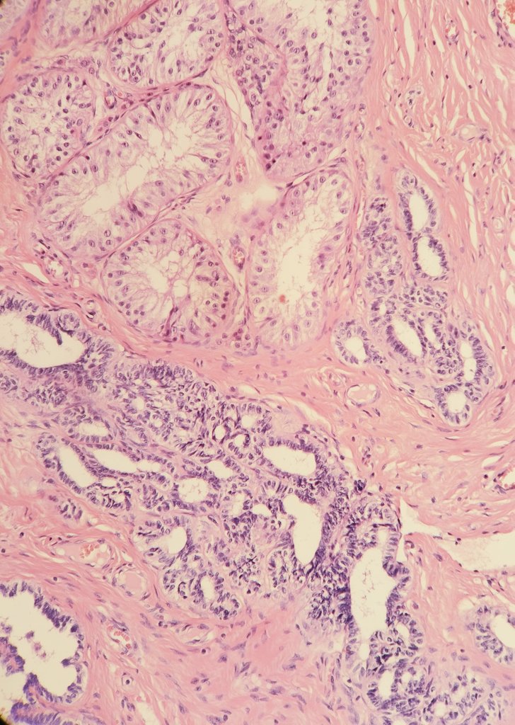
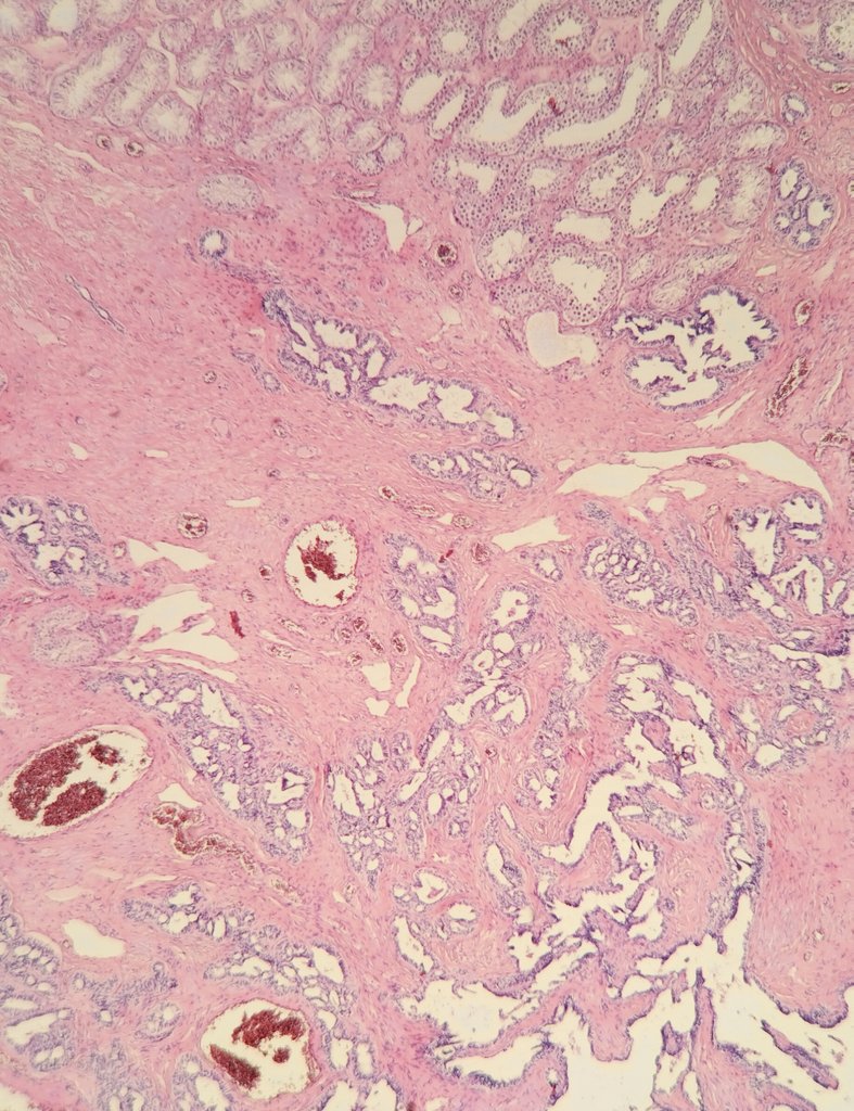
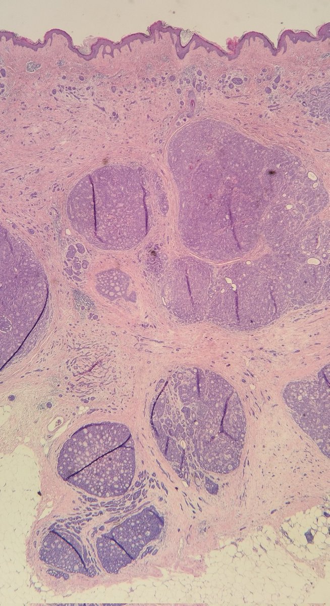
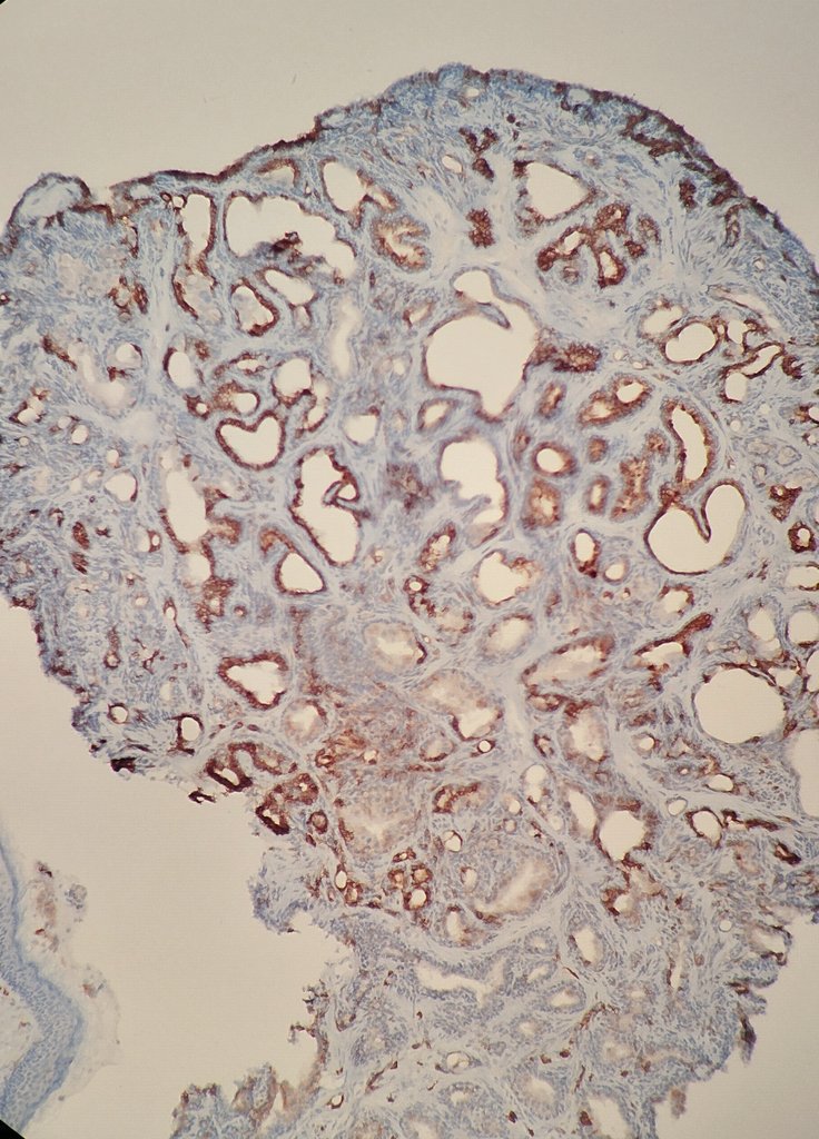
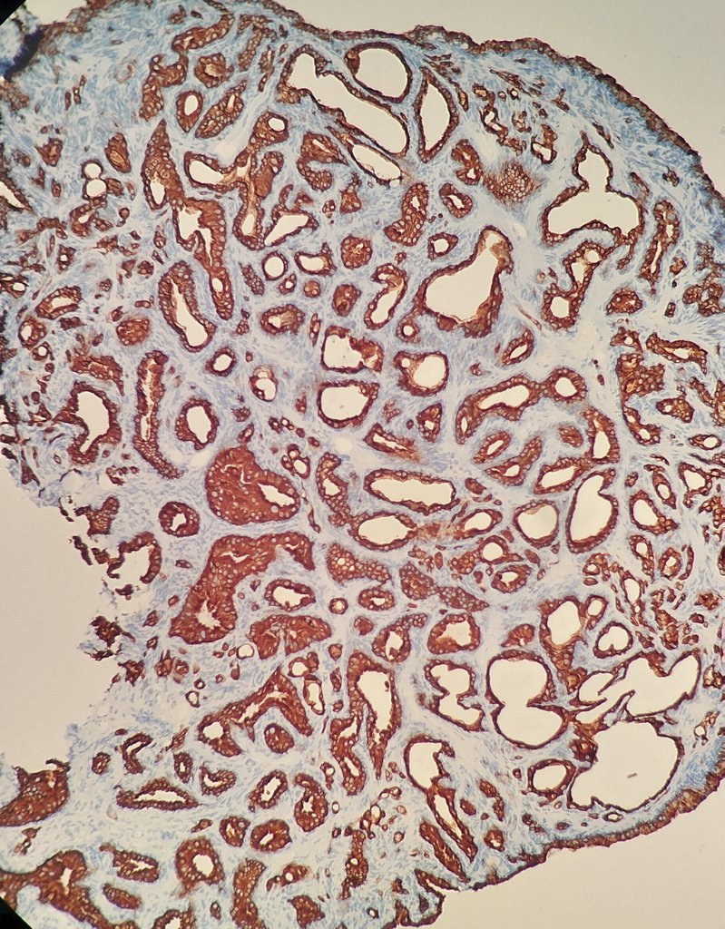
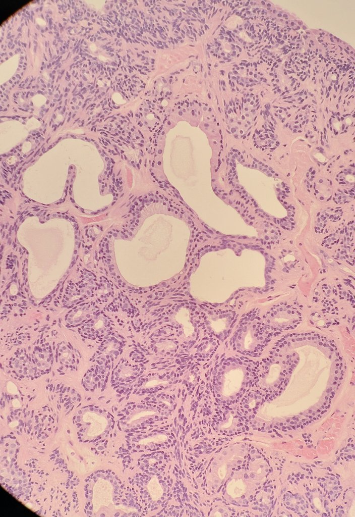
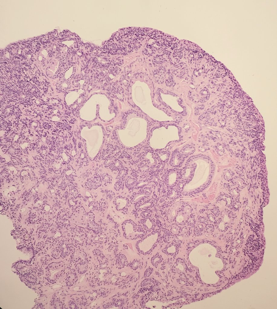
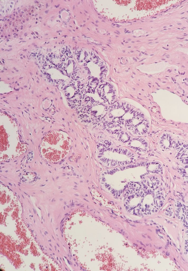
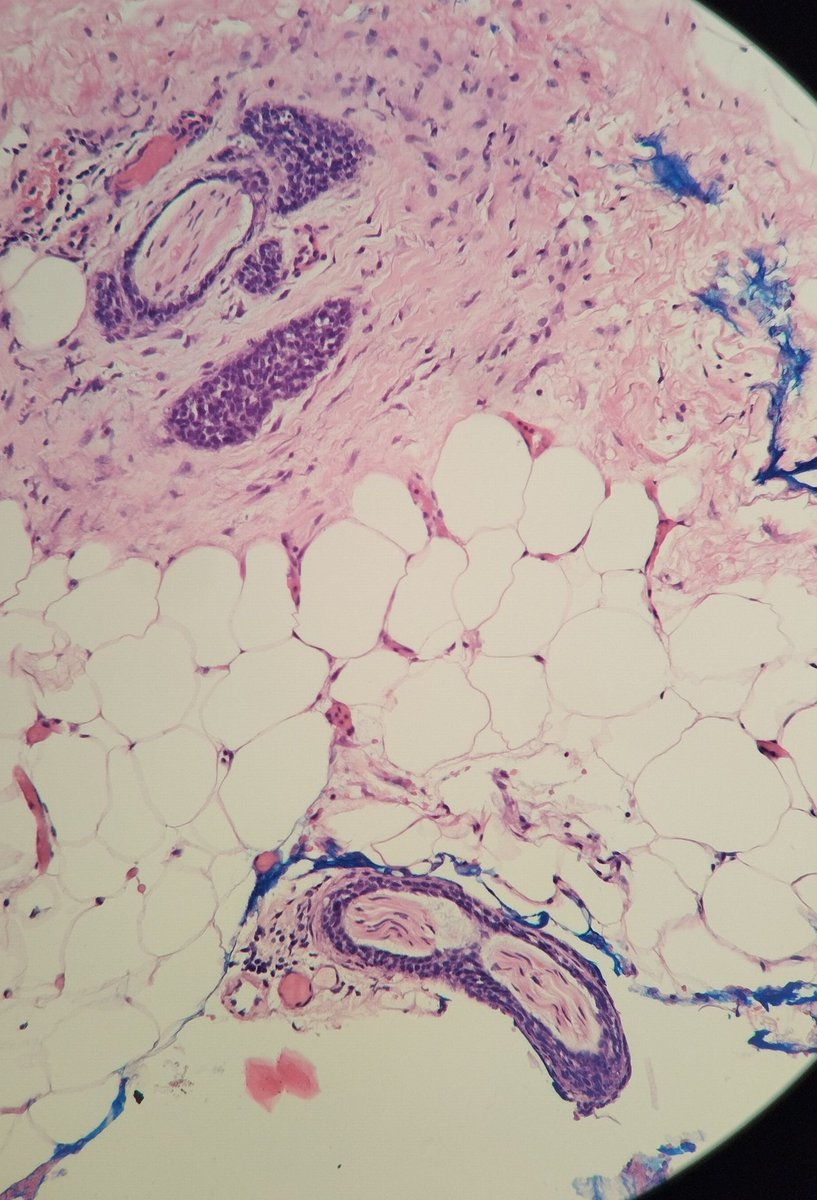
24 yo boy with testicular torsion. Within rete testis columnar cells with cribriform architecture and pseudoinvasive features are found. These findings are suggestive of adenenomatous hyperplasia of rete testis!
#GUpittfals#pathology@DrGeeONE@MirunaPopescu13@tri
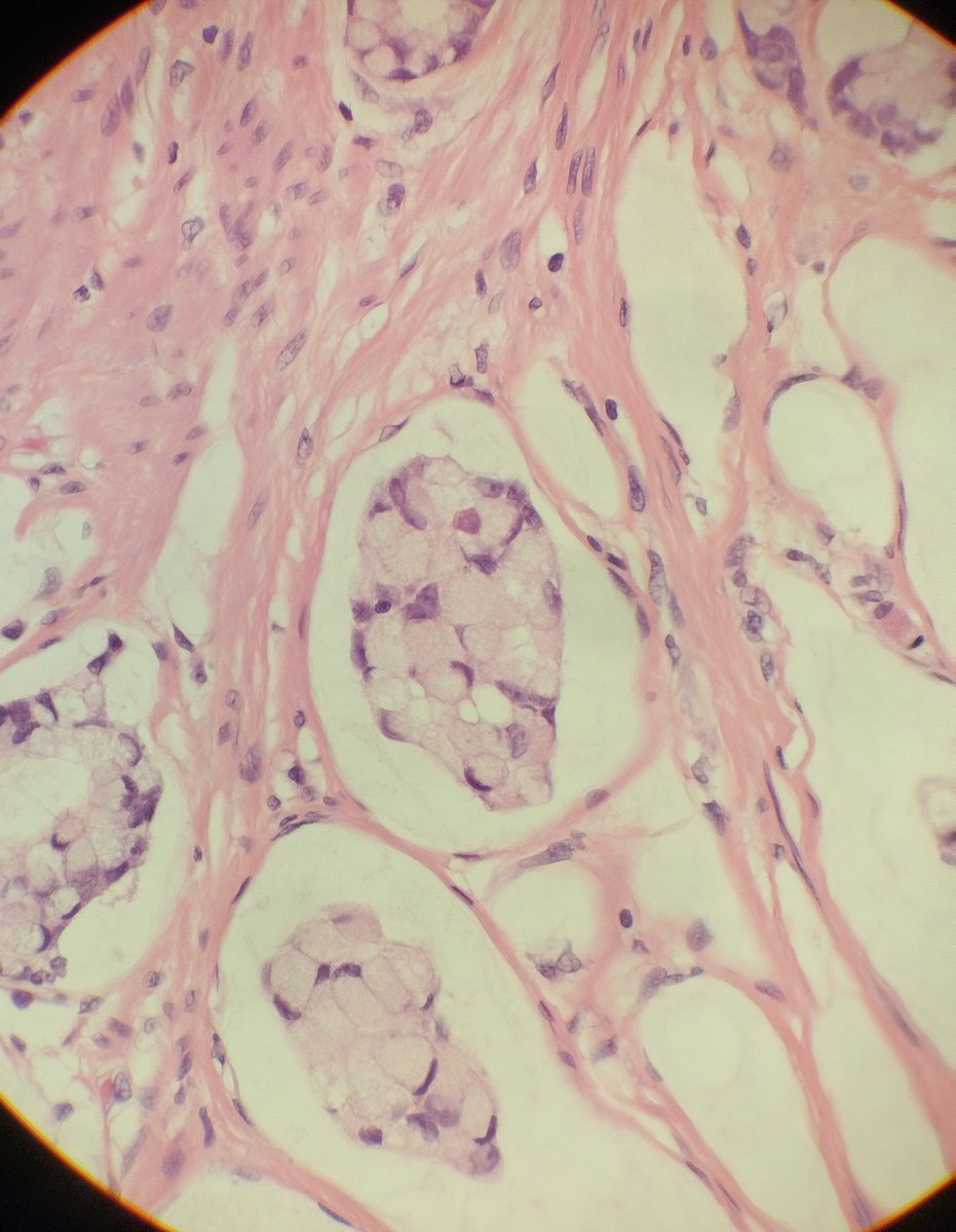
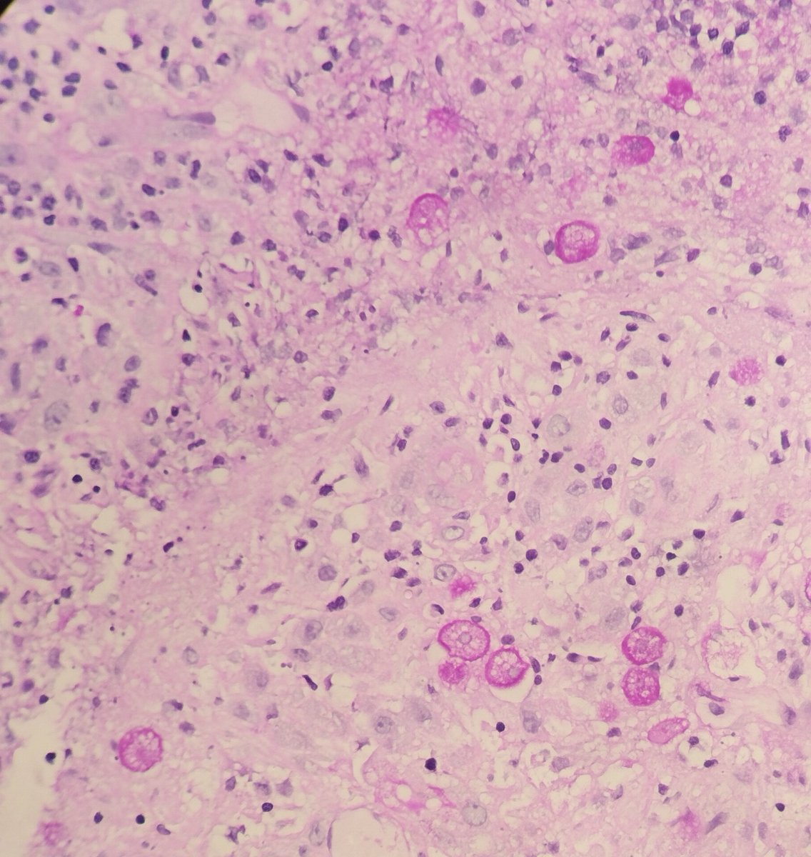
The bizarre cells are PAS positive (shown here) and negative to Grocott stain.
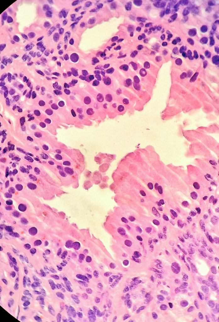
Final diagnosis: amebic liver abscess!
Shown here are Entamoeba histolytica trophozoites with ingested red blood cells
49 yo female patient with hepatic lesions suspected for metastatic colon cancer. Colonic mucosal biopsy reveals normal intestinal epithelium adjacent to necrotic material containing macrophage-like cells 📸
#PathTwitter#pathology@DrGeeONE@TristanRutland7@JMGardnerMD
Resection of the IV hepatic segment: wall of an abscess cavity lined by necrotic-inflammatory tissue containing the same macrophage-like cells. Each element presents with one or multiple eosinophilic globules in the cytoplasm
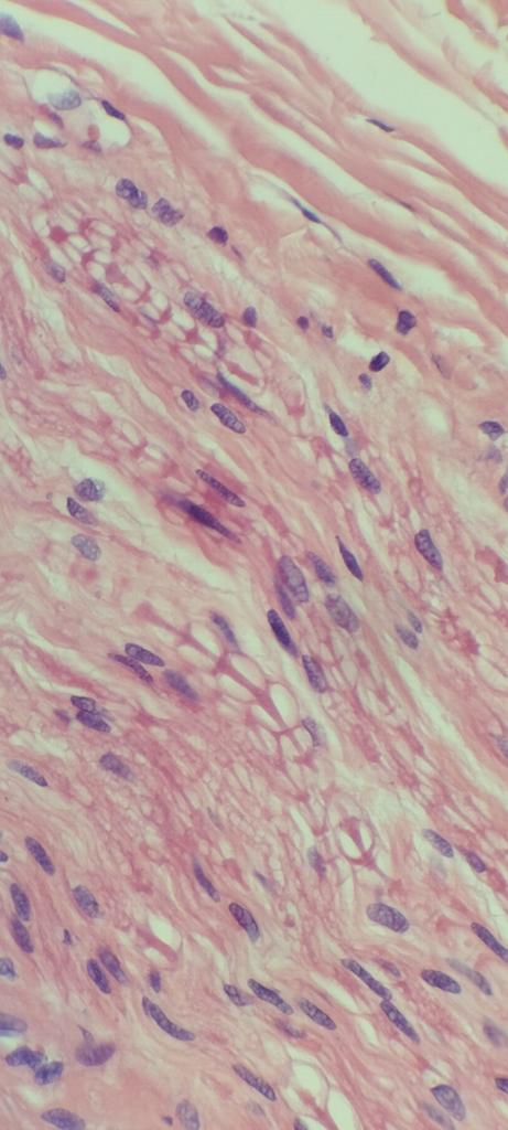
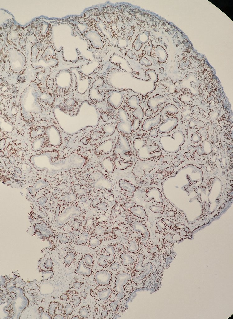
CD20 staining reveals the presence of mature B cells infiltrating the bone marrow, forming an aggregate and perisinusal Indian files, typical of Marginal Zone Lymphoma (MZL).
@JMGardnerMD@TristanRutland7 @AaronGoodman33 @hemepathguy#Pathology#PathTwitter