Okay, this is genuinely impressive.
I asked GPT-6 Astra to help me understand my own ankle pain. It gave me a full interactive 3D atlas — bones, ligaments, tendons, real motion axes, sliders for plantarflexion and inversion, and a live readout of what each ligament is doing.
One session.
Here's another exploration of what GPT-6 Astra can do with 3D human anatomy. Exploding view of the right upper extremity!
Check out yesterday's post for a carpal tunnel release surgical demo.
Try it here: https://t.co/VnNEokSCbR
1/Have MULTIPLE questions about the new criteria for MULTIPLE sclerosis?
Do you know the NEW McDonald criteria for multiple sclerosis?
This month’s @theAJNR SCANtastic has the lastest you need to know!
https://t.co/7QhXYeQP4n
Spotter of the Day - September 2026 - Question 7
History: A 70-year-old female presents with right upper quadrant pain associated with fever and leucocytosis. She reports that the discomfort has gradually worsened, and she notes reduced appetite over the past several days.
What is the diagnosis?
Check the full case here 🔗 https://t.co/chPEol7nHC
DM us "DAILY" if you want FREE access to the spotter subscription.
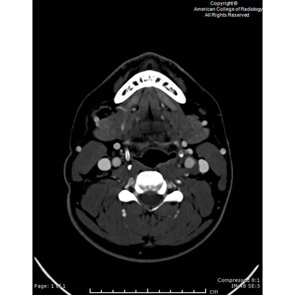
A 27-year-old male presents with acute onset of sharp pain along the left superior neck, beginning 5 days prior. #ACRCaseinPoint https://t.co/5cqdGEDRvX
🦴 How well do you know the vertebral column?
Which region of the spine contains the greatest number of vertebrae?
A. Cervical spine
B. Thoracic spine
C. Lumbar spine
D. Sacrum
The vertebral column is divided into cervical, thoracic, lumbar, sacral, and coccygeal regions. Together, these regions provide structural support, protect the spinal cord, and permit movement.
Post your answer in the comments! 👇
#Anatomy #SpineAnatomy #VertebralColumn #MusculoskeletalEducation #MedicalEducation
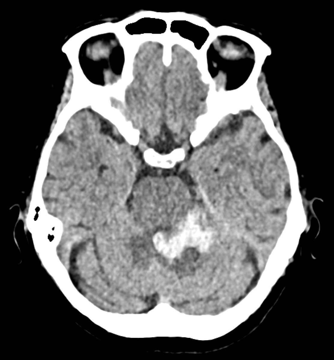
Acute non-traumatic cerebellar haemorrhage in a young patient. Nothing seen on the CT angiogram or the post-contrast MRI, but look at the ASL...
There's high signal in the occipital sinus; this is not normal. If veins light up on ASL, that means labelled arterial blood got there too fast, which usually means it skipped the capillary bed. This implies arteriovenous shunting!
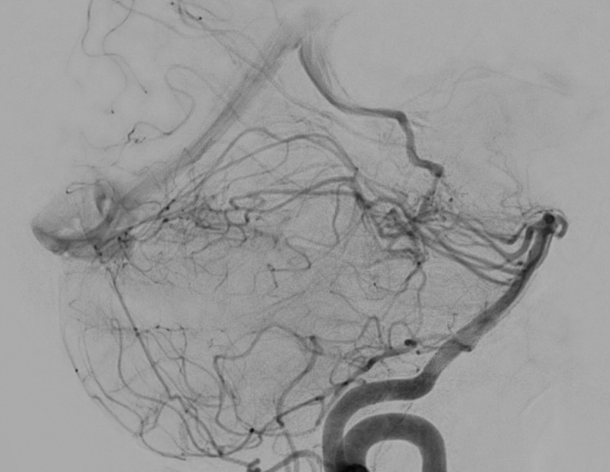
Lo and behold, the DSA shows a left perimesencephalic arteriovenous malformation supplied by basilar perforators and branches from the left superior cerebellar artery, draining into the left basal vein, vein of Galen, and straight sinus.