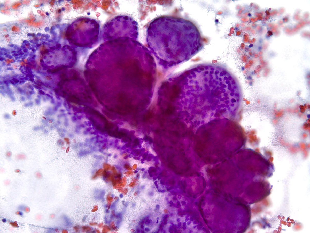
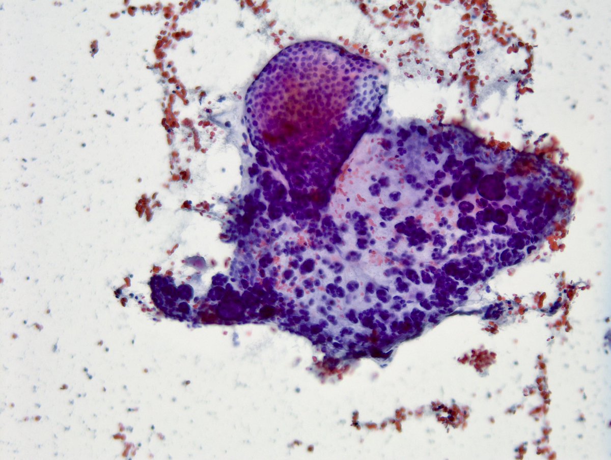
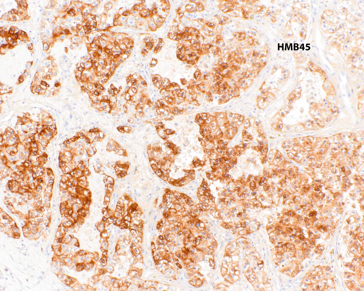
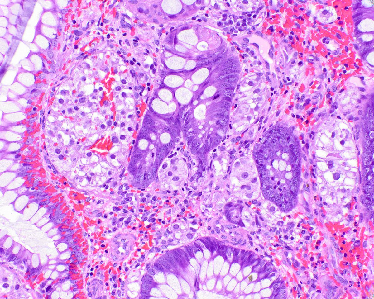
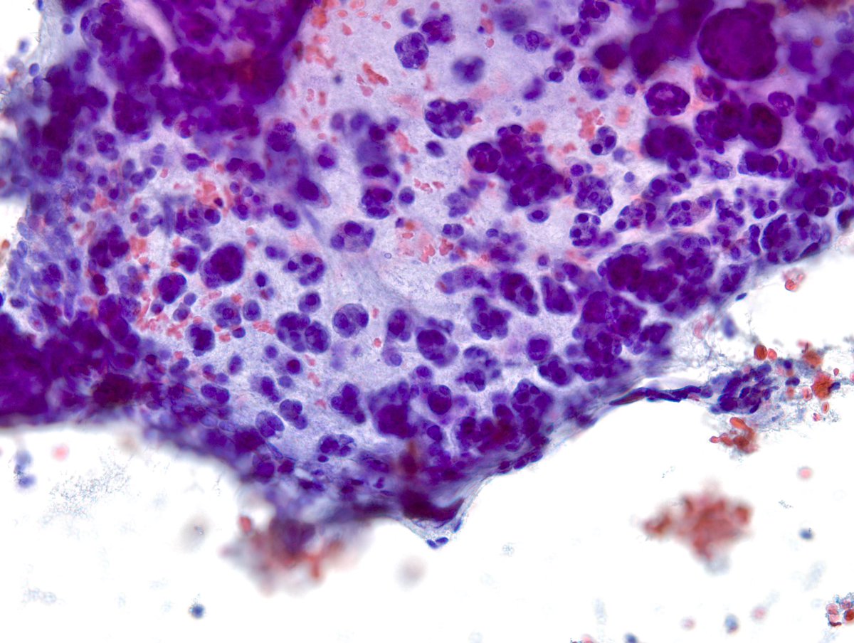
“The Sneakiest Carcinoma” to diagnose in the breast is “Lobular”, especially in a limited cellularity aspirate. Most of the cells/nuclei seen on the left are benign except for a cluster or two (esp in the middle). Same cells on a different pass.
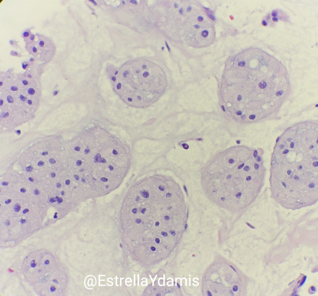
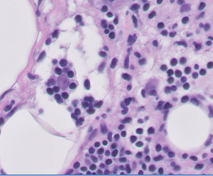
A beautiful example of “hypervacuolization” of malignant epithelial cells in a “Papillary Thyroid Carcinoma, Cystic Variant”. Don’t disregard them as simply histiocytic.
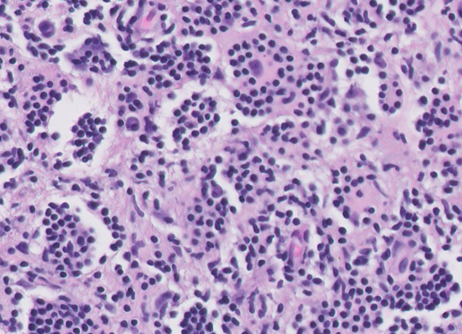
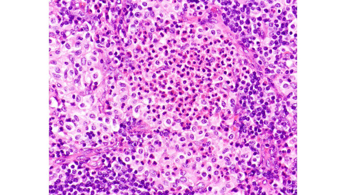
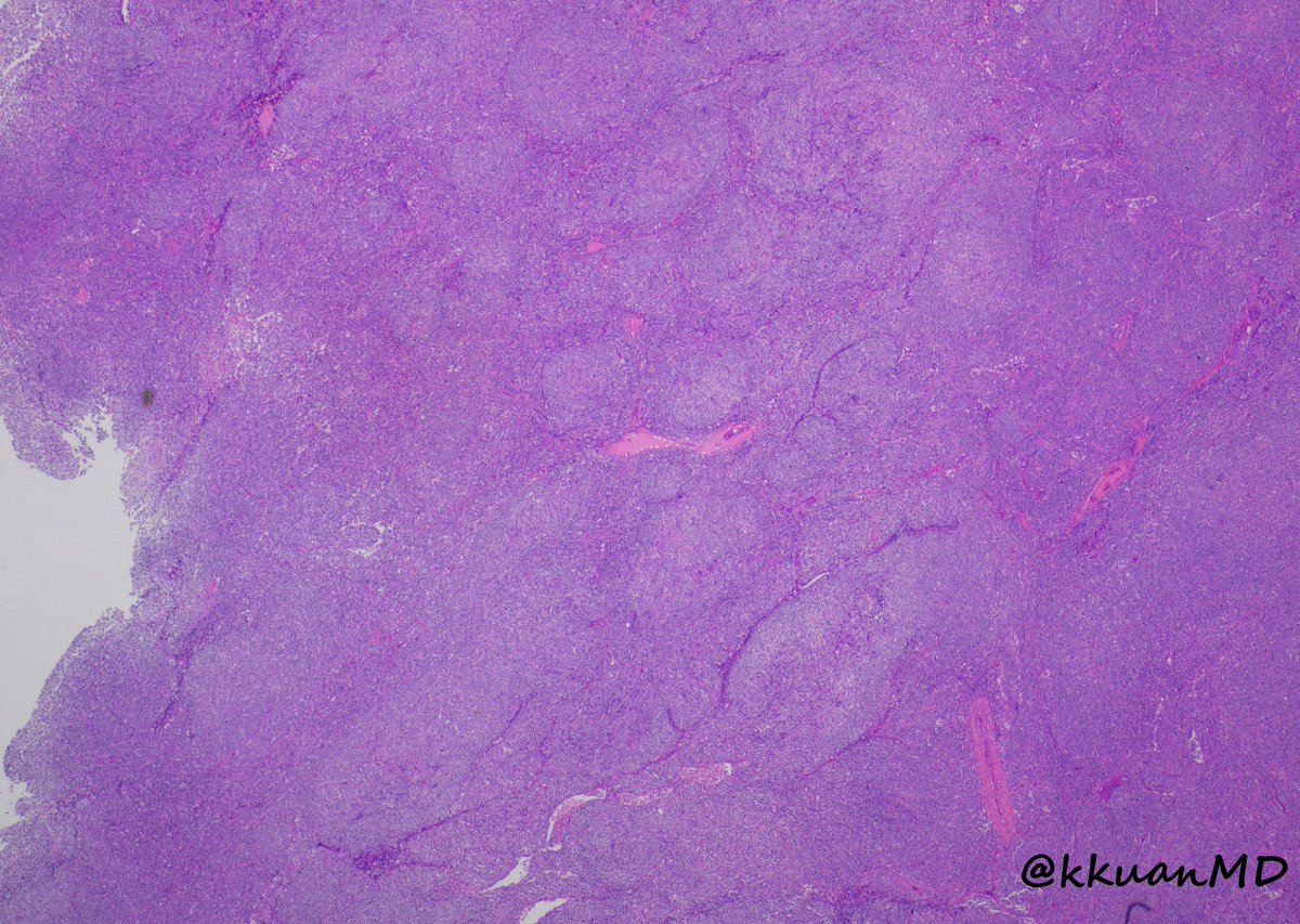
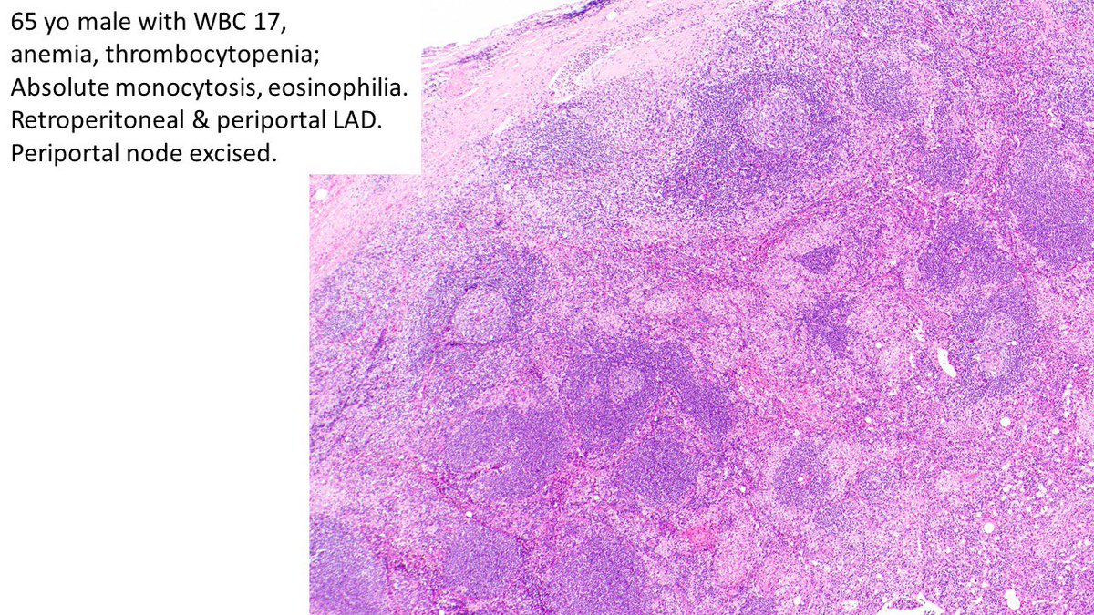
Pathologists! What’s your Dx for this periportal LN from older man with leukocytosis, anemia, thrombocytopenia, absolute monocytosis, eosinophilia and lymphadenopathy? What other work up is warranted? #hemepath
"The Scorpion and the Ants" - Thyroid FNA can be extremely challenging when facing the dilemma of "Medullary CA versus Hurthle Cell Neo". This case, despite hypercellularity, single cells and lack of nucleolar prominence, turned out to be "Hurthle Cell CA" on resection.
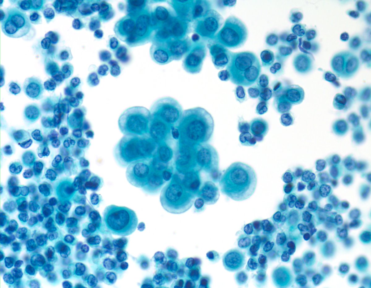
"Deviations" from usual/traditional morphology are not infrequent in Cytopathology. I always rely on the presence of discrete "macronucleoli" when diagnosing "Mesothelioma" in effusions. This case of pleural mesothelioma however appears so innocuous, lacking that feature.
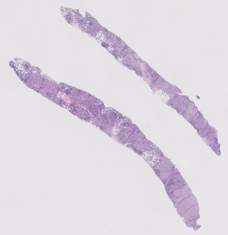
Nice liver biopsy in alpha-1 antitrypsin deficiency disease. I did not spot them initially on the H&E, but after seeing the PAS/D, I found a few candidates. Oops.
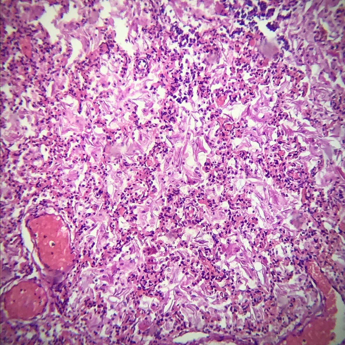
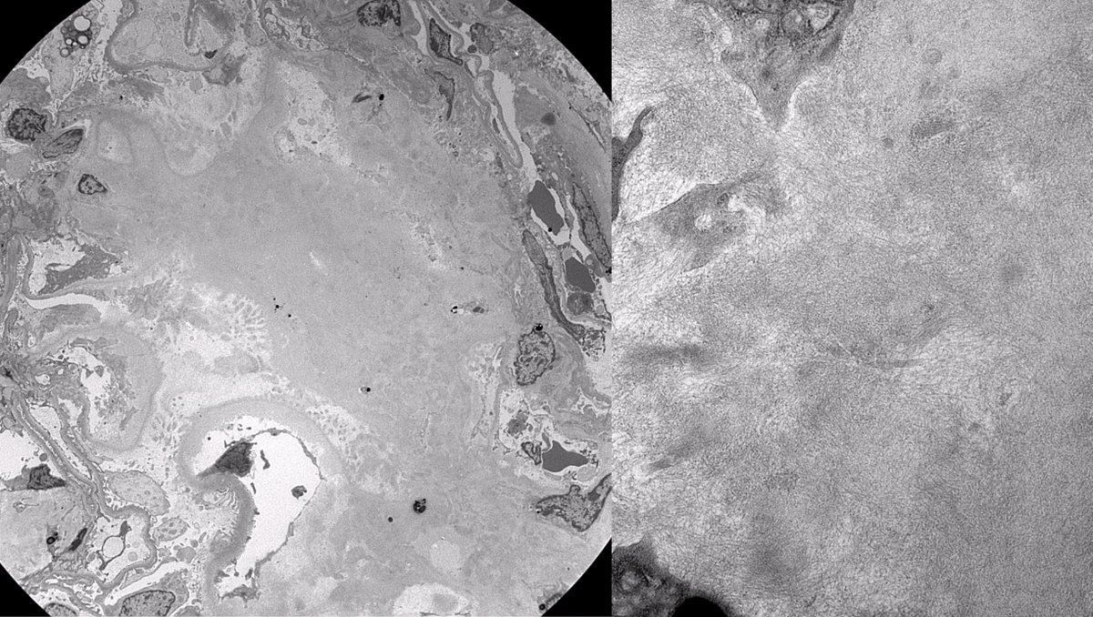
Patient with seronegative spondyloarthropathy with new onset nephrotic syndrome and hematuria. Renal bx demonstrates AA amyloidosis. Often seen in the setting of chronic inflammatory conditions. #renalpath
IMPORTANT Teaching Point - It’s NOT uncommon to have focal microfollicular (MF) proliferation (image#1) with rare colloid in “Benign” Thyroid FNAs. This “Adenomatoid Nodule” on another pass clearly shows macrofollicles. I look for at least 50% MFs for FN/SFN diagnosis.
When phenotypic “kinship or resemblance” changes markedly between the met and the primary (happens often in exfoliative effusion cytology). Impossible to call this case metastatic “Colonic Adenocarcinoma”, if not for the history and IHCs.
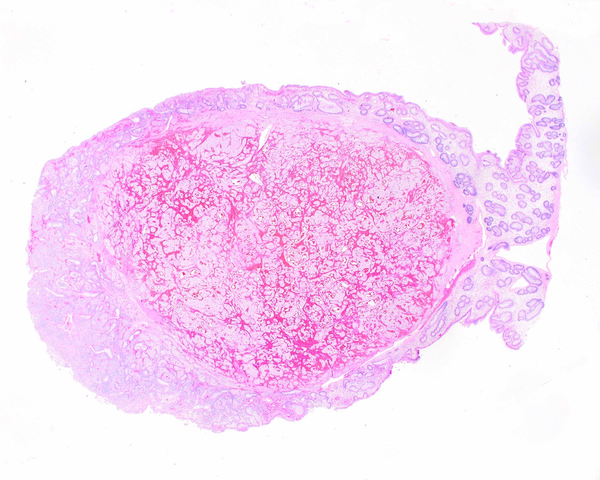
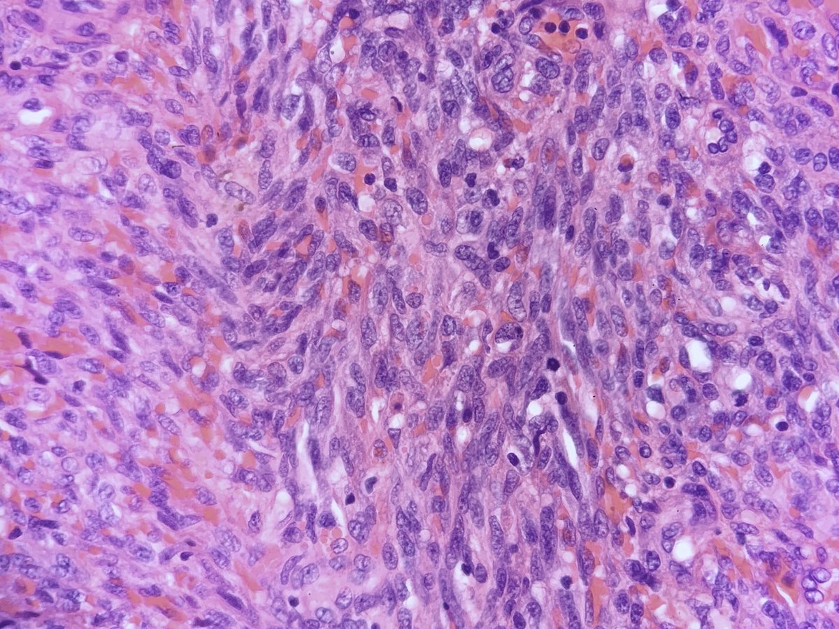
Mucosal proliferation of bland spindle cells, occasionally associated with serrated polyps, benign. Spindle cells positive for EMA, negative for S100, CD117/DOG1. Dx: Perineurioma (aka benign fibroblastic polyp). #GIPath#Surgpath#histology
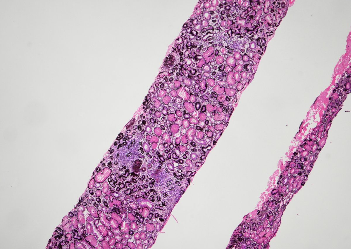
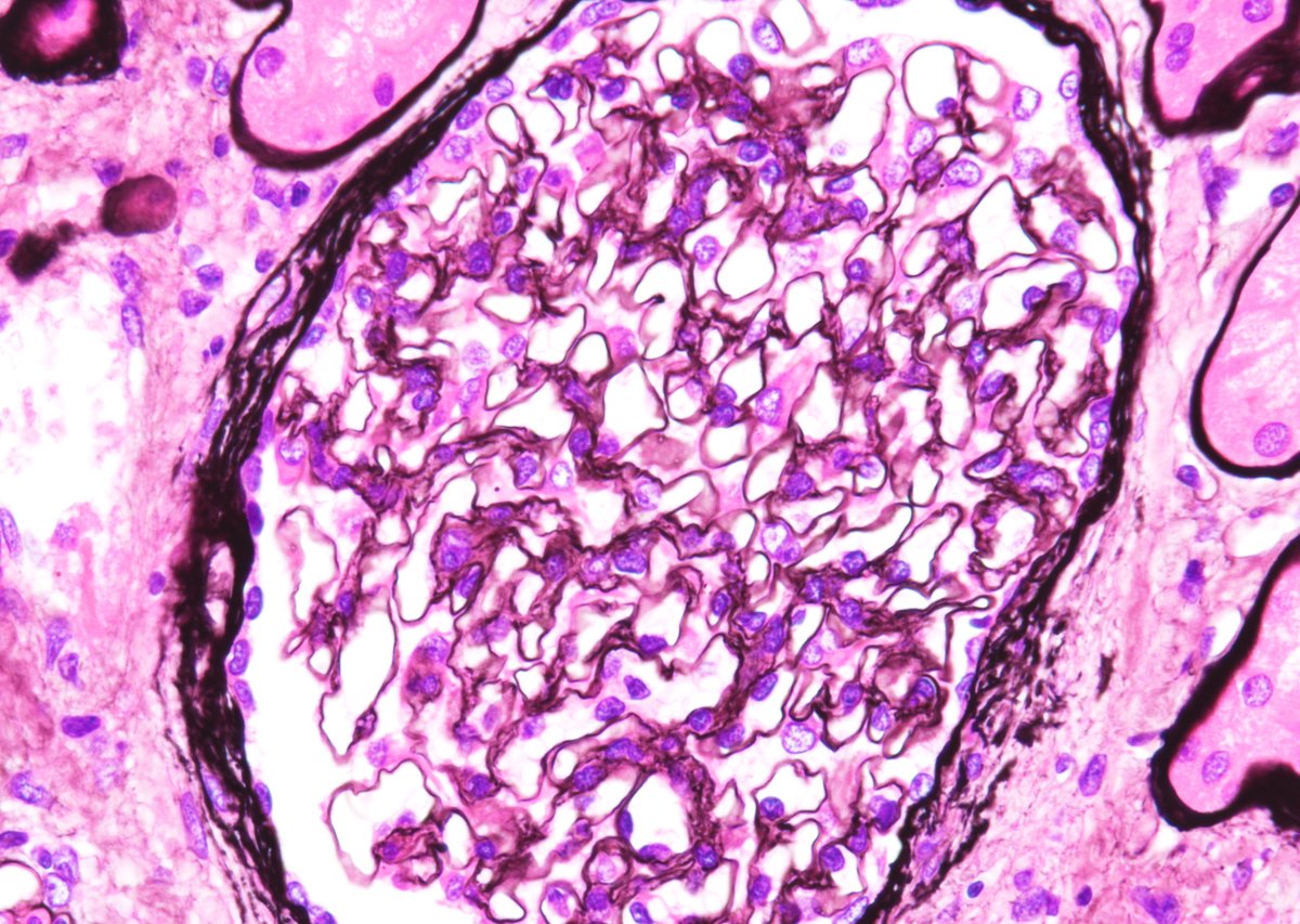
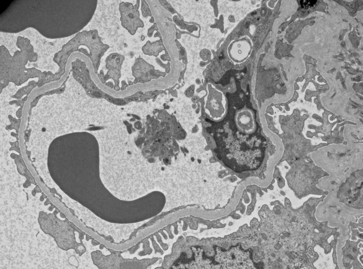
Struck down by steroids: 25 yo male with history of anabolic steroid use for over 5 years, bx'ed for proteinuria and worsening renal function. Advanced FSGS with severe parenchymal scarring (45% global and 40% segmental sclerosis), glomerulomegaly, partial FPE. JUST SAY NO!!!!
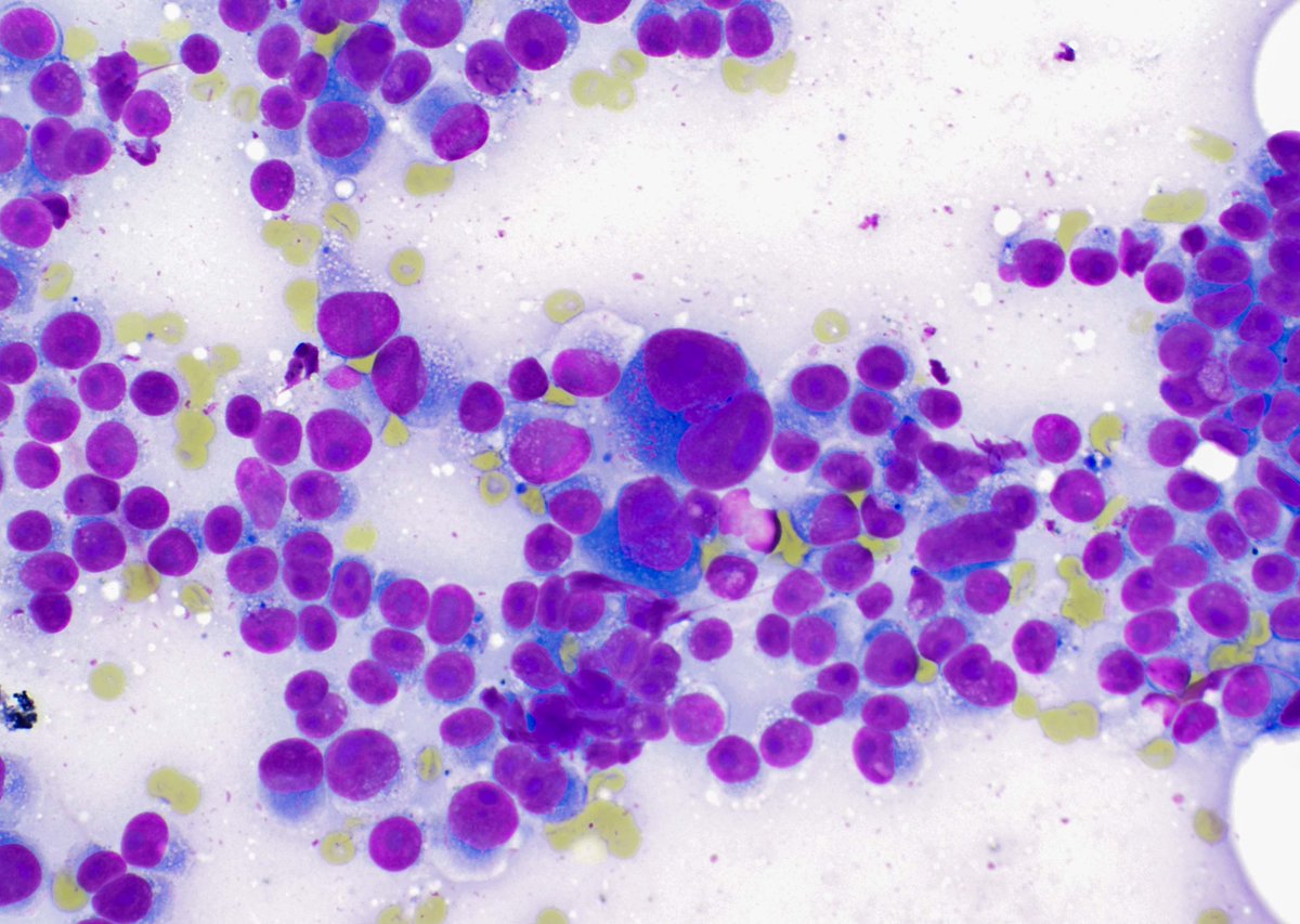
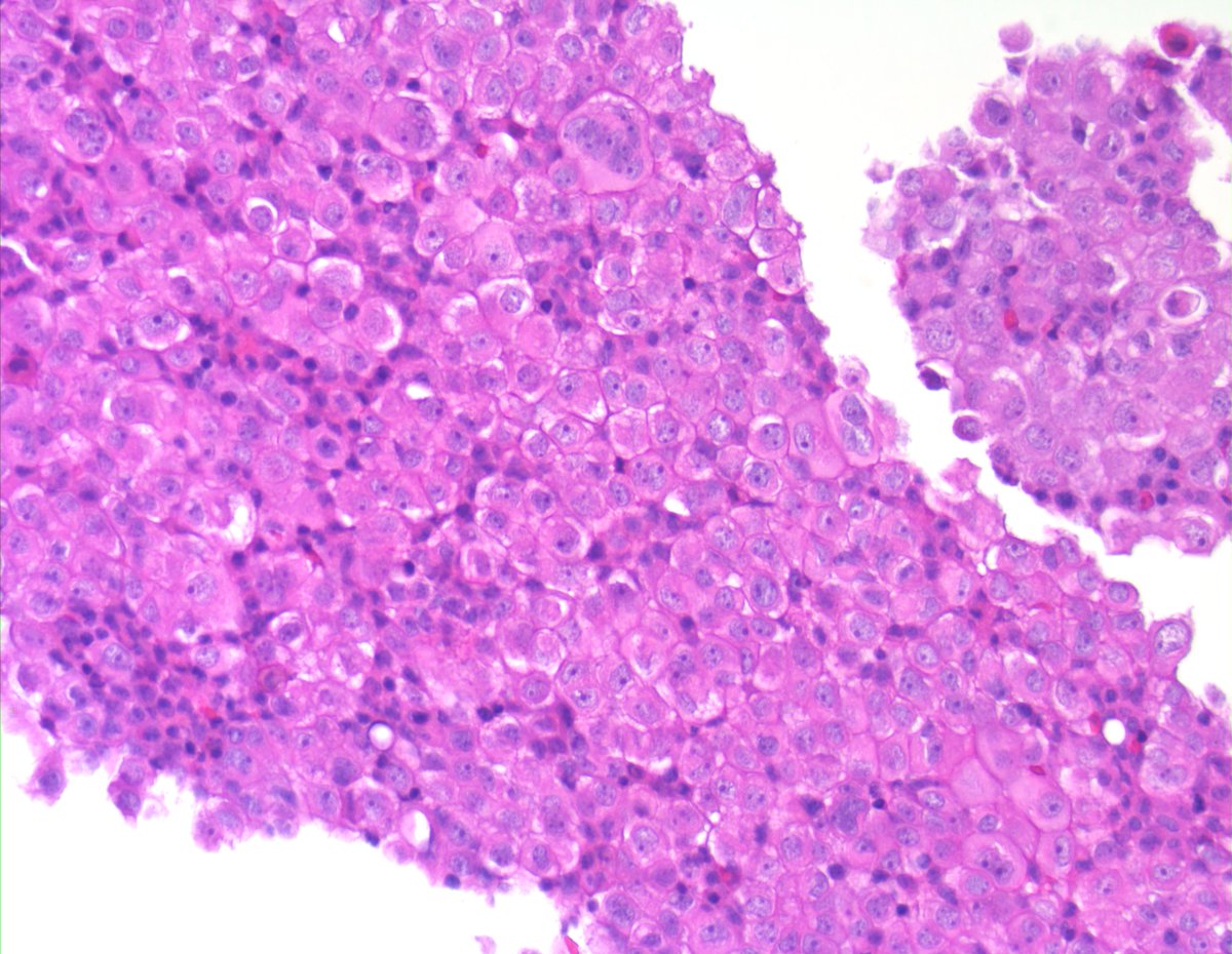
Dispersed melanoma cells in an FNA specimen. Eccentrically placed nuclei with very, very large nucleoli. Some with abundant granular and vacuolated cytoplasm. Occasional binucleate forms. Significant nuclear size variation. #cytology#cytopath