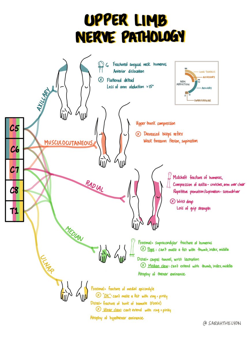
Ulnar nerve distal motor branches beautifully illustrated 👍
👉Flexor digiti minimi brevis does not typically have a paired longus muscle in humans. If present, the flexor digiti minimi longus is considered an anatomical variant.
This wrist diagram illustrates the arrangement of the carpal bones and highlights the Scapholunate (S-L) ligament, which plays a critical role in wrist stability. Key points:
Bones Involved:Scaphoid: Located on the radial side, connecting with the thumb.
Lunate: Central carpal bone that articulates with the radius.
S-L Ligament: Connects the scaphoid and lunate bones.
Vital for wrist mechanics and load distribution.
Clinical Significance: Injury to the S-L ligament can cause scapholunate dissociation, which, if untreated, can lead to instability and long-term arthritis.
This diagram is essential for understanding wrist anatomy and recognising pathologies associated with ligament injuries.
Anatomy and function of rotator cuff muscles
✅Anterior view👉🏼subscapularis
✅Posterior view 👉🏼infraspinatus, supraspinatus, teres minor
✅Lateral view👉🏼all four muscles and their anatomical relation to glenoid fossa
#shoulder#anatomy#rotatorcuff
https://t.co/wwildLCyqh
Rotator cuff (RC): a complex of 4 muscles/tendons wrapping the humeral head:
SSP👉Supraspinatus
ISP👉Infraspinatus
TM👉Teres Minor
SSC👉Subscapularis
RC tear👉the most common source of shoulder pain/disability in people >50 years
#pain#shoulder
https://t.co/clPlh4fH54
How does years of experience influence the way in which hand surgeons diagnose carpal tunnel syndrome? Find out here:
article: https://t.co/iwGTPdN6nG
podcast: https://t.co/8Yq4XUSRb4
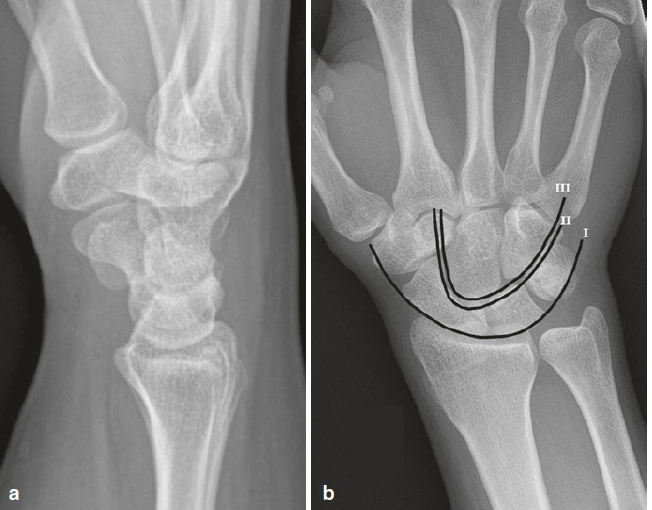
Normal wrist radiographs. (a) On the lateral view, a straight line can be drawn through the radius, lunate, and capitate. (b) On the frontal view, look for the three Gilula arcs.