New article out now #AIMMhighlights
▶️ IHC p53 in NordiQC: assay recalibration needed ▶️ Overexpression is easy; wild-type and absence are not ▶️ 1,796 assays (2007–2024) show frequent misses of key p53 patterns
@IHC_NordiQC@DrRasmusRoge@AndersonKristiB
🔵 IHC p53 performance in NordiQC: recalibration of IHC assays is needed
🔵 Overexpression is easy; wild-type and absence are not
🔵 Data from 1,796 assays (2007–2024) show that IHC assays frequently miss critical p53 patterns
@IHC_NordiQC@DrRasmusRoge@AndersonKristiB
Check out our Current Issue!!
Figure from "Repurposing HER2 IHC: Pan-HER2 and Low-HER2” by Megan L. Troxell, MD, PhD @StanfordPath
🔗 Read the full article here: https://t.co/qqjEzE2Tot
Our case report is out :) To the best of our knowledge, this is the first case report describing a benign leiomyoma forming a fistula between the uterus and the abdominal surface.
See how placental MRI may improve antenatal identification of SGA pregnancies in the future.
Here is our newest paper:
https://t.co/mWTEwwj4ua
#placenta#MRI#research
What a fantastic start to the year 2020!
The hard work has paid off and our article about placental diffusion-weighted MRI correlations with placental histology is now available online!
Fingers crossed that 2020 holds the trend!
https://t.co/9hASS3cP4I
#Pathologists- This ovary is replaced by fleshy tan tissue with yellow necrotic foci. Micro shows cellular tumor with cord-like and single file growth mimicking breast Ca. But it’s relapsed B lymphoblastic leukemia. Lymphoma & leukemia in ovaries can have this pattern. Look out!
Spot on everyone! 🔬
Moderately-Differentiated Sertoli-Leydig Cell Tumor
• Young women (often <30)
• Virilization in ~50%
• Sporadic or germline DICER1 mutations
• Lobulated at LP
• Nests and poorly-formed tubules of Sertoli cells
• Leydig cells in nests or single cells
1/2
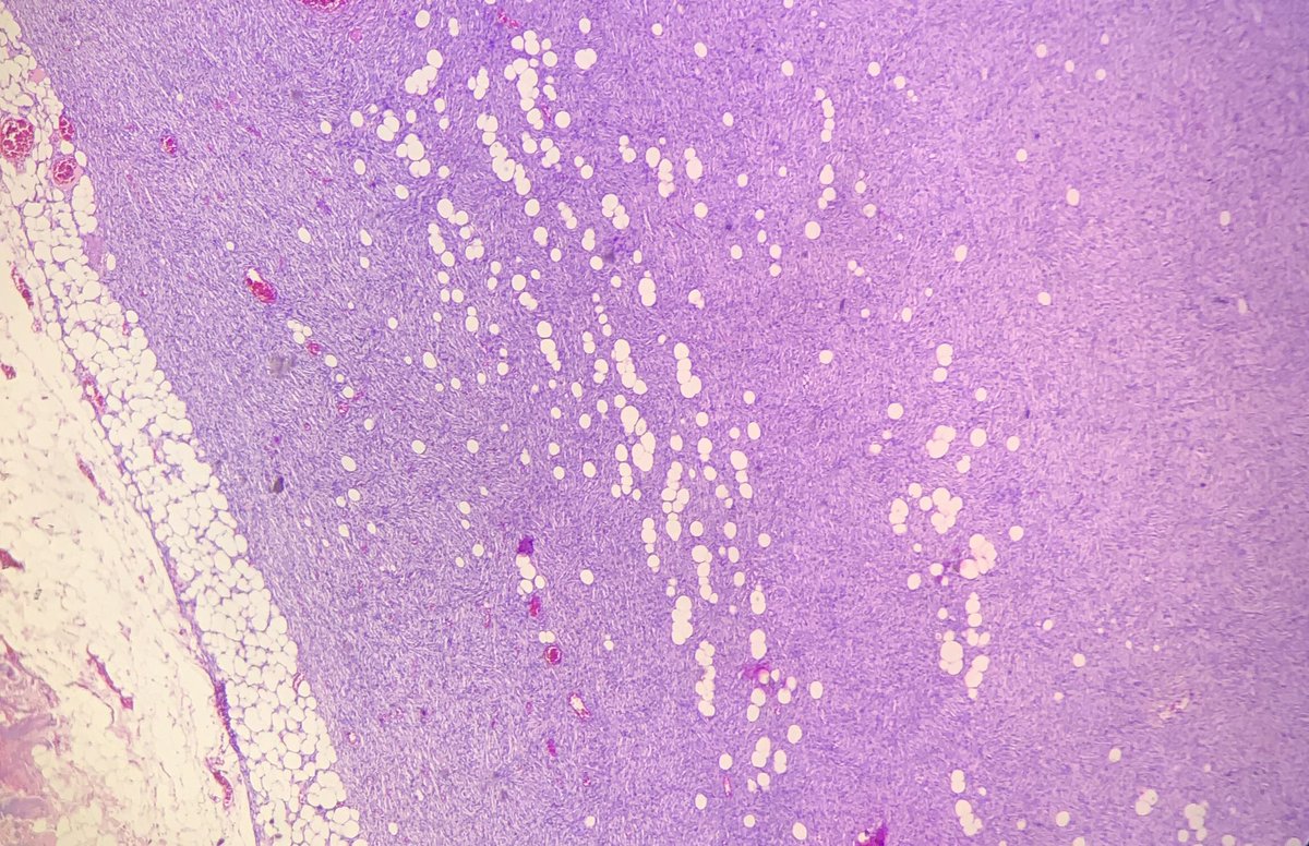
Middle aged female with soft tissue chest mass of 4 years duration.
🔅Giant Cell Tumour of Soft Tissue
-Circumscribed tumour comprising mononuclear stromal cells, osteoclast like giant cells and central area of metaplastic bone
@HENRYY_MD@DrFNA#BSTpath#Pathology
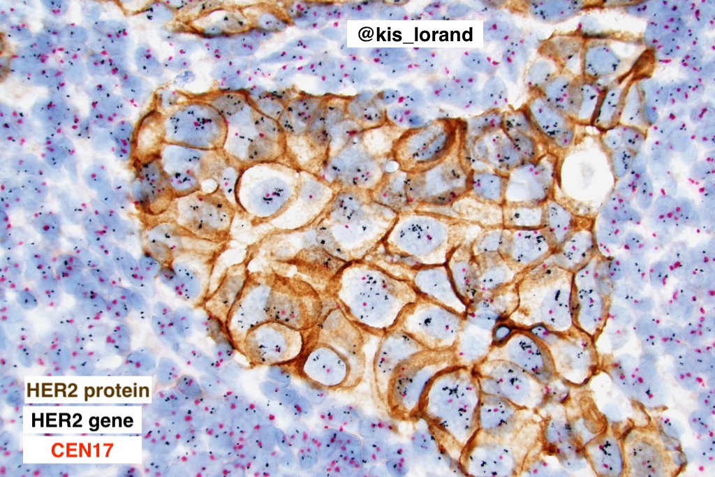
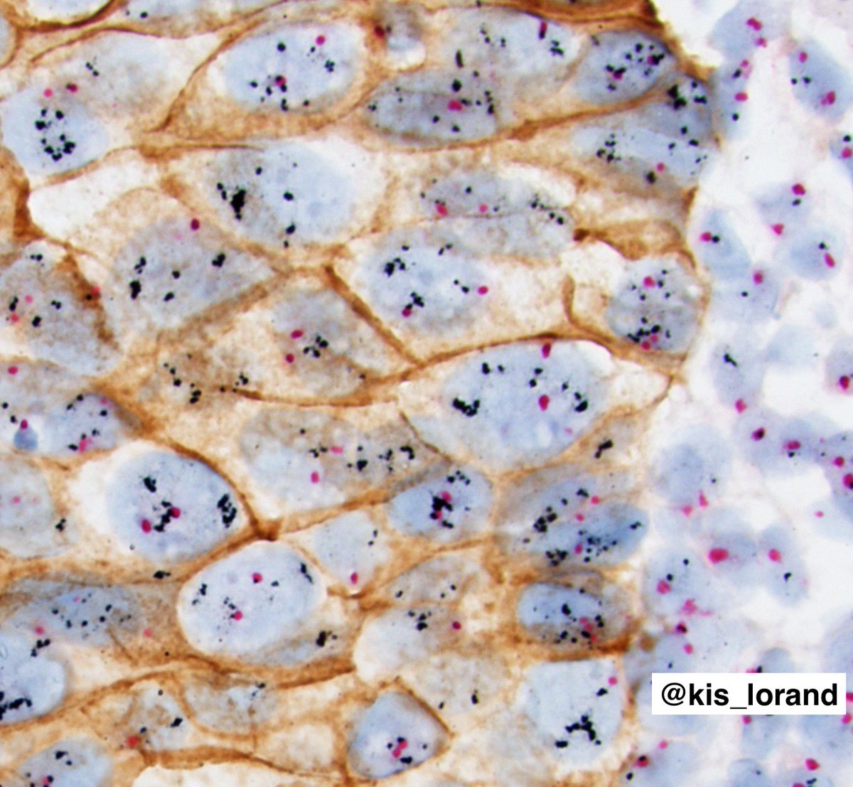
HER2-positive invasive breast carcinoma (NST).
HER2 gene-protein assay: brightfield tricolor assay (combined ICH+ISH) for the visualization of HER2 protein (brown), the HER2 gene (black), and chromosome 17 centromere (CEN17) (red).
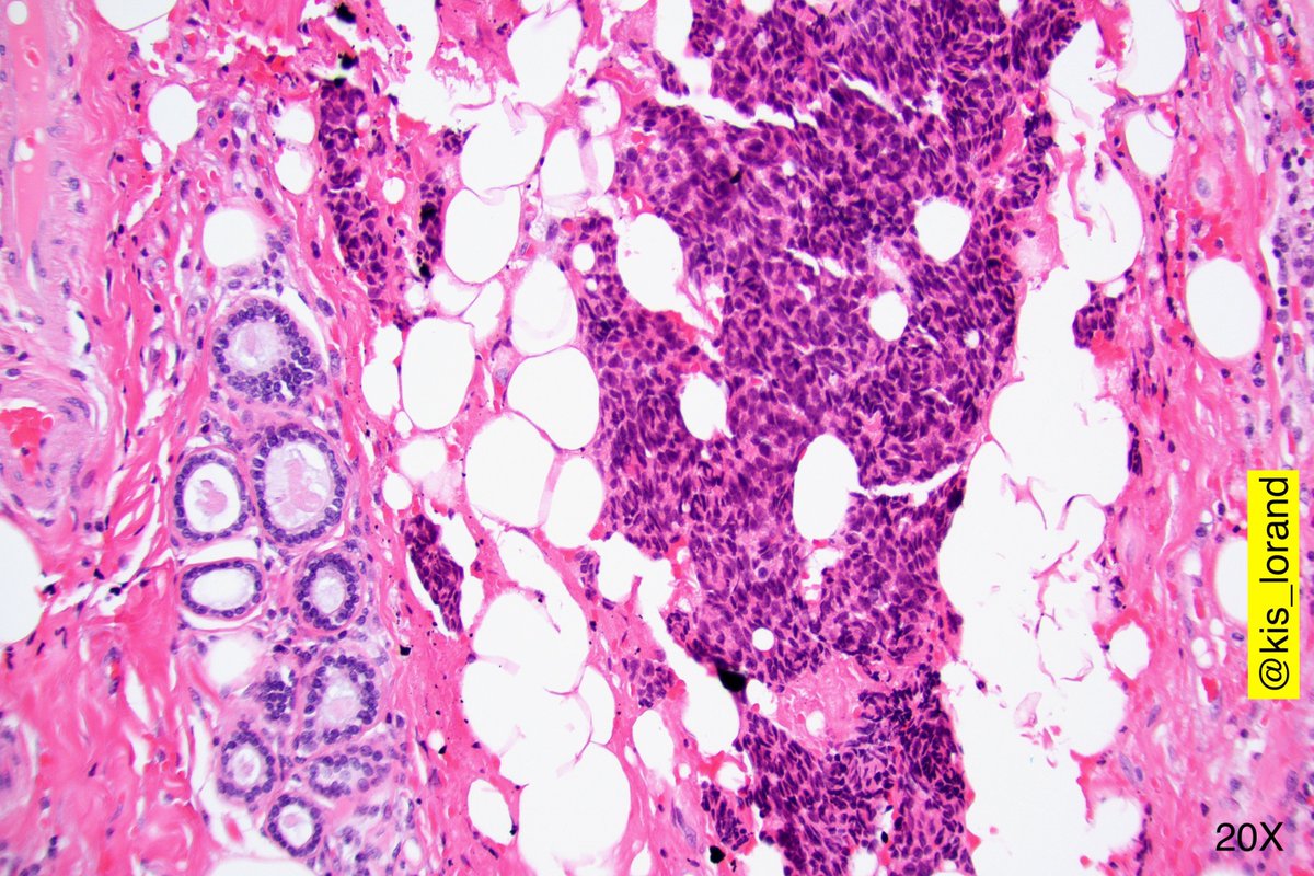
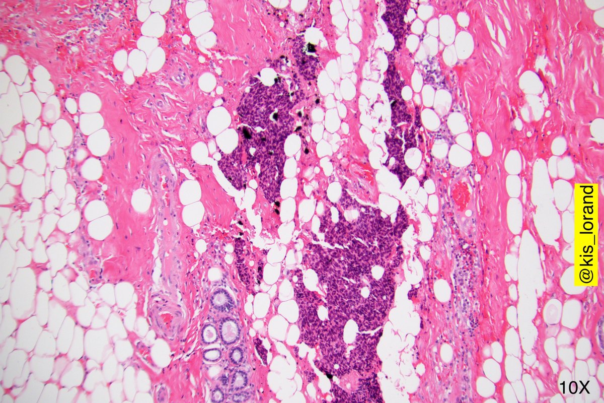
#Breastpath I leave for your toughts this 2 cm spiculated nodule sent as probably Carcinoma .Early 30s.Your favored diagnosis .DDs and IHC request .See second set of pics 😊🙏🙏